Medical Foundations 1
Chapter 5Respiratory Examination
Part 1: Focused History – Based on Chief Complaint (Begin with Open-Ended Questions)
HPI and Assoc. Symptoms
Shortness of Breath (SOB)/ Dyspnea
(the sensation of shortness of breath)
(Also seen in Chapter 6 – Cardiac Exam)
- Onset: When did the shortness of breath start? Was it sudden or gradual onset?
- Palliating/Provoking: Does anything make the shortness of breath better or worse? Any triggers (e.g. smoking, environmental triggers)? Any treatments help (e.g. puffers – such as salbutamol or ipratropium; or oxygen)?
- Focus on Exertion i.e. SOB on Exertion (SOBOE):
- Do you find you become short of breath when walking up a flight of stairs?
- How many flights of stairs? How many blocks can you walk before you need to rest?
- Do you find you are short of breath at rest? – Red Flag Symptom
- Note: Refer to New York Heart Association (NYHA) CHF classifications or MMRC Breathlessness Scale for more info.
- Focus on Position re. Orthopnea (Breathlessness sensation worse when laying down, better sitting up – occurs immediately when lying flat due to increased preload to heart): Is SOB worse when lying down, better when siting up? Are you able to lay flat? How many pillows do you sleep on?
- Focus on Paroxysmal Nocturnal Dyspnea (PND – SOB awakening patient after 1-2 hours of sleep – occurs due to ↑ volume from lymphatic resorption of edema, takes longer than orthopnea): Do you wake up in the middle of the night gasping for air or feeling like you can’t breathe? Does it get better after sitting up?
- Focus on Exertion i.e. SOB on Exertion (SOBOE):
- Severity: On a scale of 0-10, how severe is the shortness of breath?
- Associated Symptoms:
- Chest Pain (e.g. Myocardial Infarction, Pulmonary Embolism, Infection)
- Constitutional Symptoms (Fever, Chills, Night Sweats, Weight Loss e.g. Infection, cancer)
- Orthopnea, Paroxysmal Nocturnal Dyspnea (e.g. Congestive Heart Failure, CHF)
- Stridor (e.g. Large Airway obstruction)
- Cough (e.g. COPD, Asthma, Interstitial Lung Disease, etc.)
- Calf Pain (e.g. Deep Venous Thrombosis/Pulmonary Embolism)
- Edema: Swelling of tissues – commonly dependent e.g. in lower extremities (e.g. CHF)
Cough
- Onset: When did the cough start?
- Acute Cough (<3 weeks) vs. Subacute Cough (3-8 weeks) vs. Chronic Cough (>8 weeks)
- Palliating/Provoking: Does anything make the cough better or worse?
- Quality: Is the cough dry or productive (i.e. sputum)? Sputum: amount? blood? colour?
- Clear: serous, normal unless increased amounts produced
- Frothy pink: pulmonary edema
- Rust-coloured: pneumococcal pneumonia
- Brown/black: chronic pneumonia or bronchitis, lung cancer
- Yellow/green: acute respiratory tract infection, lung abscess, cystic fibrosis
- Red jelly: Klebsiella infection
- Blood (Hemoptysis): tuberculosis, cancer, pulmonary embolism – Red Flag symptom
- Severity: On a scale from 0-10, how severe is the cough?
- Timing/Duration: For how long have you had this cough? Has this ever happened before?
- Associated Symptoms:
- Shortness of breath (Consider Asthma, Chronic Obstructive Pulmonary Disease, Interstitial Lung Disease etc.)
- Worse at night? (Consider congestive heart failure)
- Sour or bitter taste in mouth? (Consider gastroesophageal reflux disease = GERD)
- Recurrent triggers, chest tightness, allergic rhinitis, eczema? (Consider asthma)
- Other Red flag symptoms in Chronic Cough: Fever, Weight Loss, Hoarseness of Voice, Smoker >20 pack-years or Smoker >45 years old
- Note: Angiotensin-Converting Enzyme (ACE) Inhibitors are an antihypertensive medication that may have the side effect of dry cough via ↑ bradykinin. Angiotensin II Receptor Blockers (ARBs) are a similar class of antihypertensive medication which do not have this side-effect.
Wheezing
- Onset: When did the wheezing start?
- Palliating/Provoking: Does anything make the wheezing better or worse? For instance, is it worse at night or during the day? Is it worse with cold exposure (seen in asthma)?
- Severity: On a scale from 0-10, how severe is the wheezing?
- Timing/Duration: For how long have you been wheezing? Has this every happened before?
- Clinical Connection: Parents often misuse the term “wheezing” in infants. As such, there is low reliability between parental report of wheeze & objective clinical assessment of wheeze on auscultation.
Chest Pain or Tightness
(Explored further in Chapter 6 – Cardiac Exam)
- Location: Where is the pain located? Can you point to it? (Angina pain is retro- or sub-sternal)
- Onset: When did the pain start? What were you doing when it started? (e.g. on exertion)
- Palliating/Provoking: Does anything make the pain better or worse?
- Worse with exertion or emotional stress? Relieved promptly by rest or nitroglycerin? (Consider Angina)
- Better or worse in certain positions? (e.g. better leaning forward – consider Pericarditis)
- Worse on inspiration? (With stabbing pain: pleuritic – suggestive of pulmonary embolism, pericarditis, pneumonia, pneumothorax, rib fracture)
- Worse with palpation? (Consider mechanical cause e.g. rib fracture, costochondritis)
- Quality: How would you describe the pain? Sharp? Pressure?
- Does it feel like an elephant is sitting on your chest? (make a fist with your hand and press to sternum – common description for angina)
- Tearing or ripping pain? (consider aortic dissection)
- Is it a tightness, a pressure or a pain? Is it sharp/stabbing? (consider pericarditis)
- Radiation: Does the pain radiate to the arm, jaw, or neck? (Cardiac)
- Note: Classical teaching is that myocardial infarction pain radiates down the left arm; however, pain radiating down right arm or both arms increases likelihood of suspected MI further (LR+ve for pain in L arm = 2.2; R arm = 7.3; Both arms = 9.7)
- Severity: How severe is the pain on a scale from 1-10?
- Timing: Does the pain come and go? How frequently? How long has it been ongoing?
- Chest Pain >30 minutes: Consider myocardial infraction, where target time from door to cath lab is <90 minutes – “time is myocardium”
- Associated Symptoms: Any Shortness of Breath? Nausea or Vomiting? Presyncope? – These can be anginal (chest pain) equivalents (e.g. a patient may present with Nausea as their main symptom for a Myocardial Infarction – be particularly mindful in patients who are elderly, have diabetes, and/or female for more atypical presentations of chest pain)
- Can’t Miss Diagnoses of Acute Chest Pain (PETMAC Mnemonic):
- Pulmonary Embolism: Pleuritic chest pain, tachycardia, SOB, DVT, Immobilization
- Esophageal Rupture: Vomiting, Chest/Epigastric Pain, Anxiety, Recent Instrumentation
- Tension Pneumothorax: Tracheal Deviation, Sudden SOB, Pleuritic Chest Pain, Recent Instrumentation
- Myocardial Infarction: Retrosternal Chest Pain worse with exertion relieved by rest or nitroglycerin, radiating to arm/jaw; Diaphoresis; Hypotension; Anginal Equivalents (Nausea, Dyspnea, Presyncope)
- Aortic Dissection/Rupture: Tearing sensation; upper back pain; Hypertension; BP ∆ > 20mmHg for R vs. L arm
- Cardiac Tamponade: SOB; Beck’s Triad (Hypotension, Jugular Venous Distension, Muffled Heart Sounds)
- Note: Pain and distress can manifest atypically in patients with limited communication and can be difficult to recognize. Nonspecific changes in vital signs, appearance and behavior, including less responsive, more withdrawn or new Behaviors that Challenge (BTC) can indicate pain and distress. Consider tools e.g. Chronic Pain Scale for Nonverbal Adults with Intellectual Disabilities1, or Non-Communicating Adult Pain Checklist2
Constitutional Symptoms
- Any unintended weight loss, fevers, loss of appetite, or night sweats?
Stridor (Pediatrics)
- Any history of choking, aspiration, cyanosis, or wheeze?
- Note: History is most important for foreign body aspiration (FBA); however, stridor is an uncommon presenting sign in FBA – choking & cough more common.
- Is the sound predominantly on breathing in (i.e. during inspiration)?
- Note: This would suggest an extra-thoracic airway obstruction.
- Is the sound harsh or more of a soft, vibrating “snoring” sound?
- Note: Soft associated with epiglottitis (pediatric emergency); harsh → viral upper respiratory tract infection (URTI) symptoms; brassy/barky cough → croup.
Past Medical History (PMHx)
- Respiratory: Chronic Obstructive Pulmonary Disease (COPD); Asthma (What puffers? Can you show me how you use them? (re. inhaler technique); Pneumonias; Lung Cancer; Previous TB diagnosis
- Cardiac: Coronary Artery disease; Cardiovascular Risk Factors – including history of Hypertension, Diabetes, Dyslipidemia, Family History (See below) and Smoking (See below); Heart Failure; Previous Deep Vein Thrombosis (DVT) or Pulmonary Embolism (PE)
Relevant Clinical Scores:
- Well’s Score for PE (Risk of pulmonary embolism based on clinical criteria)
- PERC Rule (Clinically ruling out pulmonary embolism in low-risk patients)
- YEARS Algorithm for PE (Helps rule out PE, validated in pregnant patients with clinically suspected PE)
- CURB-65 (Community-acquired pneumonia mortality, decision re. treat outpatient vs. inpatient vs. ICU)
- Light’s Criteria (Determining exudative vs. transudative pleural effusion)
Past Surgical History (PS Hx)
- History of Chest tube insertion, Lung resection; Lung transplant; Coronary Artery Bypass Graft (CABG); Cardiac Stents
Family Medical History (FMHx)
- Malignancy
- Atopic Triad – (1) Asthma; (2) Atopic Dermatitis (Eczema); (3) Allergic Rhinitis (Hay Fever)
- Cardiovascular Disease (higher risk of MI if 1st degree relative with early MI: Male < 55, Female < 65 years old)
Social Hx
Smoking
- Pack years = packs/day x years smoking
- Strong risk factor for lung malignancy and Chronic Obstructive Pulmonary Disease (COPD)
- >40 pack-year smoking history has LR+ve of 12 for COPD; <20 pack-year has LR+ve of 0.5
- Use of cigarettes? E-cigarettes? Vapes?
- Have you thought about quitting within the next 6 months? How long ago did you quit smoking?
- The benefits of smoking cessation begin with the last cigarette; quit before Age 35, life expectancy same as non-smokers; quit age 35-65, add 5 years to life expectancy; quit age 65-74, add 1 year.
- 5As approach taking a smoking Hx (Explored in Chapter 38 – Motivational Interviewing – IF)
- Strong risk factor for lung malignancy and Chronic Obstructive Pulmonary Disease (COPD)
- Pro-Comp Connection: Regarding patient’s position, do not jump to thinking in terms of “compliance” with recommendations for smoking cessation.
Alcohol
- Increases risk of aspiration
Occupational Hx
- An occupational history is particularly important in a respiratory examination
- Coal miners → coal workers’ pneumoconiosis
- Farmer → hypersensitivity pneumonitis
- Construction worker → asbestosis/mesothelioma
- Mining/foundries → silicosis
Travel Hx
- TB exposure risk
IV Drug Use Hx
- TB exposure risk
Pregnancy
- Estrogen increases risk for Deep Vein Thrombosis / Pulmonary Embolism
Medications Hx, Allergies
Inhalational Therapy (e.g. Asthma, COPD)
- Short-Acting β Agonists (SABA) – e.g. Salbutamol = Ventolin → Rescue inhaler
- Short-Acting Muscarinic Agents (SAMA) – e.g. Ipratropium = Atrovent → Rescue inhaler
- Inhaled Corticosteroids (ICS) – e.g. Fluticasone = Flovent, Budesonide = Pulmicort
- Long-Acting β Agonists (LABA) – e.g. Salmeterol = Serevent, Formoterol = Oxeze
- Long-Acting Muscarinic Agents (LAMA) e.g. Tiotropium = Spiriva, Glycopyrronium = Seebri
- Combination Inhaler (NOTE: “Triple Inhaler Therapy” = ICS + LAMA + LABA)
- LABA/ICS e.g. Advair = Salmeterol + Fluticasone; Symbicort = Formoterol + Budesonide
- LAMA/LABA e.g. Inspiolto =Olodaterol +Tiotropium; Ultibro=Indacaterol +Glycopyrronium
- Patient Education: Ask patient to show how they use it; helpful videos for different inhaler techniques you can review with patient available at Canadian Thoracic Society
Leukotriene Antagonists (LTA)
- LTAs (e.g. Montelukast = Singulair) – used for Asthma management, seasonal allergic rhinitis, and preventing exercise- induced bronchoconstriction
ACE Inhibitors
- ACE inhibitors cause ↑ bradykinin which can lead to non-productive cough
Beta Blockers
- Possible side effect of bronchospasm, particularly in patients with Hx of Asthma or COPD
Oral Contraceptives (OCP)
- Estrogen increases risk for Deep Vein Thrombosis (DVT) and Pulmonary Embolism (PE)
Environmental Triggers
- Associated with Asthma (e.g. smoking, animal dander)
Part 2: Pertinent Notes about the Physical Exam
- Patient Positioning during Respiratory exam: After taking vitals, patient should be seated, arms at side and legs uncrossed
- Draping: In both Respiratory and Cardiac examinations, draping important especially for females. Advise minimal exposure.
Part 3: Specific Maneuvers in the Physical Exam – Say, Setup, Do, Report
Notes on Vitals
Vitals – Always do HR, BP, RR, Temp
In This Exam
AFMC level Pre-clerkship 4 · Clerkship 4
- Check for abnormal respiratory rate, tachycardia and O2 saturation in particular
- CURB-65 score for pneumonia: **1 point each for C**onfusion, **U**rea >7, **R**R ≥30, BP <90/60, Age >65. If CURB-65 Score >2, recommend admit to inpatient.
- Pulsus Paradoxus (drop in BP during inspiration) >25mmHg greatly ↑ likelihood of severe asthma (LR+ 22.6, LR- 0.8) – Differential diagnosis includes cardiac tamponade – see Chapter 6 – Cardiac Exam
Inspection
Work of Breathing
AFMC level Pre-clerkship 4 · Clerkship 4
Say: If you can breathe normally, I will examine the face and torso for signs of work of breathing.
Do:
- Look for signs of increased work of breathing: tripod position, pursed lips, nasal flaring, accessory muscle use (sternocleidomastoid, scalene, trapezius, internal intercostal, or abdominal muscles), paradoxical breathing (Hoover’s sign), tracheal tug (abnormal downward movement of trachea during inspiration), suprasternal indrawing, intercostal indrawing, supraclavicular retraction, grunting (pediatrics).
- Observe if patient can finish complete sentences (not interrupted by work of breathing).
Report: There are no signs of increased work of breathing (comment on all the above out loud as you observe on inspection). You are speaking in complete sentences which is reassuring as well.
Why Check: Important in assessing dyspneic patients and assessing if a patient is in respiratory distress. The inability to speak in complete sentences demonstrates a higher degree of respiratory distress.
Rhythm of Breathing
AFMC level Pre-clerkship 3 · Clerkship 4
Say: If you can breathe normally, I will examine the face and torso for rhythm of breathing.
Do: Look at the rate, rhythm and depth of inspiratory and expiratory phases of breathing. Look for signs of dyspnea (laboured breathing) or apnea (absent breathing) in each cycle. Look for abnormal breathing patterns:
- Cheyne Stokes Breathing: Periodic breathing – deep breathing gradually becoming shallower with a period of apnea subsequent → CHF, ↑ Intracranial Pressure (ICP) Carbon Monoxide Poisoning
- Kussmaul Breathing: Rapid, deep laboured breathing → Metabolic Acidosis (e.g. Diabetic Ketoacidosis)
- Shallow breathing: Inhibited breaths → Pleuritic Chest Pain (e.g. PE, pericarditis, pneumonia, rib fracture)
- Cushing’s Triad: Suggests ↑ Intracranial pressure (ICP) → Irregular respirations, bradycardia, and widened pulse pressure (↑ Systolic BP, ↓ Diastolic BP)
Report: You have a regular rhythm of breathing which is reassuring.
Why Check: Specific breathing patterns and rhythms can help in diagnosing particular etiologies.
Assistive Equipment
Say: I will now inspect for any assistive equipment such as oxygen and IV lines.
Do: Look for any supplemental oxygen; look at any peripheral lines (e.g. IV, Foley i.e. Urinary Catheter) etc.
Report: You are on 2 litres of oxygen through nasal prongs. I don’t notice any other IV lines or catheters.
Why Check: More complete assessment in an inpatient setting
Scars & Medical Devices
AFMC level Pre-clerkship 4 · Clerkship 4
Say: I will now inspect for any signs of scars or medical implants
Do: Look for evidence of surgical scars or medical implants (e.g. pacemaker)
Report: I note no surgical scars or medical implants.
Why Check: Provides evidence for past medical and surgical history
Cachexia/ Sarcopenia
AFMC level Pre-clerkship 3 · Clerkship 4
Say: I will now inspect the torso for muscle and subcutaneous tissue wasting.
Do: Looking for signs of wasting/muscle atrophy, as well as subcutaneous tissue wasting – inspect clavicle (muscle– protrusion/prominence), shoulders (muscle– square look, protruding acromion process), scapula (muscle– bony prominences, depressions), and ribs (muscle, subcutaneous fat–bony prominences, indentation)
Report: I note no signs of cachexia on inspection of the chest
Why Check: Cachexia is a process of fat and muscle wasting; Sarcopenia is specifically a process of muscle wasting. Cachexia is important to assess for, where early intervention (e.g. nutrition, referral to dietitian) is critical. Pulmonary cachexia can occur in advanced lung disease – e.g. in COPD or Interstitial Lung Disease (ILD) such as Idiopathic Pulmonary Fibrosis (IPF) and is associated with accelerated functional decline and increased mortality. This is explored further in Chapter 41 – Adult Palliative Care Assessment (Integration Foundation)
Barrel Chest, Flail Chest
AFMC level Pre-clerkship 4 · Clerkship 4
Say: If you can breathe in and out, I will assess how the rib cage moves for changes in shape.
Do: Look for signs of barrel chest (broad and deep chest), flail chest (section of chest moving in opposite direction of rest of chest), or any bruises of the chest wall.
Report: I note no signs of bruising, barrel chest or flail chest.
Why Check: Barrel chest is associated with hyperinflation (e.g. emphysema re. pink puffers); Flail chest is when rib fractures at two points compromising chest wall rigidity – a life-threatening condition that can alter breathing mechanics, and increase risk of pneumothorax, etc.
Pectus Excavatum & Carinatum
AFMC level Pre-clerkship 4 · Clerkship 4
Say: I will now inspect the chest for sternal and chest wall deformities.
Do: Assess from anterior aspect, looking for any abnormal concave (pectus excavatum) or convex (pectus carinatum) presentation
Report: I note no pectus excavatum or carinatum on inspection.
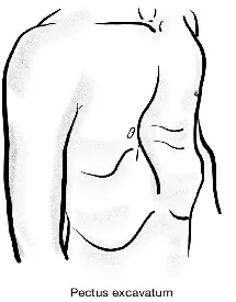
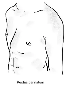
Why Check: Severe pectus excavatum or carinatum can impair both cardiac and respiratory function, painful
Scoliosis & Kyphosis
AFMC level Pre-clerkship 4 · Clerkship 4
Say: I will now inspect the back for spinal deformities.
Do:
- Assess from posterior aspect while patient standing.
- Look for abnormal lordosis (inward curvature), kyphosis (outward curvature) to spine or scoliosis (lateral curvature, common deformity).
- If you suspect scoliosis, have the patient bend forward until their spine is horizontal as this will allow for more accurate assessment of Scoliosis. This maneuver is called the Adam’s Forward Bend Test (Will learn in MF4 Ch 29 – Back Exam).
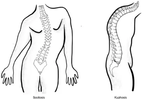
Report: I note no scoliosis or kyphosis on inspection.
What Is Normal? Cervical spine physiologically lordotic; Thoracic spine physiologically kyphotic; Lumbar spine physiologically lordotic; Sacral spine physiologically kyphotic. Excessive spinal curvature is not normal (explored in detail in MF4)
Why Check: Severe scoliosis can lead to reduced lung capacity (When Scoliosis > 50° curvature)
Central Cyanosis
AFMC level Pre-clerkship 4 · Clerkship 4
Say: I will now inspect under the tongue for signs of low oxygen levels. Can you briefly open the mouth, and lift the tongue up?
Do: Inspect under the tongue for central cyanosis
Report: I note no signs of central cyanosis; I note the tongue appears proper size, free in motion, slightly red in colour and with thin white coating layer.
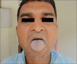
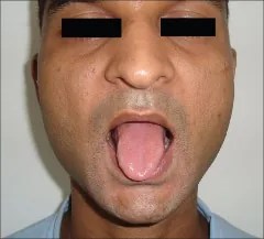
Central Cyanosis4 · Cyanosis Resolved4
Why Check: Central cyanosis indicates poor oxygenation in the lungs (specifically PaO2 <50mmHg given an Hb of 140-160g/L; PaO2 45mmHg if Hb 120 g/L). Images show tongue colouration changes before and after resolution of central cyanosis4.
Peripheral Cyanosis
AFMC level Pre-clerkship 4 · Clerkship 4
Say: I will now inspect the hands for signs of changes in circulation. Can you show the hands with palms up? And now with palms down?
Do: Hold hands lightly and inspect for peripheral cyanosis
Report: I note no signs of peripheral cyanosis.
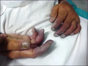
Peripheral Cyanosis5
Why Check: Peripheral cyanosis indicates inadequate circulation, as shown in image affecting digits5.
Clubbing (Schamroth Sign)
AFMC level Pre-clerkship 4 · Clerkship 4
Say: I will now inspect the fingers for signs of low oxygen levels. Can you hold the index finger nails together like this? (See picture, model for patient)
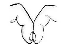
Do: Model with your own hands. Look for a visible gap between the two nail beds.
- If arthritic changes, check for a concave bump between base of nail bed and finger
Report: Schamroth’s sign is negative, with no evidence of clubbing.
Why Check: Associated with chronic hypoxia, cyanotic heart disease, lung cancer, interstitial lung disease
Tar Staining of Fingers
Say: I will now inspect the hands for signs of abnormal staining. Can you show the hands with palms up? And now with palms down?
Do: Look for tar stains on fingers (nicotine)
Report: I do not note any nicotine staining on fingers.
Why Check: If you’re concerned the patient was not being forthright about smoking
Palpation
General
AFMC level Pre-clerkship 3 · Clerkship 4
Say: I will now palpate the chest anteriorly and posteriorly for masses; please tell me if you are in discomfort.
Do: Palpate quickly around the chest area anteriorly and posteriorly; look at patient for signs of discomfort and stop the exam if the patient is in distress.
Report: I note no masses or tenderness in the chest area anteriorly or posteriorly
Why Check: Important general physical examination maneuvers, with potential for assessing mechanical causes of chest pain e.g. rib fractures, costochondritis, etc.
Tracheal Deviation
AFMC level Pre-clerkship 3 · Clerkship 4
Say: I will now palpate the trachea with two fingers for signs of movement with breathing (point on yourself to where you will palpate). I will ask you to breathe in and out. Please tell me if you are in discomfort.
Do: Make V shape with index and middle finger, above sternal notch. Instruct to breathe in, then breathe out. Look at patient for signs of discomfort and stop the exam if the patient is in distress.
Report: The trachea remains midline on inspiration and is mobile in all 4 directions.
Why Check: Tracheal deviation occurs when a pressure differential exists between Left and Right pleural cavities. It will shift away from pathology increasing pressure on one side (unilateral tension pneumothorax or pleural effusion); or towards a pathology decreasing pressure on one side (e.g. atelectasis/lung collapse).
- Can’t Miss Diagnosis: Tension pneumothorax is an emergency where a one-way valve is created, where air can get in but not out, increasing intrapleural pressure. Look for dyspnea, deviated trachea, Jugular Venous Distension, unilateral absent breath sounds, unilateral hyperresonance to percussion, and trauma. A tension pneumothorax requires emergent needle decompression (in 2nd intercostal space at Mid-clavicular line) or chest tube placement (in 5th intercostal space just anterior to mid-axillary line), often at bedside even before imaging can be completed.
Laryngeal Height (Tracheal Descent)
AFMC level Pre-clerkship 3 · Clerkship 4
Say: I will now palpate the thyroid cartilage to measure movement with normal breathing (point on yourself to where you will palpate).
Do: As patient reaches end-expiration, record maximum distance between thyroid cartilage and suprasternal notch. This is the maximum laryngeal height. Look at patient for signs of painful discomfort and stop the exam if the patient is in distress.
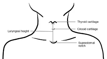
Report: The maximum laryngeal height is more than 4cm, a normal value.
Why Check: Maximum laryngeal height occurs at the end of expiration, while minimum height occurs at the end of inspiration. A maximum laryngeal height <4cm indicates hyperinflation and significant obstruction, affecting this rise in height during expiration. Important finding for obstructive airway disease.
Bilateral Chest Expansion
AFMC level Pre-clerkship 3 · Clerkship 4
(Also seen in Ch 29 – Back Exam – MF4)
Say: If you can sit up and turn to the side of the exam table, I will palpate the back for chest expansion with breathing.
Do:
- Place hands at 10th rib, press together.
- Ask patient to breathe out fully, then position hands.
- Have patient take a deep breath.
- Hands should move equidistant on each side with ≥4cm deviation bilaterally
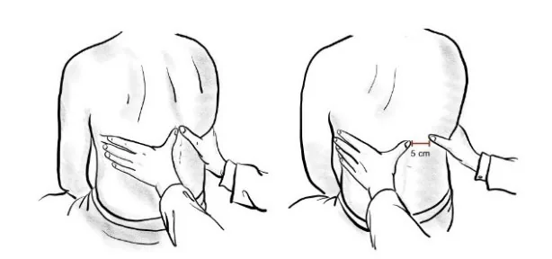
Alternative Method: Measure horizontally with measuring tape around patient at level of the xiphisternum. Note: takes longer, can comment that you would measure only if there was time in an OSCE scenario.
Report: The chest shows chest expansion of 4 cm with bilateral symmetry, a normal value.
Why Check: Bilateral ↓ expansion can occur in asthma and COPD, as well as Thoracic Scoliosis and Ankylosing Spondylitis (See Chapter 29 – Back Exam – MF4); unilateral ↓ expansion seen in pleural effusion (LR+ 8.1, LR- 0.29), pneumothorax, etc. on the affected side.
Tactile Fremitus
AFMC level Pre-clerkship 3 · Clerkship 4
Say: I will now palpate the back for air entry on each side. When you feel my hands, say “toy boat.”
Do:
- Patient crosses their arms to displace scapulae. Place medial edges of hands horizontally on the back.
- Start superiorly and move inferiorly. At each stage, assess vibration level and any differences bilaterally.
- Also, switch anteriorly and continue, to inspect upper lobes bilaterally.
Report: The chest shows bilateral resonance throughout all lung fields.
Why Check: Vibrations increased over areas of consolidation, reduced over areas of pleural effusion (LR+ 5.7, LR- 0.21) or pneumothorax (consider the mediums the sound produced is being conducted through)
Percussion
Intercostal
AFMC level Pre-clerkship 3 · Clerkship 4
Say: I will assess for dull sounds along the back & chest. Please tell me if you are in discomfort.
Setup: Patient crosses their arms to displace scapulae.
Do: Using wrist drop technique, move side to side beginning superiorly and progressing inferiorly to assess for bilateral resonance. Look at patient for signs of discomfort and stop the exam if the patient is in distress.
Report: The chest shows equal resonance bilaterally on percussion.
Why Check: Dullness to percussion increases likelihood of suspected consolidation (LR+ 3.0 for pneumonia) or pleural effusion (LR+ 8.7). Dullness at lung bases points to pulmonary edema. Hyper-resonance on percussion increases likelihood of suspected chronic airflow obstruction (i.e. hyperinflation, LR+ 5.1) or Pneumothorax.
Diaphragmatic Excursion
AFMC level Pre-clerkship 3 · Clerkship 4
Say: I will assess how the diaphragm moves up and down along the back with breathing.
Do:
- Start below the level of the scapula on one side.
- Patient should expire and hold while you percuss down until transition from resonant to dull.
- Patient can now inspire and hold while you percuss further down until dull again.
- Measure distance and repeat on other side.
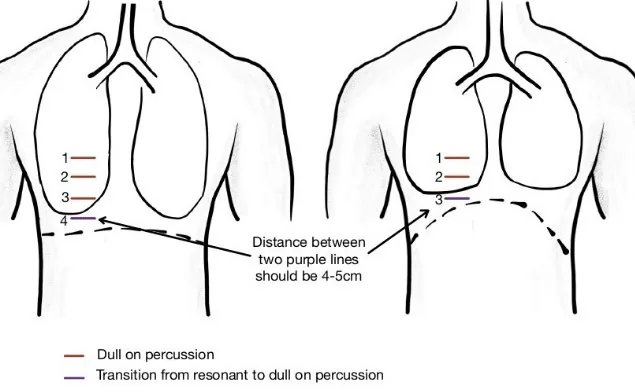
What is Normal: 4-5 cm
Report: Diaphragmatic excursion is 4-5cm bilaterally.
Why Check: Unilateral changes can indicate a pneumothorax or pleural effusion re filling up pleural space. Alternate method: Reverse order i.e. percuss while patient inspires first, then percuss while patient expires.
Auscultation
Listening to Breathing
- General- [3, 4]
- Stridor - [4, 4]
- Pleural Rub- [1, 4]
- Stridor (Peds) - [1, 3]
- Wheeze (Infant) - [1, 2]
Say: I will now listen to the chest and back – breathe in and out each time you feel the stethoscope. Please pause if you become light-headed.
Setup: Patient crosses arms to displace scapulae.
Do: Hold stethoscope still when listening (don’t want to only hear rustling of moving it around).
Report: I note air entry bilaterally with vesicular breath sounds and no adventitious sounds.
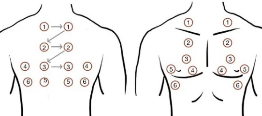
What is Normal? Normal auscultation depends on listening location
- (1) Trachea: Bronchial – harsh, high-pitched, heard through full inspiration and expiration
- (2) Sternum: Bronchovesicular – less harsh, heard through full inspiration, short and softer on expiration
- (3) Everywhere else: Vesicular - gentle, low-pitched, quiet on inspiration and nearly silent on expiration
Why Check: Assessing for Adventitious (Abnormal) Sounds, including:
| Sound | Respiratory Phase | Description | Significance |
|---|---|---|---|
| Fine Crackles | Inspiration | Discontinuous, soft, high-pitched, more per breath, popping sound. | Distal lower airway process (e.g. pulmonary fibrosis, congestive heart failure) |
| Coarse Crackles | Inspiration alone; or Inspiration + early expiration | Discontinuous, loud, low-pitched, fewer per breath, bubbling sound. | Proximal lower airway process (e.g. pneumonia, pulmonary edema, adult respiratory distress syndrome) |
| Stridor | Inspiration | Loud, musical sound, constant pitch, louder over neck. | Upper airway obstruction (e.g. croup, foreign body aspiration, epiglottitis, bacterial tracheitis, retropharyngeal abscess, peritonsillar abscess) |
| Pleural Rub | Expiration alone; or Expiration + Inspiration | Loud, grating or rubbing sound; can be crackling. | Pleural inflammation (e.g. pleural effusion, pulmonary embolism, pleuritis) |
| Wheeze | Expiration alone; or Expiration + Inspiration | Continuous, high-pitched, musical sound. | ↓ Distal lower airway diameter (e.g. asthma, chronic obstructive pulmonary disease) |
| Rhonchus | Expiration alone; or Expiration + Inspiration | Continuous, low-pitched, coarse sound; may clear after cough. | ↓ Proximal lower airway diameter by mucous secretions, lesions or foreign bodies (e.g. pneumonia, cystic fibrosis) |
-
Bronchial breath sounds over lung (normal over trachea) points to consolidation
-
Diminished breath sounds greatly increases likelihood for suspected pleural fluid (LR+ 18.6)
-
Early Crackles increases likelihood of suspected obstructive disease (LR+ 14.6), and particularly for severe obstruction in patients with chronic airflow limitation (LR+ 20.8; LR- 0.1)
-
Stridor: Sensitivity of 80.4% & specificity of 59.5% for Foreign Body Aspiration6. May be the earliest sign worrisome for airway compromise.
-
Note - Pediatrics: Wheezing in Infants: Respiratory exam of wheezing in infants shows good inter-rater reliability, but respiratory status overall is most reliable.7
-
Issue: Adventitious sounds on auscultation is “the most ambiguous and confusing nomenclature in all of physical diagnosis” where clinicians use diverse terms to describe similar sounds8.
-
Modification for Patients with Limited Mobility or Supine Patients: For auscultation of the lungs, auscultate anteriorly, then roll onto a lateral decubitus position to auscultate lung bases posteriorly.
-
Modification for Patients with Obesity: Auscultate directly over exposed skin, to ensure breath sounds can be appreciated.
Consolidation Testing (Egophony, Whispered Pectoriloquy, Bronchophony
AFMC level Pre-clerkship 1 · Clerkship 3
Part 1 – Egophony:
- Say: When you feel the stethoscope, please say “EEEE” while I listen with the stethoscope.
- Do: Place stethoscope in same locations as general auscultation. Hearing change to AAAA is abnormal.
Part 2 – Whispered Pectoriloquy:
- Say: When you feel the stethoscope, please whisper “1,2,3,4” while I listen with the stethoscope.
- Do: Place stethoscope in same locations as general auscultation. Hearing the whisper is abnormal.
Part 3 – Bronchophony:
- Say: When you feel the stethoscope, please say “toy boat” in a normal tone of voice while I listen with the stethoscope.
- Do: Place stethoscope in same locations as general auscultation. Hearing loud(er) voice is abnormal.
Report: I note evidence of consolidation. If positive – specify which lobe(s) you heard abnormal findings in.
Why Check: Check if you suspect consolidation in a particular lobe from tactile fremitus, percussion, etc. Though uncommonly used in clinical practice, Egophony does moderately increase the likelihood of suspected pneumonia (LR+ 4.1). Reduced vocal resonance (bronchophony) increases the likelihood of pleural effusion (LR+ 6.5, LR- 0.27).
Forced Expiratory Time (FET)
AFMC level Pre-clerkship 1 · Clerkship 3
Say: I will now listen to the trachea (point on yourself to where you will auscultate) with maximum effort breathing. Please take in a deep breath as much as possible, and then exhale as fast as possible with the mouth wide open.
Do:
- Place bell of stethoscope over trachea. Listen to a cycle of normal breathing. Monitor your clock
- Listen until end inspiration. Start recording time at this point.
- Patient then exhales with an open mouth as quickly as possible. Once airflow is inaudible, this is the end point of forced expiratory time.
Report: The recorded forced expiratory time is less than 9 seconds, a normal value.
Why Check: Forced Expiratory Time (FET) is a simple, inexpensive, sensitive and fairly reproducible test of airway obstruction (defined as FEV1/FVC < 70%). FET >9 seconds increases the likelihood for suspected Obstructive Airway Disease (e.g. COPD) with LR+ 6.7. In practice, pulmonary function testing (e.g. spirometry) is commonly used.
Helpful Stethopedia Videos to Review
- Video 1 – Respiratory Exam OSCE Style (Link: https://videopress.com/v/ZMrJEAXn)
- Video 2 – Respiratory Exam Special Tests (Link: https://videopress.com/v/PBR0adsa)
- Video 3 – Auscultation (Link: https://videopress.com/v/610xm8AP)
- Video 4 – Percussion (Link: https://videopress.com/v/Igbh9peS)
Part 4: Relevant Likelihood Ratios – from Rational Clinical Examination JAMA series
Clinical Question 1: Does this patient have Pneumonia?9
| Procedure | LR+ (95% CI) | LR- (95% CI) |
|---|---|---|
| Egophony | 4.1 (2.1-7.8) (about +15%) | 0.9 (0.9-1.0) (no change: 95% CI includes 1) |
| Cachexia | 4.0 (1.7-9.6) (about +15%) | 0.9 (0.8-1.0) (no change: 95% CI includes 1) |
| Bronchial breath sounds | 3.3 (2.0-5.6) (about +15%) | 0.9 (0.8-1.0) (no change: 95% CI includes 1) |
| Percussion dullness | 3.0 (1.7-5.2) (about +15%) | 0.9 (0.8-1.0) (no change: 95% CI includes 1) |
| Diminished breath sounds | 2.3 (1.9-2.8) (about +15%) | 0.8 (0.7-0.9) (about −15%) |
| Absence of rhinorrhea | 2.2 (1.5-3.2) (about +15%) | 0.8 (0.7-0.9) (about −15%) |
| Temperature >37.8°C | 2.0 (1.5-2.6) (about +15%) | 0.7 (0.6-0.8) (about −15%) |
| RR >28/min | 2.0 (1.4-2.8) (about +15%) | 0.8 (0.7-0.9) (about −15%) |
| Abnormal mental status | 1.9 (1.2-3.0) (about +15%) | 0.9 (0.9-1.0) (no change: 95% CI includes 1) |
| Absence of sore throat | 1.8 (1.3-2.5) (about +15%) | 0.7 (0.6-0.9) (about −15%) |
| Crackles | 1.8 (1.2-2.7) (about +15%) | 0.8 (0.7-0.9) (about −15%) |
| HR >100 beats/min | 1.6 (1.4-1.7) (about +15%) | 0.8 (0.7-0.9) (about −15%) |
| Wheezes | 0.8 (0.7-0.9) (about −15%) | 1.1 (1.0-1.1) (no change: 95% CI includes 1) |
| Heckerling score, 0 or 1 findings | 0.3 (0.2-0.4) (about −30%) | N/A |
| Heckerling score, 2 or 3 findings | 1.0 (0.9-1.2) (no change: 95% CI includes 1) | N/A |
| Heckerling score, 4 or 5 findings | 8.2 (5.8-11.5) (about +30%) | N/A |
Heckerling Score (referred to above)6:
- 1 Point each for: Temperature >37.8°C, HR >100 beats/minute, Crackles, Decreased breath sounds, and Absence of Asthma
Clinical Question 2: Does this patient have Severe Asthma?9
*Note: For description of Pulsus Paradoxus maneuver, see Chapter 6 – Cardiac and Hematological Examination
| Procedure | LR+ (95% CI) | LR- (95% CI) |
|---|---|---|
| Pulsus Paradoxus > 10 mmHg | 2.7 (1.7-4.3) (about +15%) | 0.5 (0.4-0.7) (about −15%) |
| Pulsus Paradoxus > 20 mmHg | 8.2 (1.7-40.3) (about +30%) | 0.8 (0.7-0.9) (about −15%) |
| Pulsus Paradoxus >25 mmHg | 22.6 (1.4-363.2) (about +45%) | 0.8 (0.8-0.9) (about −15%) |
Clinical Question 3: Does this patient have a significant Deoxyhemoglobin Level (>2.38 g/dL)?9
| Procedure | LR+ (95% CI) | LR- (95% CI) |
|---|---|---|
| Central Cyanosis | 7.4 (1.5-36.8) (about +30%) | 0.2 (0.1-0.5) (about −30%) |
Clinical Question 4: Does this patient with no prior diagnosis have Obstructive Airway Disease (FEV1/FVC < 70%)?10
| Procedure | LR+ (95% CI) | LR- (95% CI) |
|---|---|---|
| Wheezing | 4.4 (1.6-12) (about +15%) | 0.88 (0.84-0.92) (about −15%) |
| Maximum Laryngeal Height < 4cm | 4.2 (2.3-7.9) (about +15%) | 0.7 (0.5-0.9) (about −15%) |
| Decreased breath sounds | 2.6 (1.9-3.6) (about +15%) | 0.66 (0.49-0.69) (about −15%) |
| Forced Expiratory Time >9s | 6.7 (2.1-21) (about +30%) | N/A |
| Forced Expiratory Time 6-9 s | 1.8 (0.77-4.0) (no change: 95% CI includes 1) | N/A |
| Forced Expiratory Time <6s | 0.6 (0.5-0.8) (about −15%) | N/A |
| Forced Expiratory Time >6s and patient >60yo | 3.4 (2.2-5.2) (about +15%) | N/A |
| Forced Expiratory Time >6s and patient <60yo | 2.1 (1.3-3.5) (about +15%) | N/A |
| Forced Expiratory Time <6s and patient >60 yo | 0.33 (0.23-0.47) (about −30%) | N/A |
| Forced Expiratory Time <6s and patient <60yo | 0.57 (0.34-0.95) (about −15%) | N/A |
| Smoking >40 Pack Years | 12 (2.7-50) (about +45%) | N/A |
| Smoking 20-40 Pack Years | 0.8 (0.4-1.6) (no change: 95% CI includes 1) | N/A |
| Smoking <20 Pack Years | 0.5 (0.3-0.9) (about −15%) | N/A |
| Overall Clinical Prediction: Moderate-severe disease | 5.6 (3.1-10) (about +30%) | N/A |
| Overall Clinical Prediction: Mild disease | 2.3 (0.55-9.7) (no change: 95% CI includes 1) | N/A |
| Overall Clinical Prediction: No disease | 0.59 (0.51-0.68) (about −15%) | N/A |
Clinical Question 5: Does this patient have a Pulmonary Embolism?9
| Procedure | LR+ (95% CI) | LR- (95% CI) |
|---|---|---|
| Sudden dyspnea | 2.4 (2.0-2.9) (about +15%) | 0.3 (0.2-0.3) (about −30%) |
| Unilateral calf pain or swelling | 2.3 (1.8-3.0) (about +15%) | 0.9 (0.8-1.0) (no change: 95% CI includes 1) |
| Syncope | 2.0 (1.6-2.5) (about +15%) | 0.9 (0.8-1.0) (no change: 95% CI includes 1) |
| RR >30/min | 2.0 (1.5-2.8) (about +15%) | 0.9 (0.8-0.9) (about −15%) |
| Hemoptysis | 2.0 (1.4-2.7) (about +15%) | 1.0 (0.9-1.0) (no change: 95% CI includes 1) |
| Systolic BP < 100 mmHg | 1.9 (1.1-3.0) (about +15%) | 1.0 (0.9-1.0) (no change: 95% CI includes 1) |
| New gallop (S3 or S4) | 2.7 (1.0-7.0) (no change: 95% CI includes 1) | 0.8 (0.6-1.0) (no change: 95% CI includes 1) |
| Pleural friction rub | 1.5 (0.6-3.8) (no change: 95% CI includes 1) | 1.0 (0.8-1.1) (no change: 95% CI includes 1) |
| Accessory muscle use | 1.5 (0.6-3.6) (no change: 95% CI includes 1) | 0.9 (0.8-1.1) (no change: 95% CI includes 1) |
| Hypoxemia (PaO2 <80 mmHg) | 1.2 (1.0-1.4) (no change: 95% CI includes 1) | 0.6 (0.3-1.2) (no change: 95% CI includes 1) |
| Pulse >100/min | 1.2 (0.9-1.5) (no change: 95% CI includes 1) | 0.9 (0.8-1.1) (no change: 95% CI includes 1) |
| Crackles | 1.2 (0.8-1.7) (no change: 95% CI includes 1) | 0.8 (0.5-1.4) (no change: 95% CI includes 1) |
| Left parasternal heave | 1.2 (0.1-29.9) (no change: 95% CI includes 1) | 1.0 (1.0-1.0) (no change: 95% CI includes 1) |
| Increased A-a gradient | 1.1 (1.0-1.2) (no change: 95% CI includes 1) | 0.3 (0-2.4) (no change: 95% CI includes 1) |
| Loud P2 | 1.1 (0.4-3.1) (no change: 95% CI includes 1) | 1.0 (0.8-1.2) (no change: 95% CI includes 1) |
| Cyanosis | 1.0 (0.1-8.3) (no change: 95% CI includes 1) | 1.0 (0.9-1.1) (no change: 95% CI includes 1) |
| Chest wall tenderness | 0.8 (0.6-1.1) (no change: 95% CI includes 1) | 1.1 (1.0-1.1) (no change: 95% CI includes 1) |
| Elevated neck veins | 0.8 (0.1-6.3) (no change: 95% CI includes 1) | 1.0 (0.9-1.1) (no change: 95% CI includes 1) |
| Temp >38C | 0.4 (0.3-0.7) (about −30%) | 1.1 (1.0-1.2) (no change: 95% CI includes 1) |
| Pulse rate <90/min | 0.3 (0.1-0.8) (about −30%) | 1.8 (1.3-2.5) (about +15%) |
| Wheezes | 0.2 (0.1-0.4) (about −30%) | 1.1 (1.1-1.1) (about +15%) |
| Orthopnea | 0.1 (0-0.2) (about −45%) | 1.1 (1.1-1.1) (about +15%) |
| Wells Score, high probability, 7 or more points | 5.0 (2.5-10) (about +15%) | N/A |
| Wells score, moderate probability, 2-6 points | 1.7 (1.5-2.0) (about +15%) | N/A |
| Wells score, low probability, 0-1 points | 0.2 (0.1-0.4) (about −30%) | N/A |
Wells Score for Pulmonary Embolism9:
| Finding | Score if Present |
|---|---|
| Clinical signs and symptoms of Deep Vein Thrombosis (DVT) | +3 |
| Pulmonary Embolism (PE) #1 diagnosis OR equally likely | +3 |
| Heart Rate >100 beats/minute | +1.5 |
| Immobilization at least 3 days OR surgery in previous 4 weeks | +1.5 |
| Previous, objectively diagnosed PE or DVT | +1.5 |
| Hemoptysis | +1 |
| Malignancy with treatment within 6 months or palliative | +1 |
Clinical Question 6: What is the potential clinical significance of an Abnormal Breathing Patterns?9
| Procedure | LR+ (95% CI) | LR- (95% CI) |
|---|---|---|
| Absent Accessory Muscle Use, detecting normal diaphragmatic stretch (patients with COPD acute exacerbation) | 4.4 (1.7-11.8) (about +15%) | 0.2 (0-2.6) (no change: 95% CI includes 1) |
| Paradoxical abdominal movements, predicting diaphragm weakness (patients with dyspnea & neuromuscular disease) | 3.2 (1.7-5.9) (about +15%) | 0.1 (0-1.1) (no change: 95% CI includes 1) |
| Asynchronous Breathing, predicting hospital death or intubation (patients with chronic airflow obstruction with acute respiratory symptoms) | 3.2 (1.3-7.8) (about +15%) | 0.5 (0.2-1.0) (no change: 95% CI includes 1) |
| Orthopnea, detecting low LV Ejection Fraction (patients with known COPD) | 2.7 (1.5-4.9) (about +15%) | 0.04 (0-0.7) (about −45%) |
Clinical Question 7: What is the potential clinical significance of Clubbing?11
Key Idea: Clubbing is a portent of serious disease, therefore worthy of examination. That said, inter-observer agreement is only fair to moderate, and accuracy of techniques to detect clubbing is not well established. For example, using the Schamroth sign is not a validated evidence-based maneuver, but is a quick bedside test for clubbing. Instead, here is the prevalence of Hyponychial Angle Exceeding Upper Limit of Normal in various conditions:
| Condition | Prevalence, % (95% CI) |
|---|---|
| Pneumonia | 23 (11-36) |
| HIV disease | 16 (0-32) |
| Cirrhosis | 16 (0-32) |
| COPD | 15 (6-23) |
| Chronic Hepatitis | 14 (0-29) |
| Bronchogenic Carcinoma | 12 (0-27) |
| Pulmonary Hypertension | 10 (0-20) |
| Acquired Valvular Heart Disease | 9 (3-15) |
| Congestive Heart Failure | 9 (4-15) |
| Ischemic Heart Disease | 9 (5-14) |
| Solid Tumor Malignancy | 6 (1-11) |
Clinical Question 8: What is the potential clinical significance of Percussion of the Chest?9
| Procedure | LR+ (95% CI) | LR- (95% CI) |
|---|---|---|
| Percussion Dullness, detecting pneumonia (patients with fever & cough) | 3.0 (1.7-5.2) (about +15%) | 0.9 (0.8-1.0) (no change: 95% CI includes 1) |
| Percussion Dullness, detecting any abnormality on CXR | 3.0 (1.4-6.3) (about +15%) | 0.9 (0.9-1.0) (no change: 95% CI includes 1) |
| Hyper-resonance, detecting Chronic airflow obstruction | 5.1 (1.7-15.6) (about +30%) | 0.7 (0.5-1.0) (no change: 95% CI includes 1) |
| Diaphragm excursion <2cm, detecting Chronic airflow obstruction | 5.3 (0.8-35.0) (no change: 95% CI includes 1) | 0.9 (0.7-1.1) (no change: 95% CI includes 1) |
| Auscultatory dullness, detecting any abnormality on CXR | 1.7 (1.0-3.0) (no change: 95% CI includes 1) | 0.8 (0.6-1.1) (no change: 95% CI includes 1) |
| Auscultatory dullness, detecting pleural fluid | 18.6 (9.8-35.2) (about +45%) | 0.04 (0-0.1) (about −45%) |
Clinical Question 9: What is the potential clinical significance of Auscultation of the Lung Fields?9
| Procedure | LR+ (95% CI) | LR- (95% CI) |
|---|---|---|
| Crackles (Pulmonary fibrosis in asbestos workers) | 5.9 (2.0-17.2) (about +30%) | 0.2 (0.1-0.5) (about −30%) |
| Crackles (Left atrial pressure elevated >12mmHg in patients with cardiomyopathy) | 3.4 (1.6-7.2) (about +15%) | 0.7 (0.6-1.0) (no change: 95% CI includes 1) |
| Crackles (Myocardial infarction in patients with chest pain) | 2.1 (1.6-2.8) (about +15%) | 0.8 (0.7-1.0) (no change: 95% CI includes 1) |
| Crackles (Pneumonia in patients with cough & fever) | 1.8 (1.2-2.7) (about +15%) | 0.8 (0.7-0.9) (about −15%) |
| Crackles, Early (Severe obstructive disease in patients with chronic airflow limitation) | 20.8 (3.0-142.2) (about +45%) | 0.1 (0-0.4) (about −45%) |
| Crackles, Early (Obstructive disease in patients with crackles) | 14.6 (3.0-70) (about +45%) | 0.4 (0.1-1.4) (no change: 95% CI includes 1) |
| Wheeze, Unforced (Obstructive disease) | 2.8 (1.5-5.0) (about +15%) | 0.8 (0.7-0.9) (about −15%) |
| Wheeze, with Methacholine Challenge (Asthma) | 6.0 (1.5-24.3) (about +30%) | 0.6 (0.4-0.9) (about −15%) |
| Diminished breath sounds (Pleural effusions in mechanically ventilated patients) | 4.3 (2.8-6.5) (about +15%) | 0.6 (0.5-0.8) (about −15%) |
| Diminished breath sounds (Asthma during methacholine challenge) | 4.2 (1.9-9.5) (about +15%) | 0.3 (0.1-0.6) (about −30%) |
| Diminished breath sounds (Pneumonia in patients with cough & fever) | 2.3 (1.9-2.8) (about +15%) | 0.8 (0.7-0.9) (about −15%) |
| Bronchial breath sounds (Pneumonia in patients with cough & fever) | 3.3 (2.0-5.6) (about +15%) | 0.9 (0.8-1.0) (no change: 95% CI includes 1) |
| Egophony (Pneumonia in patients with cough & fever) | 4.1 (2.1-7.8) (about +15%) | 0.9 (0.9-1.0) (no change: 95% CI includes 1) |
Clinical Question 10: Does this patient have a Pleural Effusion?12
| Finding | Sensitivity (95% CI) | Specificity (95% CI) | LR+ (95% CI) | LR- (95% CI) |
|---|---|---|---|---|
| Dullness to Conventional percussion (Pooled) | 73 (61-82) | 91 (88-93) | 8.7 (2.2-33.8) (about +30%) | 0.31 (0.03-3.3) (no change: 95% CI includes 1) |
| Asymmetric Chest Expansion | 74 (60-85) | 91 (86-94) | 8.1 (5.2-12.7) (about +30%) | 0.29 (0.19-0.45) (about −30%) |
| Auscultatory Percussion (Pooled) | 77 (71-83) | 92 (89-94) | 7.7 (2.4-25.1) (about +30%) | 0.27 (0.07-1.0) (no change: 95% CI includes 1) |
| Reduced Vocal Resonance (Bronchophony) | 76 (63-87) | 88 (83-92) | 6.5 (4.4-9.6) (about +30%) | 0.27 (0.17-0.43) (about −30%) |
| Reduced Tactile Vocal Fremitus | 82 (70-91) | 86 (80-90) | 5.7 (4.0-8.0) (about +30%) | 0.21 (0.12-0.37) (about −30%) |
| Diminished Breath Sounds – Study 2 | 88 (76-95) | 83 (77-88) | 5.2 (3.8-7.1) (about +30%) | 0.15 (0.07-0.30) (about −30%) |
| Diminished Breath Sounds –Study 1 | 42 (33-53) | 90 (86-93) | 4.3 (2.8-6.6) (about +15%) | 0.64 (0.54-0.76) (about −15%) |
| Pleural Friction Rub | 5.3 (1.1-14.6) | 99 (96-100) | 3.9 (0.80-18.7) (no change: 95% CI includes 1) | 0.96 (0.90-1.0) (no change: 95% CI includes 1) |
| Crackles | 56 (42-69) | 62 (55-68) | 1.5 (1.1-2.0) (about +15%) | 0.71 (0.52-0.97) (about −15%) |
References
-
pediatric-pain.ca. Accessed March 15, 2019. http://pediatric-pain.ca/wp-content/uploads/2013/04/CPSNAID.pdf ↩
-
Lotan M, et al. Measurement properties of the Non-Communicating Adult Pain Checklist (NCAPC): a pain scale for adults with intellectual and developmental disabilities, scored in a clinical setting. Res Dev Disabil. 2010;31:367-75. ↩
-
Recommendations. Choosing Wisely Canada. Accessed August 1, 2022. https://choosingwiselycanada.org/recommendations/#list ↩ ↩2
-
Das J, Katyal A, Naunwaar D. Dapsone-induced methemoglobinemia in a patient of leprosy. Indian J Dermatol. 2015;60(1):108. ↩ ↩2 ↩3
-
Tripathy S, Rath B. Symmetric peripheral gangrene: catch it early! J Emerg Trauma Shock. 2010;3(2):189-190. ↩ ↩2
-
Even L, Heno N, Talmon Y, Samet E, Zonis Z, Kugelman A. Diagnostic evaluation of foreign body aspiration in children: a prospective study. J Pediatr Surg. 2005;40(7):1122-1127. ↩ ↩2
-
Stevens MW, et al. Interrater agreement in the clinical evaluation of acute pediatric asthma. J Asthma. 2003;40(3):311-315. ↩
-
McGee S. Evidence Based Physical Diagnosis. 4th ed. Elsevier; 2017:268. ↩
-
McGee S. Evidence Based Physical Diagnosis. Saunders; 2012:806-819. ↩ ↩2 ↩3 ↩4 ↩5 ↩6 ↩7 ↩8
-
Does the clinical examination predict airflow limitation update. In: Rational Clinical Examination Series. Chapter 12. 2009:160. ↩
-
Does this patient have clubbing – update. In: Rational Clinical Examination Series. Chapter 14. 2009:172. ↩
-
Wong CL, et al. Does this patient have a pleural effusion? JAMA. 2009;301(3):309-317. ↩