Medical Foundations 4
Chapter 28Ankle and Foot Examination
Part 1: Focused History – Based on Chief Complaint (Begin with Open-Ended Questions)
HPI
Pain Assessment
- Onset:
- Acute onset of pain, disability immediate with injury (e.g. acute trauma, concern for fracture) (see Mechanism of Injury below).
- Delayed onset of pain, able to weight-bear immediately after injury (e.g. ligamentous injury).
- Acute onset with no associated injury (e.g. septic arthritis).
- Insidious onset gradually worsening over time (e.g. overuse injury, osteoarthritis).
- Pediatric patients: Is the patient in an active growth spurt → increased risk of overuse injuries
- Provoking:
- Aggravating postures: Ankle dorsiflexion or plantarflexion; foot inversion or eversion
- Aggravating activities: Weightbearing (Any weightbearing vs. forefoot-specific); heel strike; walking; running; jumping up vs. landing from jump; worsens with activity
- Footwear: New? Tight vs. loose-fitting? Soft vs. firm support? High heels? (Alters biomechanics)
- Palliating:
- Relieving postures and activities: Non-weightbearing; improves with activity (e.g. Plantar fasciitis)
- Previous treatment: POLICE (Protect, Optimally Load, Ice, Compress, Elevate)? Physio? Analgesics?
- Rehab Program: Ask patient to demonstrate exercises performed in rehab program (e.g. ROM exercises, strengthening exercises, proprioceptive retraining). Inadequate rehab program can contribute to persistent ankle pain (>3-6 weeks), ↓ ROM, limiting function with ↑ activity.
- Supportive measures: Use of protective tape, ankle braces, cast? Supportive footwear (e.g. orthotics)?
- Investigations: Type (x-rays, Ultrasound, MRI, etc.) and results (if available)
- Quality: Deep ache, sharp vs. dull? – Ask patient what words they would use
- Region: Poorly localized (e.g. Tarsal Tunnel Syndrome) vs. well-localized (e.g. Deltoid Ligament Injury)
- Ankle: Anterior, Posterior, Medial or Lateral – helps organize differential diagnosis
- Foot: Rearfoot, Midfoot or Forefoot – helps organize differential diagnosis
- Radiation: For example, radiating along course of tendon (e.g. Tibialis Posterior Tendinopathy); vs. peripheral nerve (e.g. Tarsal Tunnel Syndrome)
- Severity: Immediately after injury, vs. subsequently. Has it improved over time with rest?
- Was patient able to continue their activity, e.g. after the sport completed, noticed pain got worse?
- Timing: Pain at rest (e.g. waking patient up at night); constant vs. intermittent
- Associated Symptoms:
- Joint Inflammatory Symptoms: Swelling, erythema, instability
- Immediate Swelling (<2 hours) – suggests hemarthrosis (joint bleed – e.g. fracture)
- Swelling day after injury (e.g. 6-24 hrs) – effusion (e.g. ligamentous injury)
- Audible snap/crack/tear, “pop” felt – less diagnostic for acute ankle injury (vs. ACL tear in knee injury)
- Clicking, Locking – may indicate intra-articular abnormality (e.g. fracture)
- Sensory neuropathy e.g. paresthesias or numbness – assess distribution e.g. Peripheral nerve, Dermatomal or Spinal level (See Ch 31 – Screening Neuro Exam)
- Weakness: Weakness in specific motion may indicate a muscle tear; if concern for a neurologic cause may need to do a screening neuro exam (See Ch 31 – Screening Neuro Exam)
- Joint Inflammatory Symptoms: Swelling, erythema, instability
Ottawa Ankle Rule1
- Clinically Significant Ankle Fractures: Difficult to distinguish fracture from moderate-severe ligament sprain given similar mechanism of injury, severe pain and inability to bear weight.
- Ottawa Ankle/Foot Rules:
- For a patient with acute ankle/foot injury, Ankle Radiographic Series is needed if:
- Bone tenderness at Posterior edge/tip of Lateral Malleolus (LR+ 2.4, LR- 0.4)
- Bone tenderness at Posterior edge/tip of Medial Malleolus (LR+ 4.8, LR- 0.6)
- Inability to bear weight (i.e. unable to take 4 steps) both immediately (LR+ 2.6, LR- 0.5) and in the emergency department (LR+ 2.5, LR- 0.3)
- For a patient with acute ankle/foot injury, Foot Radiographic Series is needed if:
- Bone tenderness at Base of 5th Metatarsal (LR+ 2.9, LR- 0.1)
- Bone tenderness at Navicular (LR+ 0.4)
- Inability to bear weight (i.e. unable to take 4 steps) both immediately and in the emergency department
- Note: A negative Ottawa Ankle Rule significantly reduces the likelihood of a clinically significant ankle (LR+ 1.5, LR- 0.1) or midfoot fracture (LR+ 2.1, LR- 0.1). If a patient fails the criteria, the need for X-ray can be left to clinical judgment (e.g. if patient uncooperative; concern for intoxication; other distracting painful injuries; sensory neuropathy in legs; gross swelling inhibiting palpation of bony tenderness)
- For a patient with acute ankle/foot injury, Ankle Radiographic Series is needed if:
- Calculator1: https://www.mdcalc.com/ottawa-ankle-rule#next-steps
Mechanism of Injury
- If injury occurred during an activity, ask open-ended questions about mechanism of injury e.g. “run me through what happened, play-by-play”. Try to visualize what happened. Assess low energy vs. high energy force injury and the forces involved (e.g. direct vs. indirect forces and their direction).
- If assessing an athlete, helpful to know/learn about their sport to understand common injuries and the demands of the position they play in the sport.
- Deltoid ligament sprains commonly occur due to eversion injuries.
- Talofibular/Calcaneofibular ligament sprains commonly occur due to inversion injuries.
Red Flags
- Pain: Night pain; rest pain; pain unrelated to activity; inability to weight bear; pain with passive movement of joint; poor response to treatment
- Constitutional Symptoms: Fever, night sweats, weight loss, anorexia
- Acute Compartment Syndrome (Can occur minutes-hours after acute injury, particularly closed fracture with compartments intact) → Surgical emergency (fasciotomy relieves compartment pressure)
- 6 Ps of compartment syndrome: pain, poikilothermia, paresthesia, paralysis, pulselessness, pallor.
- Pain: Out of proportion; not improving with analgesia; at rest; with passive muscle stretching
- LATE Findings of acute compartment syndrome: Pallor, paresthesia, paralysis, and pulselessness
- High Risk Fracture of Lower Limb: Tibial diaphyseal fracture
- Fullness or distension of involved compartment (typically anterior or lateral leg)
- Inflammatory Arthritis:
- Intra-articular features: New onset joint swelling/effusion; morning stiffness >30 mins (vs. <30 mins in Osteoarthritis); improves during day and with activity (vs. worsens in osteoarthritis); distribution (Monoarthritis = 1, Oligoarthritis = 2-4, Polyarthritis = 5+ joints, symmetrical vs. asymmetrical)
- Extraarticular features: Psoriasis, Inflammatory Bowel Disease; Uveitis/Iritis; Photosensitivity rash; Raynaud’s Phenomenon; Urethritis; Diarrheal Illness
- Septic Arthritis: Acute atraumatic ankle pain with impaired weightbearing due to pain. Look for adjacent skin infection (vs. hematogenous seeding e.g. in a patient with sepsis), fevers and chills with a warm, erythematous, swollen joint and painful restricted range of motion in all directions.
- Neurovascular Damage: Neurologic deficit; open fracture; skin devitalized (e.g. severe crush or shearing injury); vascular compromise (e.g. no pulses); foot drop
- Non-Accidental Injury (Pediatrics): Delay seeking medical attention. Hx not fitting with injury. Unwitnessed injury. Bruises over soft tissue areas. History of injuries/neglect. Fracture types (e.g. metaphyseal)
Achilles (Posterior Ankle) Pain
- Common Illness Presentations:
- Midportion Achilles Tendinopathy: Gradual onset pain near midportion of Achilles tendon first noticed on waking and at start of activity, associated with morning stiffness.
- Insertional Achilles Tendinopathy (forms Enthesis Organ with retrocalcaneal bursa and calcaneus): Pain with repeated excessive loaded dorsiflexion inferior to midportion of Achilles tendon. In sports medicine, generally co-occurs with retrocalcaneal bursitis (see below).
- Retrocalcaneal Bursitis: Acute, irritable pain with low-load activities (e.g. heel raises) or end-range compression in dorsiflexion, associated with soft-tissue swelling medial and lateral to Achilles at posterosuperior calcaneus. In sports medicine, generally occurs with Insertional Achilles tendinopathy (see above), but in inflammatory arthropathy may be an isolated infectious bursitis.
- Superficial Calcaneal Bursitis (Bursa between calcaneal prominence or Achilles tendon and skin): Acute, irritable pain with visible swelling and discoloration of skin, often at posterolateral calcaneus. Caused by excess friction (e.g. heel tabs, poorly fitting shoes, stiff skates, etc.).
- Posterior Impingement Syndrome (Soft tissue/bony impingement between posterior tibia and superior calcaneus): Pain at posterior ankle worse with plantarflexion. May be gradual onset of pain with repetitive loading stresses in maximum plantarflexion or can be sudden onset with forceful hyper-plantarflexion causing posterior talar process fracture.
- Os Trigonum Syndrome (Part of posterior impingement syndrome – due to persistence of lateral ossification center on posterior talus; often asymptomatic; occurs with impingement of Os trigonum between calcaneus and posterior tibia during maximum plantarflexion).
- Plantaris involvement: Proximal medial Achilles pain, in a patient with repeated soleus strains (low calf pain that comes and goes) and intolerant of dorsiflexion loading.
- Can’t Miss Diagnosis Illness Presentations:
- Acute Achilles Tendon Rupture: Acute onset of severe pain with inability to function, commonly following an explosive movement (e.g. in basketball). Patient reports hearing a noise like a gunshot or feeling like they were kicked on the tendon. Patient may be able to weakly plantarflex in NON-weightbearing position (by recruiting intact accessory plantar flexors).
- Plantaris Tendon Rupture: Acute onset of pain on medial side of upper Achilles midportion during sprinting. Patient may report immediate disabling pain (e.g. 3-5 days), then able to run again.
Anterior Ankle Pain
- Common Illness Presentations:
- Anterior Tibiotalar Impingement of Ankle (bony or soft tissue trapped between tibia and talus in dorsiflexion): Gradual onset anterior ankle pain, initially poorly localized becoming localized to anterior ankle and foot, worse with dorsiflexion activity (e.g. running, kicking). May develop ankle stiffness.
- Tibialis Anterior Tendinopathy (Tendon primary dorsiflexor of foot and helps adduct and invert foot): Gradual onset anterior ankle pain worse with walking up stairs or running, with associated stiffness and swelling and possible crepitus. May be associated with tight shoelaces (compressing the tendon).
- Can’t Miss Diagnosis Illness Presentations:
- Anteroinferior Tibiofibular Ligament (AITFL)/Syndesmosis Injury: Acute onset of severe pain and disability that persists (>3-6 weeks) after injury – commonly an external rotation + axial loading injury, occasionally from hyper-dorsiflexion. Pain and disability more severe than a lateral ankle sprain, with less swelling in a syndesmotic injury vs. ankle sprain and bruising more proximal to ankle joint vs. sprain.
- Maisonneuve Fracture (Urgent referral to orthopedics): Acute severe ankle injury, unable to bear weight with forceful external rotation of ankle with foot in pronation or supination. Defined by complete rupture of medial (deltoid) ligament and/or AITFL/syndesmosis disruption and/or medial malleolus fracture, where patient may also have a proximal fibular fracture (Chapter 26 – Knee Exam).
Medial Ankle Pain
- Common Illness Presentations:
- Medial (Deltoid) Ligament Injury (Eversion injury less common, as more stable joint/ligaments medially vs. lateral): Acute onset of pain with forced eversion injury (e.g. direct trauma to lateral foot/ankle), with tenderness distal to medial malleoli/ligament attachment. Commonly presents with rapid onset swelling of medial aspect. Commonly occur with fracture (e.g. medial malleolus, talar dome). Often not able to bear weight, depending on severity of injury/ ligament laxity with tear.
- Tibialis Posterior Tendinopathy: Insidious onset of medial ankle pain behind medial malleolus along line of tendon to insertion on navicular bone of medial midfoot, with history of overuse (e.g. run, walk, jump). May be associated with crepitus. Swelling more-so a late finding around medial malleolus, where early swelling may indicate tenosynovitis (e.g. Rheumatoid arthritis) or substantial tendon injury.
- Flexor Hallucis Longus Tendinopathy: Insidious onset of medial ankle pain worse with toe-off or forefoot weightbearing and landing from jumps, with history of overuse with history of excessive toe flexion/plantarflexion, tenosynovitis (e.g. in Rheumatoid Arthritis) or tendon tear. Often associated with posterior impingement syndrome (see Achilles Pain, above). May be associated with crepitus, and in severe cases a trigger first toe (unable to flex hallux with foot in plantarflexion).
- Tarsal Tunnel Syndrome (Compression of posterior tibial nerve in tarsal tunnel behind medial malleolus): Insidious onset of poorly localized medial ankle neuropathic pain along plantar surface of medial part of heel that may radiate into toes, with associated paresthesias or numbness of same region. Pain worse with activity and at night, and relieved by rest, getting up and moving or massaging foot.
- Medial Calcaneal Nerve Entrapment (Branch of posterior tibial nerve, superficially innervates heel): Insidious onset of poorly localized medial ankle neuropathic pain along inferomedial calcaneus that may radiate into arch of foot. Pain worse with running and relieved by rest.
- Can’t Miss Diagnosis Illness Presentations:
- Osteochondritis Dessicans of Talar Dome (Separation of articular cartilage fragment): Acute onset of pain that persists (>3-6 weeks) after injury with compressive force on inversion injury. Pain is often of medial ankle. May have unremitting ankle aching, locking or catching with ↓ Range of Motion despite appropriate rehab for ankle sprain. Swelling, stiffness, catching/locking may worsen with ↑ activity.
- Tibialis Posterior Tendon Rupture/Dislocation (Rare): Acute onset of medial ankle pain that persists (>3-6 weeks) after injury with forceful ankle dorsiflexion and inversion. With dislocation, patient may be unable to bear weight, with swelling/bruising around medial malleolus, and tenderness of tibialis posterior tendon. With tendon rupture, patient’s pain may extend from navicular to medial malleolus.
Lateral Ankle Pain
- Common Illness Presentations:
- Lateral Ligament Injury (Inversion injury more common vs. eversion - lateral joint/ligaments have relative instability vs. medial joint/ligaments): Acute onset of pain with inversion and plantarflexion injury with tenderness distal to lateral malleoli/ligament attachment. Commonly presents with rapid onset swelling of lateral aspect (may be delayed x hours). May be able to bear weight initially or may stop immediately depending on severity of injury/ ligament laxity with tear.
- ATFL (most common inversion injury, tears first as taut in plantarflexion).
- CFL (commonly tears after ATFL, looser in plantarflexion).
- PTFL (uncommon tear unless frank ankle dislocation).
- Peroneal Tendinopathy: Lateral ankle pain worse with activity, relieved by rest. May be acute or recurrent ankle inversion injury, or gradual onset with history of overuse with excess eversion/pronation of foot and soft footwear. May have swelling of peroneal tendons, crepitus, and tight calf muscles.
- Sinus Tarsi Syndrome (Osseous canal from antero-inferior to lateral malleolus, to posterior to medial malleolus with synovial tissue prone to synovitis/inflammation with injury): Poorly localized lateral ankle pain often just anterior to lateral malleolus, often worse in morning, improving with warming up. May be acute or recurrent ankle inversion injury, or gradual onset with history of overuse with excessive subtalar pronation of foot. May also occur with inflammatory or osteoarthritis. May have associated difficulty walking on uneven ground, ankle/foot stiffness, and feeling unstable on hindfoot.
- Anterolateral Impingement (e.g. of ATFL): Insidious onset of lateral ankle pain localized to anterior lateral malleolus, with intermittent catching sensation and history of ankle inversion injury.
- Talus Stress Fracture: Insidious onset of lateral ankle pain at posterior ankle or just anterior to lateral malleolus (similar site of pain for sinus tarsi syndrome, above), worse with weight-bearing and running. May occur as an acute injury with excessive subtalar pronation and plantarflexion).
- Lateral Ligament Injury (Inversion injury more common vs. eversion - lateral joint/ligaments have relative instability vs. medial joint/ligaments): Acute onset of pain with inversion and plantarflexion injury with tenderness distal to lateral malleoli/ligament attachment. Commonly presents with rapid onset swelling of lateral aspect (may be delayed x hours). May be able to bear weight initially or may stop immediately depending on severity of injury/ ligament laxity with tear.
- Can’t Miss Diagnosis Illness Presentations:
- Peroneal Tendon Rupture/Dislocation: Acute onset of lateral ankle pain that persists (>3-6 weeks) after injury with forceful passive dorsiflexion (e.g. falling forward over a ski) and associated rupture of retinaculum from posterior edge of lateral malleolus (allowing tendon to move outside of its groove).
- Avulsion Fracture of Base of 5th Metatarsal: See Forefoot Pain, below.
Rearfoot Pain (Calcaneus, talus)
- Common Illness Presentations:
- Plantar Fasciitis: Gradual onset pain of inferomedial aspect of heel that is worse in morning (e.g. getting out of bed) and with prolonged standing, improves with activity but returning as ache post-activity (where pain may also be present when non-weightbearing e.g. going to bed at night). Pain associated with overuse of plantar fascia and with excessive pronation or supination.
- Heel Fat Pad Syndrome (absorbs heel strike shock): Lateral heel pain that worsens with weightbearing activities. Pain may be acute onset (e.g. jump from height onto heel) or gradual (e.g. poor heel cushioning, repetitive stop/start/direction changes).
- Lateral Plantar Nerve Entrapment (Baxter’s Nerve – branch of posterior tibial nerve, innervates medial ankle/inferior heel): Insidious onset of pain radiating from medial inferior aspect of heel to medial ankle. Uncommon to have paresthesias or numbness. May be confused with plantar fasciitis (similar location).
- Pediatrics: Sever’s Disease (Calcaneal apophysitis): Common, self-limited condition in children aged 8-14 years presenting with gradual, activity-related pain with heel tenderness and positive calcaneal squeeze test. Not associated with swelling of Achilles tendon insertion, typically does not limit ankle dorsiflexion.
- Can’t Miss Diagnosis Illness Presentations:
- Calcaneal Stress Fracture: Insidious onset posterior heel pain worse with weightbearing (e.g. running)
Midfoot Pain (Cuneiforms, cuboid, navicular)
- Common Illness Presentations:
- Navicular Stress Fracture: Insidious onset of poorly localized midfoot ache typically radiating along longitudinal arch or dorsum of foot. Pain worse with activity, improves quickly with rest.
- Midtarsal Joint Sprains – Includes:
- Dorsal calcaneocuboid ligament injury: Acute pain in lateral midfoot following inversion injury, with pain worse with foot inversion, with swelling at dorsolateral aspect of calcaneocuboid joint.
- Bifurcate ligament injuries: Acute pain in lateral midfoot following ankle sprain or injury (e.g. violent dorsiflexion, forceful plantarflexion, or inversion injury), with pain worse with combined forefoot supination and plantarflexion, and may have swelling over ligament.
- Tibialis Posterior Tendinopathy: See Medial Ankle Pain – above.
- Pediatrics – Accessory Navicular Bone (present in about 10 percent of population): Chronic medial midfoot pain onset in adolescence often associated with pes planus (“flat feet” – collapse of longitudinal arch). Patients may have bilateral accessory navicular bones. Often asymptomatic, with painful onset with trauma (e.g. foot or ankle sprain), chronic irritation (e.g. from shoes rubbing against accessory navicular bone) or overuse activity. X-rays show smooth cortical margins of navicular bone (versus irregular cortical margins in a navicular avulsion fracture).
- Can’t Miss Diagnosis Illness Presentations:
- Lisfranc Joint Fracture/Dislocation (Tarsometatarsal joint injury): Acute midfoot pain worse with forefoot weightbearing (unable to run on toes, pain with push-off phase of walk/run) and may have swelling of dorsal midfoot and plantar arch ecchymosis (may appear days after injury). Occurs with low-velocity indirect longitudinal force while foot plantarflexed and slightly rotated (e.g., longitudinal compression, fall into hole, stepping off curb) or with direct crush injury (e.g., vehicle collision). If midfoot pain >5 days after injury, suspect Lisfranc joint injury. This is a can’t miss diagnosis because of instability of mid-foot significantly impacts function (e.g., separation of 1st and 2nd Metatarsals → unstable foot arch).
Forefoot Pain (Metatarsal, Phalanges)
- Common Illness Presentations:
- Stress Fracture of 1st-4th Metatarsal (Most common: 2nd metatarsal): Insidious onset of forefoot pain with training errors (↑ load, altered training conditions, new footwear); pain localized to fracture site
- MTP Joint Synovitis: Gradual onset of forefoot pain, worse with forefoot weightbearing, especially mid-stance and toe-off phase of gait. Occurs with excess pressure on MTP joint synovium over time.
- Turf Toe (1st MTP Joint sprain): Acute onset of localized pain worse with weightbearing or movement of big toe, with occasional swelling and redness of 1st MTP. Occurs with forced hyperextension to 1st MTP joint, with patients wearing soft-flexible footwear.
- Hallux Limitus (Restricted passive dorsiflexion of hallux at 1st MTP): Deep ache of 1st MTP joint, aggravated by walking (especially high heels) or activities with forefoot weightbearing, with exotosis (bump) on dorsum of 1st MTP joint. Occurs with trauma, excess foot pronation, inflammatory arthritis.
- Hallux Valgus (Static subluxation 1st MTP Joint): Lateral deviation of great toe with medial deviation of 1st MTP. Bony exotosis of 1st MTP joint, often with overlying bursitis. Initially asymptomatic, but can become painful at medial eminence, pain improved with removing shoes or wearing soft wide-toed shoes. Occurs with constricting footwear (e.g. high heels), excess pronation, ↑ length 1st MTP and Hallux.
- Sesamoid Injury: Acute, localized pain worse with forefoot weightbearing, with potential antalgic gait to avoid painful toe-off phase. Associated with swelling overlying medial or lateral sesamoid. Occurs with forefoot weightbearing (e.g. dance, sprint), traumatic dorsiflexion of hallux, or landing after jump.
- Plantar Plate Tear (Plantar plates stabilize MTP Joint): Acute, localized pain under MTP, with potential swelling extending to dorsal aspect of MTP joint. Occurs with repetitive abnormal forefoot loading.
- Morton’s Neuroma (Swelling of nerve and scar tissue from compression of interdigital nerve – most common between 3rd and 4th metatarsals): Gradual onset of forefoot pain radiating into the toes with associated pins/needles and numbness. Pain worse with forefoot weight-bearing activities and narrow fitting footwear. May be associated with clicking sensation. Exam shows pain reproduced on metatarsal squeeze, where a palpable click may be felt on metatarsal squeeze as well.
- Pediatrics – Iselin’s Disease (Traction Apophysitis of 5th Metatarsal): Self-limited condition in children during a rapid growth spurt due to inflammation of the apophysis at base of 5th metatarsal (insertion of peroneus brevis tendon). Patients present with gradual, activity related pain (e.g. ankle inversion activities such as running, jumping, gymnastics, soccer, basketball) with localized pain at base of 5th metatarsal and pain on resisted plantarflexion and eversion of the foot.
- Pediatrics – Freiberg’s Infraction (avascular necrosis of metatarsal head): Gradual onset forefoot pain occurring commonly in adolescents > 12 years old, aggravated by standing on the forefoot (e.g. ballet dancing), with localized tenderness and swelling of affected metatarsal and MTP joint (most commonly 2nd metatarsal and 2nd MTP joint).
- Can’t Miss Diagnosis Illness Presentations:
- Avulsion Fracture of Base of 5th Metatarsal: Acute onset of lateral ankle and foot pain that persists (>3-6 weeks) after inversion injury with painful weightbearing. Often associated with lateral ligament sprain, with bony tenderness at base of 5th metatarsal which should prompt x-ray (see Ottawa Ankle Rule).
- Jones Fracture (5th metatarsal proximal diaphysis): Acute onset of lateral foot pain from acute inversion/ plantarflexion or overuse stress fracture – can’t miss diagnosis re. risk of delayed union or non-union.
Past Medical History (PMHx)
- Previous Ankle Injury: e.g. Previous Achilles tendon rupture; previous rupture of another tendon.
- Recent localized tendon corticosteroid injection: ↑ risk tendon rupture.
- Osteoporosis: ↑ risk of stress fracture.
- Risk Factors for Septic Arthritis: Immunosuppression (including diabetes mellitus and HIV); Pre-existing joint disease (e.g. Rheumatoid arthritis: LR+ 2.5, LR- 0.45); Advanced Age (>80 years old – LR+ 3.5); Skin infection (LR+ 2.8, LR- 0.76)
- Lab Markers for Septic Arthritis:
- Peripheral – CRP >100 mg/L (LR+ 1.6, LR- 0.44), WBC >10 (LR+ 1.4), ESR >30mm/h (LR+ 1.3)
- Synovial fluid – WBC >100 (LR+ 28, LR- 0.71), WBC >50 (LR+ 7.7, LR- 0.42), WBC >25 (LR+ 2.9, LR- 0.32), PMN cells ≥90% (LR+ 3.4, LR- 0.34)
- Lab Markers for Septic Arthritis:
- Bleeding Disorder History: ↑ risk of serious pathology on injury, including ↑ risk compartment syndrome.
- Risk Factors for Osteoarthritis: Age (>50 years); Female Gender; Obesity; Heavy manual labour; History of joint injury
- Psoriasis: Risk factor for Psoriatic Arthritis.
- Autoimmune disorders (↑ risk for Inflammatory Arthritis) – particularly Spondyloarthropathies e.g. Psoriatic arthritis, Uveitis, Ankylosing Spondylitis, Inflammatory Bowel Disease, Reactive Arthritis.
- Risk Factors for Gout: Obesity, Hypertension, Dyslipidemia, Diabetes Mellitus, Chronic Kidney Disease.
Relevant Clinical Scores/Classifications
- Ottawa Ankle Rule – Rules out clinically significant foot and ankle fractures
- Weber Classification – Classifies fractures of lateral malleolus based on radiographic findings
- Salter-Harris Classification – Classifies pediatric fractures involving the growth plate
- PEG Scale – Chronic pain assessment in primary care patients to monitor treatment response over time
Past Surgical History (PS Hx)
- Previous Ankle/Foot Surgery: e.g. Achilles Tendon repair, Open Reduction Internal Fixation (e.g. Maisonneuve Fracture)
- Palliative Radiation (e.g. for Painful Bony Metastasis in cancer)
- Risk Factors for Septic Arthritis: Recent joint surgery (LR+ 6.9, LR- 0.78)
Family Medical History (FMHx)
- Gout
- Autoimmune disorders (↑ risk for Inflammatory Arthritis) – particularly Spondyloarthropathies e.g. Psoriasis, Uveitis, Ankylosing Spondylitis, Inflammatory Bowel Disease, Reactive Arthritis
Social Hx
Substance Use
- Smoking: Risk factor for Osteoporosis, Avascular Necrosis
- Alcohol: Risk factor for Gout; and for Osteoporosis (>3 units/day), Avascular Necrosis
- IV Drug Use: Risk factor for Septic Arthritis
Occupational Hx
- Are you working right now? What do you do for work? What other kinds of work have you done in the past? Do you have benefits through work such as drug coverage (e.g. access to Physiotherapy coverage)?
- If injury occurred at work → separate Workplace Safety and Insurance Board (WSIB) process.
- Standing occupational work: ↑ risk plantar fasciitis
Sporting History
- History of Sports Played and Select ↑ Risks:
- Ballet ↑ Risks: Lisfranc Joint Injury; Stress fracture (e.g. Calcaneal, Metatarsal); Tendinopathy (e.g. Flexor hallicus longus); Impingement (e.g. Posterior, Anterior Tibiotalar); Freiberg’s infraction.
- Dancing ↑ Risks: Posterior Impingement Syndrome; Peroneal Tendinopathy; Sesamoid Injury; Freiberg’s infraction.
- Running/Sprinting ↑ Risks: Achilles Injury; Plantar fasciitis; Plantaris Tendon injury; Stress fractures (e.g. Calcaneal, Navicular); Tendinopathy (Tibialis Anterior/Posterior); Impingement (e.g. Posterior)
- Racquet Sports (e.g. Tennis, Squash) ↑ Risks: Achilles Injury; Sesamoid Injury; Tibialis Anterior Tendinopathy
- Track and Field (e.g. hurdling, high/long/triple-jump) ↑ Risks: Stress fracture (Talus, Calcaneus, Navicular); Flexor Hallucis Longus Tendinopathy; Midtarsal Joint Sprains
- Gymnastics ↑ Risks: Lisfranc Joint Injury; Stress fracture (e.g. Talus); Midtarsal Joint Sprains
- Basketball and Volleyball ↑ Risks: Lisfranc Joint Injury; Achilles Injury (basketball); Ankle Injury (volleyball); Lateral ligament injury; Peroneal Tendinopathy; Impingement (e.g. Anterior Tibiotalar), Sesamoid Injury (basketball)
- Soccer ↑ Risks: Lisfranc Joint Injury; Impingement (e.g. Posterior, Anterior Tibiotalar); Lateral Ligament Injury; Talus Stress Fracture; Midtarsal Joint Sprains; 1st MTP Joint sprain (e.g. on artificial turf)
- North American Football ↑ Risks: Lisfranc Joint Injury; Ankle Syndesmosis Injury; Lateral Ligament Injury; 1st MTP Joint sprain (e.g. on artificial turf)
- Current level of sport played (e.g. recreational, competitive, league level) and desired future sporting activity level; Sports position played (some positions may place ankle under ↑ loading stress)
- Recent return to play ↑ risk of Achilles Injury
- Current training history (e.g. for patients with overuse injuries) – quantity and quality of training; any recent changes to routines (e.g. training days per week, intensity, duration of training sessions)?
- Recent change of equipment (e.g. footwear for running – note shoe type, age, wear pattern)
- Overtraining Syndrome–excess fatigue, recurrent illness, ↓ motivation, persistent soreness/stiffness
Dietary History
- Inadequate nutrition for an athlete can contribute to overtraining syndrome
- If concern (e.g. diagnosis of Stress Fracture), assess for Relative Energy Deficiency in Sport (RED-S) – Impaired physiological functioning including but not limited to metabolic rate, menstrual function, bone health, immunity, protein synthesis, and cardiovascular health caused by relative energy deficiency (balance of dietary energy intake vs. energy expenditure required for health, growth and activities)
ADLs and IADLs
- Common activities impacted by ankle pain: Running; walking; jumping; going up and down stairs (as screened for in the GALS exam)
- Use of Mobility Aids: e.g. cane, walker, crutches – Any recent changes?
Medications Hx, Allergies
Inflammatory Arthritis
- Gout:
- Acute Flare: NSAIDs; Colchicine
- Urate-Lowering Therapy: Allopurinol, Febuxostat (safe in renal disease)
- Inflammatory Arthritis (e.g. Rheumatoid Arthritis, Spondyloarthropathies):
- Disease-Modifying Antirheumatic Drugs (DMARDs): Methotrexate, Sulfasalazine, Hydroxychloroquine, Leflunomide, Azathioprine
- Biologic DMARDs:
- TNF-α inhibitors: Etanercept, adalimumab, infliximab, golimumab, etc.
- Others: Abatacept, rituximab, tocilizumab, sarilumab, anakinra
- Acute Flare: Glucocorticoids (e.g. Prednisone)
Other Medications
- Fluoroquinolones (e.g. Ciprofloxacin) - ↑ risk of Tendinopathy or Tendon rupture
Analgesics
- PO: NSAIDs, Acetaminophen, Muscle Relaxant (e.g. Baclofen), Opioids – ask pain severity before versus after med, frequency and dosing.
- Topical: NSAIDs (E.g. Diclofenac), Lidocaine, etc.
- Joint Injections: Intra-articular corticosteroid injections (and/or hyaluronic acid injection e.g. in Osteoarthritis). Diagnostic injections with lidocaine – ask about pain relief immediately after (effect of lidocaine) vs. sustained pain response (effect of corticosteroid) – Foot/ankle injections often done with surface landmarking or with ultrasound guidance.
Part 2: Physical Examination of Ankle & Foot:
- Note: If ankle/foot exam unremarkable, consider examination of knee. If suspecting radiculopathic pain, may need to examine back and perform screening neurological exam (Chapter 31 – Screening Neuro Exam).
General Assessment:
Gait Assessment
Gen - [3, 4] Trendelenburg [2, 4]
- Components of Gait Cycle:
- Stance phase (60% of gait cycle): initial contact (heel strike) → mid-stance → pre-swing (toe-off)
- Swing phase (40% of gait cycle): initial swing → mid-swing → terminal swing
- Note: Former terminology in brackets re. heel-strike and toe-off.
- Quality of Gait: Smoothness of movement (legs, arm swing, pelvic tilt), symmetry, normal stride length, ability to turn quickly
- Abnormal Gaits to Assess for: Antalgic Gait (shortened stance phase on affected side, typically indicates pain with weight-bearing)
- Additional Step in Ankle/Foot Exam to assess gait: Ask patient to walk on: (1) toes; (2) heels – assess dorsiflexion strength; (3) Tandem gait (walking in a line with one foot in front of the other)
Why Check: Gait assessment is included as part of all lower limb assessments (Back, Hip, Knee, Ankle). The likelihood of clinically significant ankle fracture (i.e. bone fragment >3mm, may require plaster immobilization) is increased by inability to weight bear (i.e. unable to take 4 steps) immediately after the injury (LR+ 2.6, LR- 0.5) and in the ED (LR+ 2.5, LR- 0.3). This is a part of the Ottawa Ankle Rule.
Inspection (SEADS)
AFMC level Pre-clerkship 3 · Clerkship 4
- Key idea: Feet should be inspected both on weight bearing and non-weight bearing positions. Inspect with adequate exposure, at least up to the knee.
With Patient Standing (Weightbearing), comment on:
- Splaying of the toes → suggests collapse of transverse arch
- Pes planus “flat feet” → suggests collapse of longitudinal arch
- Pes cavus → suggests high longitudinal arch
- Note: Arches physiologically create space for soft tissues to act as a shock absorber to ↓ wear and tear, maintain foot orientation, prevent over-eversion, etc.
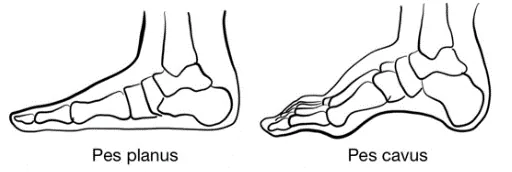
- Hindfoot valgus (Normal: 5-10°)
- “Too Many Toes” Sign: examiner stands behind patient with patient’s feet together. “Too Many Toes” sign is positive when >2 toes visible → suggests severe pes planus, or posterior tibialis dysfunction
With Patient Non-Weightbearing, comment on:
- Swelling:
- Loss of normal depressions anterior to malleoli → Suggests True Ankle Swelling
- Swelling posterior to lateral malleolus → Suggests Peroneal Tenosynovitis
- Swelling posterior to medial malleolus → Suggests Posterior Tibial Tenosynovitis
- Swelling at back of heel → Suggests Retrocalcaneal Bursitis or Retroachilleal Bursitis
- Swelling of the forefoot → Can be seen in Inflammatory Arthritis
- Erythema
- Atrophy
- Deformities:
- Hallux valgus, bunions or bunionettes
- Mallet Toes → Irreversible flexion of DIPs
- Hammer Toes → Irreversible flexion of PIPs
- Claw Toes → Irreversible flexion of both PIPs and DIPs

- Skin Changes or Scars:
- Callous formation → Abnormal stance or gait
- Inspect sole of the foot as well
- Additional Step: Footwear Inspection: Most patients wear out central or outer corners of heel of shoe
- Medial heel of shoe worn out → pronation of the foot
- Medial forefoot of shoe worn out → over-pronation of foot
- Lateral forefoot of shoe worn out → over-supination of foot
Palpation, including assessment for ankle and MTP effusions
AFMC level Pre-clerkship 3 · Clerkship 4
- Commenting on: TESTCA – Tenderness, Effusions, Swelling, Temperature, Crepitus, & Atrophy
- Note: In a screening exam, palpate all areas. In clinical encounters, depending on where the patient complains of pain, may focus palpation on area causing patient’s symptoms.
- Ottawa Ankle Rule: Palpate posterior edge/tip of lateral malleolus, posterior edge/tip of medial malleolus, base of 5th metatarsal, and navicular bone
- Palpate Laterally:
- Fibula
- Lateral Malleolus and Lateral Malleolar Bursa
- Lateral Ligaments (Posterior and Anterior Talofibular, Calcaneofibular)
- Peroneal Tendons
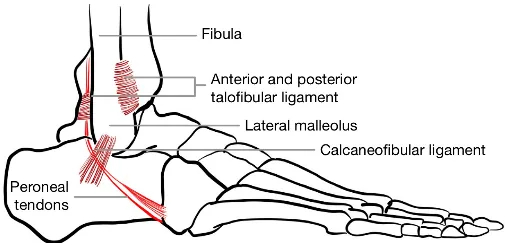
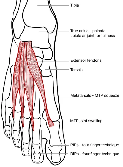
- Palpate Anteriorly:
- Shin (Tibia)
- Tibiotalar joint
- True Ankle Joint: Cup hands around ankle. Palpate tibiotalar joint space anteriorly for fullness. Try to ballot fluid between your thumbs.
- Extensor Tendons
- Tarsal Bones: includes 5 bones of midfoot (cuboid, medial/intermediate/lateral cuneiforms, and navicular) and 2 bones of hindfoot (talus and calcaneus)
- Metatarsal Bones
- MTP Joint Swelling: Palpate 1-2 cm proximal to the web-space with four-finger technique (tips of your thumbs on top of joint, while fingers stabilize joint underneath).
- MTP Squeeze: Grasp across forefoot/ MTPs with one hand and squeeze. Pain suggests synovitis (Inflammatory Arthritis) or Intermetatarsal Bursitis
- PIP and DIP joints: Palpate with two fingers of one hand stabilizing at sides of joint, while the index finger and thumb of other hand palpate from top and bottom.
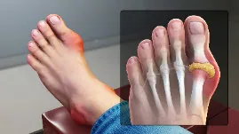
Podagra (Gout of 1st MTP)3
- Palpate Medially:
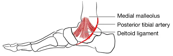
- Medial Malleolus
- Deltoid Ligament
- Posterior Tibialis (pulse)
- Palpate Posteriorly:
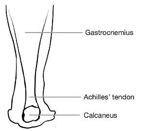
- Gastrocnemius Muscle
- Musculotendinous Junction
- Achilles Tendon and Enthesis
- Calcaneus
- Palpate Sole:
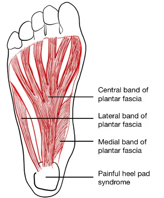
- Plantar Fasciitis:
- Apply pressure to medial calcaneus tuberosity (insertion of plantar fascia), checking for tenderness
- Tense plantar fascia by passive dorsiflexion of toes and assess for signs of pain
- Painful Heel Pad Syndrome: Palpate the centre of the heel, and assess for signs of pain
- Plantar Fasciitis:
Why Check: Palpation can be very helpful to determine source of pain. Palpate for tenderness and swelling in a systematic manner, to assess for potential fractures and other abnormalities. The likelihood of clinically significant ankle fracture (i.e. bone fragment >3mm, may require plaster immobilization) is increased by tenderness over posterior medial malleolus (LR+ 4.8, LR- 0.6), and by tenderness over posterior lateral malleolus (LR+ 2.4, LR- 0.4). The likelihood of clinically significant midfoot fracture is increased by tenderness at base of 5th metatarsal (LR+ 2.9, LR- 0.1), and decreased by tenderness at navicular bone (LR+ 0.4). These are all a part of the Ottawa Ankle Rule. The likelihood of a suspected Achilles tendon rupture is moderately increased by a palpable gap in the Achilles tendon (LR+ 6.8, LR- 0.3). Gout classically presents with initial flare the first metatarsophalangeal joint (known as podagra) or knee, but may also present in other MTP joints, ankle, and midfoot.
Range of Motion (ROM)
AFMC level Pre-clerkship 3 · Clerkship 4
- Approach: Perform Active ROM, and then Passive ROM if Active ROM is limited. These can be integrated with examiner guiding the ankle/foot beyond patient’s active ROM.
- Setup: Have knee flexed to relax calves, and foot in a neutral position (90 degrees flexion).
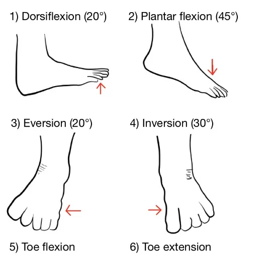
- Tibiotalar Joint (True Ankle): Ankle Dorsiflexion (15-25°); Ankle Plantarflexion (40-50°)
- Subtalar Joint (between talus and calcaneum): Ankle Inversion (Up to 30°); Ankle Eversion (Up to 20°)
- Midtarsal Joint: Stabilize heel with one hand while grasping and manipulating the mid and forefoot with your other hand - Inversion (30°); Eversion (20°); Adduction; Abduction
- Toes: Flexion; Extension
Why Check: Assessing passive range of motion if active range of motion is limited helps distinguish between ROM limitations caused by pain, tendon rupture etc. and those caused by structural limitation. Asymmetry with passive range of motion may indicate arthritis (bony end-feel), joint effusion (e.g. septic arthritis or gout with soft end-feel), or internal derangements such as fracture.
Power Assessment
AFMC level Pre-clerkship 3 · Clerkship 4
- Approach: Resisted Isometric Testing i.e. patient resists force applied by clinician.
- Assess: Ankle Dorsiflexion and Plantarflexion; Ankle Inversion and Eversion; Toe Flexion and Extension
Reporting: For each muscle group tested, assess per the Medical Research Council Power Grading. If it is all 5/5, you can say “Power was 5/5 bilaterally in both upper and lower limbs” and report specific deficits. NOTE: This scale is detailed in Chapter 31 – Screening Neurological Examination
Special Tests:
Stability Testing
Say: I will examine the ankle for signs of instability by applying pressure to the ankle in different directions. Please let me know if you feel pain during the test.
Do: Stabilize lower leg with one hand anteriorly, and grasp the hindfoot.
- Apply eversion force and check for excessive movement
- Apply inversion force and check for excessive movement
- Anterior Drawer Test: Flex foot to 20 degrees plantarflexion, stabilize lower leg with one hand anteriorly and grasp the calcaneus. Pull heel forward and check for excessive movement.
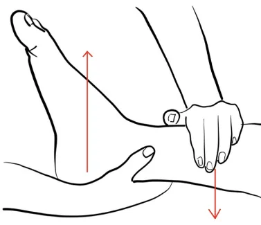
Report: I note no excess movement with ankle joint stability testing – Deltoid Ligament, Lateral Ligaments, and Anterior Talofibular Ligaments are stable.
Why Check:
- Excess movement with foot eversion suggests a Deltoid Ligament Tear. The Deltoid ligament is strongly attached to Medial Malleolus, where over-eversion injury can cause avulsion fracture of medial malleolus.
- Excess movement with foot inversion suggests Lateral Ligament Tear (e.g. calcaneofibular ligament tear).
- Excess movement (Normal: less than 5mm) with anterior drawer test suggests an Anterior Talofibular Ligament (ATFL) Tear – ATFL most common ligament involved in ankle sprain from over-inversion injury.
Tinel’s Sign
AFMC level Pre-clerkship 1 · Clerkship 3
Say: I will examine the Tarsal Tunnel in the ankle by gently tapping an area on the inside of the ankle for 30 seconds. Let me know if you feel pins and needles in the foot during the test.
Do: Locate the tarsal tunnel area, located just posterior to the medial malleolus. Tap over tarsal tunnel for 30 seconds. Paresthesias radiating to first three toes indicates a possible tarsal tunnel syndrome.
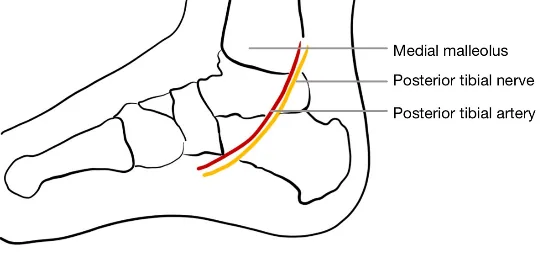
Report: No paresthesia reported, and Tinel’s Test is negative.
Why Check: If paresthesia radiating to first three toes reported → possible tarsal tunnel syndrome.
Thompson Calf-Squeeze Test
Say: I will examine the Achilles Tendon for tears by squeezing the calf muscle. I will ask you to lie on the stomach for this test with the legs relaxed. Please let me know if you feel pain.
Setup: Patient prone with foot hanging off exam table or kneeling in a chair with foot hanging off chair.
Do: Squeeze calf, looking for normal foot plantarflexion.
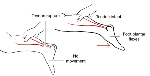
Report: Calf-Squeeze produces normal foot plantarflexion – Thompson Calf-Squeeze test is negative.
Why Check: The likelihood of suspected Achilles Tendon Rupture is significantly increased with positive Thompson Calf Squeeze Test (LR+ 13.5), and significantly decreased with a negative Thompson Calf-Squeeze Test (LR- 0.05). A positive test is where the foot fails to plantarflex with calf squeeze.
Helpful Videos to Review:
- Video 1: Ankle and Foot Exam – McMaster MSK Examination Series (Link: ANKLE AND FOOT EXAMINATION - McMASTER UNIVERSITY)
- Video 2 and 3: Bates’ Visual Guide to the Physical Exam – Anatomy Review and Examining the Ankles and Feet (Link: https://www.medportal.ca/ug/educational-resources/bates/lists/bates/musculoskeletal)
- Video 4: Dr. Janice Harvey’s Joint Assessment Made Easy – Ankle Assessment (Link: https://www.medportal.ca/ug/educational-resources/joint-assessment-made-easy)
Part 3: Relevant Likelihood Ratios – from Rational Clinical Examination JAMA series
Clinical Question 1: Does this patient have a Clinically Significant Ankle Fracture?4
Note: Clinically significant defined as bone fragment >3mm in breadth (size that may require plaster immobilization)
| Procedure | LR+ (95% CI) | LR- (95% CI) |
|---|---|---|
| Tenderness over posterior medial malleolus | 4.8 (2.6-9.0) (about +15%) | 0.6 (0.6-0.7) (about −15%) |
| Inability to bear weight immediately after injury | 2.6 (2.2-3.1) (about +15%) | 0.5 (0.4-0.6) (about −15%) |
| Inability to bear weight 4 steps in ER | 2.5 (2.2-2.8) (about +15%) | 0.3 (0.2-0.4) (about −30%) |
| Tenderness over posterior lateral malleolus | 2.4 (1.9-2.8) (about +15%) | 0.4 (0.3-0.5) (about −30%) |
| Ottawa Ankle Rule | 1.5 (1.2-1.8) (about +15%) | 0.1 (0-0.2) (about −45%) |
Clinical Question 2: Does this patient have a Clinically Significant Midfoot Fracture?4
Note: Clinically significant defined as bone fragment >3mm in breadth (size that may require plaster immobilization)
| Procedure | LR+ (95% CI) | LR- (95% CI) |
|---|---|---|
| Tenderness at base of 5th metatarsal | 2.9 (2.5-3.3) (about +15%) | 0.1 (0.1-0.2) (about −45%) |
| Inability to bear weight 4 steps in ER | 1.1 (0.8-1.4) (no change: 95% CI includes 1) | 0.9 (0.8-1.1) (no change: 95% CI includes 1) |
| Inability to bear weight immediately after injury | 1.0 (0.5-2.3) (no change: 95% CI includes 1) | 1.0 (0.8-1.3) (no change: 95% CI includes 1) |
| Tenderness of navicular bone | 0.4 (0.2-0.9) (about −30%) | 1.1 (1.0-1.2) (no change: 95% CI includes 1) |
| Ottawa Ankle Rule | 2.1 (1.3-3.3) (about +15%) | 0.1 (0-0.2) (about −45%) |
Clinical Question 3: Does This Patient have an Achilles Tendon Rupture?5
| Procedure | Sensitivity | Specificity | LR+ | LR- |
|---|---|---|---|---|
| Thompson Calf Squeeze Test | 0.96 | 0.93 | 13.5 (about +45%) | 0.05 (about −45%) |
| Palpable Gap in Achilles Tendon | 0.73 | 0.89 | 6.8 (about +30%) | 0.3 (about −30%) |
| Knee Flexion Test | 0.88 | 0.86 | 6.2 (about +30%) | 0.1 (about −45%) |
References
-
Ottawa Ankle Rule. MDCalc. Accessed March 12, 2022. https://www.mdcalc.com/ottawa-ankle-rule#next-steps ↩ ↩2
-
Recommendations. Choosing Wisely Canada. Accessed July 22, 2022. https://choosingwiselycanada.org/recommendations/#list ↩
-
Wiki images. Scientific Animations. Accessed May 2, 2023. https://www.scientificanimations.com/wiki-images/ ↩
-
McGee S. Evidence Based Physical Diagnosis. Saunders; 2012:835. ↩ ↩2
-
McGee S. Evidence-Based Physical Diagnosis. 4th ed. Elsevier; 2012:chap 64. ↩