Medical Foundations 4
Chapter 27Hip Examination
Part 1: Focused History – Based on Chief Complaint (Begin with Open-Ended Questions)
HPI
Pain Assessment
- Onset:
- Acute onset of pain, disability immediate with injury (e.g. acute trauma, concern of structural damage) (see Mechanism of Injury below)
- Delayed onset of pain following injury (e.g. tendon strain, cartilage contusion, minor soft tissue tear)
- Acute onset with no associated injury (e.g. septic arthritis)
- Insidious onset gradually worsening over time (e.g. overuse injury, osteoarthritis)
- Pediatric patients: Is the patient in an active growth spurt → increased risk of overuse injuries
- Provoking:
- Aggravating postures (e.g. worse with Hip flexion/rotation → Intra-articular pathology)
- Aggravating activities (e.g. worse with prolonged standing, weightbearing → Osteoarthritis)
- Pain not worse climbing steps or walking down slopes (LR- 0.47); Pain on initial steps after rest (LR- 0.31); Pain on walking (LR- 0.25-0.58) moderately reduces the probability of Hip osteoarthritis.
- Pain that is not relieved by sitting moderately reduces the probability of Hip osteoarthritis (LR- 0.24)
- Palliating:
- Relieving postures/activities: Relieved by rest?
- Improved with hip abduction, external rotation and slight hip flexion → Intra-articular pathology – this position maximizes joint space volume
- Previous treatment: POLICE (Protect, Optimally Load, Ice, Compress, Elevate)? Physio? Analgesics?
- Investigations: Type (x-rays, Ultrasound, MRI, etc.) and results (if available)
- Relieving postures/activities: Relieved by rest?
- Quality: Deep ache, sharp vs. dull? – Ask patient what words they would use
- Region: Anterior vs. Lateral vs. Posterior/Buttock Pain – can differentiate causes (see below)
- C-Sign: When patient points to the pain, the patient may cup the anterolateral hip with thumb and forefinger in a “C” shape → suggests Intra-articular hip pain
- Radiation:
- To or from the low back (low back pain may refer to hip)
- To or from the knee (knee pain may refer to the hip)
- Sciatica pain (radiating down back of affected leg) – Sciatica has Sensitivity of 95%, Specificity of 88% for detecting a herniated disk.
- Severity: Immediately after injury, vs. subsequently. Has it improved over time with rest?
- Was patient able to continue their activity, e.g. after the sport completed noticed pain got worse?
- Timing: Pain at rest (e.g. waking patient up at night); Constant vs. intermittent
- Associated Symptoms:
- Mechanical Symptoms: Locking, clicking, giving way (e.g. labral tear, ligamentum teres tear)
- Acute Trauma: Snap, crack, tear or other sensation at time of injury?
- Joint Inflammatory Symptoms: Swelling, erythema, instability
- Sensory Neuropathy e.g. paresthesias or numbness – assess distribution e.g. Peripheral nerve, Dermatomal or Spinal level (See Ch 31 – Screening Neuro Exam)
- Weakness: Weakness in specific motion may indicate a muscle tear; if concern for a neurologic cause may need to do a screening neuro exam (See Ch 31 – Screening Neuro Exam)
Red Flags
- Pain: Night pain; Pain at rest; Pain unrelated to activity; Inability to weight bear; Pain with passive movement of joint; Poor response to treatment
- Constitutional Symptoms: Fever, night sweats, weight loss, anorexia
- Acute Compartment Syndrome (Can occur minutes-hours after acute injury, particularly closed fracture with compartments intact) → Surgical emergency (fasciotomy relieves compartment pressure)
- 6 Ps of compartment syndrome: pain, poikilothermia, paresthesia, paralysis, pulselessness, pallor.
- Pain: Out of proportion; not improving with analgesia; at rest; with passive muscle stretching
- LATE Findings of acute compartment syndrome: Pallor, paresthesia, paralysis, and pulselessness
- Fullness or distension of involved compartment (typically anterior or lateral leg)
- Inflammatory Arthritis:
- Intra-articular features: New onset joint swelling/effusion; Morning stiffness >30 mins (vs. <30 mins in Osteoarthritis); Improves during day and with activity (vs. worsens in osteoarthritis); distribution (Monoarthritis = 1, Oligoarthritis = 2-4, Polyarthritis = 5+ joints, symmetrical vs. asymmetrical)
- Extraarticular features: Psoriasis, Inflammatory Bowel Disease; Uveitis/Iritis; Photosensitivity rash; Raynaud’s Phenomenon; Urethritis; Diarrheal Illness
- Septic Arthritis: Acute atraumatic hip pain with impaired weightbearing due to pain. Look for adjacent skin infection (vs. hematogenous seeding e.g. sepsis), fevers/chills with warm, erythematous, swollen joint (may be absent due to depth of hip structures) and painful restricted ROM in all directions.
- Neurologic Symptoms: Paresthesias (pins and needles), numbness, weakness
- Non-Accidental Injury (Pediatrics): Delay seeking medical attention. Unwitnessed, history not fitting with injury. Bruises over soft tissue areas. History of injury/neglect. Types of fracture (e.g. metaphyseal)
- Elderly: Trauma/falls; inability to weight-bear; functional decline.
Mechanism of Injury
- If injury occurred during an activity/event, ask open-ended questions about the mechanism of injury e.g. “run me through what happened, play-by-play”. Try to visualize what happened e.g. did the patient have their foot planted, was hit and twisted to fall onto their lateral hip (raising concern for a femoral neck fracture – can’t miss diagnosis for lateral hip pain). Assess low energy vs. high energy force injury, and the forces involved (e.g. direct vs. indirect forces and their direction).
- If assessing an athlete, helpful to know/learn about their sport to understand common injuries and the demands of the position they play in the sport.
- Elderly: Fall from standing height as predominant mechanism of hip fracture in the elderly.
Anterior Hip Pain
- Common Illness Presentations:
- Labral Tear: Often an insidious onset (may occur after trauma or repetitive motion), dull or sharp groin pain worse with sitting, rising from seat, getting in/out of car, or leaning forward. Pain may radiate to lateral hip, anterior thigh or buttock. Patients may describe catching, painful clicking, or popping with activity.
- Femoroacetabular Impingement (FAI): Often occurs in young and physically active patients, presenting with insidious onset pain in the groin that may radiate to lateral hip and anterior thigh. It is worse with sitting, rising from a seat, getting in/out of a car, or leaning forward. Patients may also describe clicking, locking, catching, stiffness, decreased range of motion, or giving way.
- Osteoarthritis: Often occurs in older adults, with gradual onset of an aching, constant, deep pain with joint stiffness worse after prolonged sitting (may be initially relieved by sitting), standing or weightbearing, climbing stairs, and initial steps after a period of rest.
- Worst pain in medial thigh moderately increases the likelihood of hip OA (LR+ 7.8).
- Groin pain with hip adduction/abduction moderately increases the likelihood of hip OA (LR+ 5.7).
- Iliopsoas Bursitis: Commonly provoked when extending hip from a flexed position, with associated intermittent catching, snapping or popping of hip.
- Stress Fracture of Hip: Post-trauma or with repetitive weight-bearing exercises, worse with activity with pain in anterior hip or groin. Usually insidious onset.
- Can’t Miss Diagnosis Illness Presentations:
- Septic Arthritis: Described above (see Red Flags).
- Referred Pain: Important to consider intra-abdominal or intra-pelvic causes of anterior hip/groin pain
- GI (e.g. appendicitis R-sided; diverticulitis L-sided; IBD, inguinal hernia, bowel obstruction)
- Genitourinary (e.g. nephrolithiasis, pyelonephritis, ureteritis, urinary retention)
- Utero-ovarian (e.g. Ectopic pregnancy, Ovarian Torsion, Ruptured Ovarian Cyst, Tubo-Ovarian Abscess, Ovarian Cancer, Pelvic Inflammatory Disease, Endometriosis)
- Testicular (e.g. Testicular torsion, epididymitis, epididymo-orchitis) or Prostate (e.g. prostatitis)
- Avascular Necrosis of the Hip: May occur in middle-older age adults with history of smoking, alcohol use, systemic corticosteroid use, hemoglobinopathies, chemotherapy, metabolic syndrome or obesity. Presents with antalgic gait, pain with range of motion and limited range of motion.
- Pediatrics: Slipped Capital Femoral Epiphysis (SCFE): Typically a gradual onset anterior hip pain in adolescents (age 12-15 years) with ↑ BMI. Commonly presents with a limp and may have associated knee pain. Bilateral in some cases. A SCFE is a can’t miss diagnosis due to to risk of Avascular Necrosis (AVN) of Femoral Head. If a patient with a SCFE is unable to weight-bear due to pain or instability, it is an Orthopedic Emergency.
- Pediatrics: Legg-Calve-Perthes Disease (LCP – Idiopathic Avascular Necrosis of Hip): Typically an acute or insidious onset in childhood (age 4-8 years). Commonly presents with a limp, activity-related pain (e.g. jumping, running), and may have associated pain in thigh or knee. Bilateral in some cases.
- Pediatrics: Avulsion Fracture of Pelvis: Typically occurs in adolescent athletes due to sudden, forceful muscle contraction (e.g. sprinting, jumping, swinging a bat, or kicking). Commonly presents with an audible pop at time of injury, where the patient may have swelling, ecchymosis, point tenderness over fracture site, and an antalgic gait or not be able to weight bear. Avulsion fracture sites may include the Anterior Superior Iliac Spine (ASIS – sartorius muscle attachment), Anterior Inferior Iliac Spine (AIIS – rectus femoris muscle attachment) or Ischial Tuberosity (hamstring muscle attachment – presents with buttock pain).
Lateral Hip Pain (usually not caused by intra-articular hip pathologies)
- Common Illness Presentations:
- Greater Trochanteric Pain Syndrome: Lateral hip pain over greater trochanter aggravated by ambulation. Often atraumatic and insidious onset from repetitive use (e.g. long-distance run), prolonged sitting, post-fall, or with weight bearing. May not be able to sleep on affected side, and have difficulty rising from a chair, but no difficulty putting on socks and shoes (an issue in Hip OA).
- Umbrella Term: Includes Trochanteric bursitis; Iliotibial band thickening, Gluteal muscle tendinopathy or tear.
- Osteoarthritis: As noted above.
- Greater Trochanteric Pain Syndrome: Lateral hip pain over greater trochanter aggravated by ambulation. Often atraumatic and insidious onset from repetitive use (e.g. long-distance run), prolonged sitting, post-fall, or with weight bearing. May not be able to sleep on affected side, and have difficulty rising from a chair, but no difficulty putting on socks and shoes (an issue in Hip OA).
- Can’t Miss Diagnosis Illness Presentations:
- Fracture of Neck of Femur: Fall directly onto lateral hip, with twisting mechanism where patient foot planted and body rotates, with sudden onset of hip pain, inability to walk. Leg may appear shortened and externally rotated. Insufficiency fractures may have no clear trauma history.
- Polymyalgia Rheumatica (PMR): Acute onset of aching pain and stiffness of shoulders, hip girdle, neck and torso in a patient >50 yo. Pain may begin as unilateral, progresses to bilateral. Pain is worst on arising in morning with significant morning stiffness that may progress to last into afternoon and worsen with prolonged inactivity (e.g. after car ride). Pain in hips commonly in lateral hips and groin that radiates to posterior thighs. Pain in shoulders commonly limiting active shoulder abduction.
Posterior Hip, Buttock Pain
- Common Illness Presentations:
- Piriformis Syndrome (entrapment of sciatic nerve by piriformis muscle): Buttock pain aggravated by sitting or walking ± radiation down ipsilateral posterior thigh from sciatic nerve compression. Worse with active external hip rotation.
- Ischiofemoral Impingement (pressure of quadratus femoris muscle and nerve between proximal femur at lesser trochanter and ischial tuberosity): Gradual onset, deep buttock pain worse with activities with a long stride (e.g. running)
- Sacroiliac (SI) Joint Dysfunction: Posterior hip pain with tenderness over SI joint.
- Osteoarthritis: Posterior hip pain on squatting moderately increases likelihood of OA (LR+ 6.1).
- Can’t Miss Diagnosis Illness Presentations:
- Referred Pain: Important to consider intra-abdominal or intra-pelvic causes of posterior hip/buttock pain (see Anterior Hip Pain above)
Past Medical History (PMHx)
- Previous hip injuries
- Risk Factors for Septic Arthritis: Immunosuppression (including diabetes mellitus and HIV); Pre-existing joint disease (e.g. Rheumatoid arthritis: LR+ 2.5, LR- 0.45); Advanced Age (>80 years old – LR+ 3.5); Skin infection (LR+ 2.8, LR- 0.76)
- Lab Markers for Septic Arthritis:
- Peripheral – CRP >100 mg/L (LR+ 1.6, LR- 0.44), WBC >10 (LR+ 1.4), ESR >30mm/h (LR+ 1.3)
- Synovial fluid – WBC >100 (LR+ 28, LR- 0.71), WBC >50 (LR+ 7.7, LR- 0.42), WBC >25 (LR+ 2.9, LR- 0.32), PMN cells ≥90% (LR+ 3.4, LR- 0.34)
- Lab Markers for Septic Arthritis:
- Risk Factors for Osteonecrosis: Systemic Lupus Erythematosus, Sickle Cell Disease, HIV Infection
- Risk Factors for Hip Osteoarthritis: Age (>50 years); Female Gender; Obesity; Heavy manual labour; History of joint injury; Femoroacetabular Impingement (FAI); Developmental Dysplasia of Hip (DDH); Slipped Capital/Femoral Epiphysis (SCFE), Perthes disease (Idiopathic osteonecrosis of femoral head)
- Bleeding Disorder History: ↑ risk of serious pathology on injury, including ↑ risk compartment syndrome
- Osteoporosis (Risk factor for occult hip fractures)
- Psoriasis: Risk factor for Psoriatic Arthritis.
- Autoimmune disorders (↑ risk for Inflammatory Arthritis) – particularly Spondyloarthropathies (e.g. Psoriatic arthritis, Uveitis, Ankylosing Spondylitis, Inflammatory Bowel Disease, Reactive Arthritis)
- Giant Cell Arthritis (GCA): Associated with ↑ risk of Polymyalgia Rheumatica (PMR)
Relevant Clinical Scores:
- Kocher Criteria: Septic arthritis vs. transient synovitis in pediatric inflamed hip
- PEG Scale – Chronic pain assessment in primary care patients to monitor treatment response over time
Past Surgical History (PS Hx)
- History of Surgical Intervention: Arthroscopic surgery; Open-Reduction-Internal-Fixation
- Palliative Radiation (e.g. for painful bony metastasis in cancer)
- Risk Factors for Septic Arthritis: Hip/Knee prosthesis AND overlying skin infection (LR+ 15.0, LR- 0.77); recent joint surgery (LR+ 6.9, LR- 0.78); history of hip or knee prostheses (LR+ 3.1, LR- 0.73)
Family Medical History (FMHx)
- Hip Osteoarthritis and Hip Pain (↑ risk of Femoroacetabular Impingement – FAI – if present in other family members)
- Presence of family history of arthritis mildly increases the probability of hip osteoarthritis (LR+ 2.1, LR- 0.79)
- Autoimmune disorders (↑ risk for Inflammatory Arthritis) – particularly Spondyloarthropathies (e.g. Psoriasis, Uveitis, Ankylosing Spondylitis, Inflammatory Bowel Disease, Reactive Arthritis).
Social Hx
Substance Use
- Smoking: Risk factor for Avascular Necrosis of Hip and for Osteoporosis
- Alcohol: Risk factor for Gout, Osteoporosis (>3 units/day), and Avascular Necrosis
- IV Drug Use: Risk factor for Septic Arthritis
Occupational Hx
- Are you working right now? What do you do for work? What other kinds of work have you done in the past? Do you have benefits through work such as drug coverage (e.g. access to physiotherapy coverage)?
- Length of time spent sitting, amount of hip flexion and rotation, degree of manual labour at work
- If injury occurred at work → separate Workplace Safety and Insurance Board (WSIB) process.
Sporting History
- History of sports played: Sports associated with ↑ hip injury risk includes dancing, gymnastics, martial arts, tennis and hockey (repeated combined hip flexion, abduction and adduction, and loaded rotational or twisting movements)
- Current level of sport played (e.g. recreational, competitive, league level) and desired future sporting activity level; Sports position played (some positions may place hip under ↑ loading stress)
- Current training history (e.g. for patients with overuse injuries) – quantity and quality of training; any recent changes to routines (e.g. risk factor for injury)?
- Recent change of equipment (e.g. footwear for running – note shoe type, age, wear pattern)
- Overtraining Syndrome: Excess fatigue, recurrent illness, ↓ motivation, persistent soreness/stiffness
Dietary History
- Inadequate nutrition for an athlete can contribute to overtraining syndrome
- If concern (e.g. diagnosis of Stress Fracture), assess for Relative Energy Deficiency in Sport (RED-S) – Impaired physiological functioning including but not limited to metabolic rate, menstrual function, bone health, immunity, protein synthesis, and cardiovascular health caused by relative energy deficiency (balance of dietary energy intake vs. energy expenditure required for health, growth and activities)
ADLs and IADLs
- Common activities impacted by hip pain: Getting in and out of a car; Getting out of a chair; Putting on socks and shoes (difficulty common in Hip Osteoarthritis); Running; Walking; Going up and down stairs (as screened for in the GALS exam)
Medications Hx, Allergies
Inflammatory Arthritis
- Gout:
- Acute Flare: NSAIDs, Colchicine
- Urate-Lowering Therapy: Allopurinol, Febuxostat (safe in renal disease)
- Inflammatory Arthritis (e.g. Rheumatoid Arthritis, Spondyloarthropathies):
- Disease-Modifying Antirheumatic Drugs (DMARDs): Methotrexate, Sulfasalazine, Hydroxychloroquine, Leflunomide, Azathioprine
- Biologic DMARDs:
- TNF-α inhibitors: Etanercept, adalimumab, infliximab, golimumab, etc.
- Others: Abatacept, rituximab, tocilizumab, sarilumab, anakinra
- Acute Flare: Glucocorticoids (e.g. Prednisone)
Other Medications
- Fluoroquinolones (e.g. Ciprofloxacin) - ↑ risk of Tendinopathy or Tendon rupture
- Glucocorticoids (e.g. Prednisone) - ↑ risk of Osteonecrosis of Hip
Analgesics
- PO: NSAIDs (concern re. GI bleed, Hypertension, Renal impairment), Acetaminophen, Muscle Relaxant (e.g. Baclofen), Opioids – ask pain severity before versus after med, frequency and dosing (e.g. concern for Acetaminophen overdose >4g/day in healthy patient, >3g/day in elderly patient, >2g/day in liver disease)
- Topical: NSAIDs (E.g. Diclofenac), Lidocaine, etc.
- Joint Injections: Intra-articular corticosteroid injections, Diagnostic injections with lidocaine – ask about pain relief immediately after (effect of lidocaine) vs. sustained pain response (effect of corticosteroid) – Hip injection under image guidance (e.g. ultrasound)
Part 2: Physical Examination of the Hip Joint:
- Note: If hip exam unremarkable or you suspect referred pain, consider examination of back and knee.
General Assessment:
Gait Assessment
Gen - [3, 4] Trendelenburg [2, 4]
- Components of Gait Cycle:
- Stance phase (60% of gait cycle): initial contact (heel strike) → mid-stance → pre-swing (toe-off)
- Swing phase (40% of gait cycle): initial swing → mid-swing → terminal swing
- Note: Former terminology in brackets re. heel-strike and toe-off.
- Quality of Gait: Smoothness of movement (legs, arm swing, pelvic tilt), symmetry, normal stride length, ability to turn quickly, symmetry.
- Abnormal Gaits to Assess for:
- Antalgic Gait: Stance phase shortened on affected side, usually indicating pain on weight-bearing
- Why: Patient is trying to reduce the time spent weight-bearing on the affected limb
- Absence of limp moderately decreases probability of Hip Osteoarthritis (LR- 0.35)
- Trendelenburg Gait:
- Unilateral: During stance phase, if hip abductors weak on standing side, pelvis will drop to opposite side. The trunk may compensate by lurching towards side of weakened abductor muscles, with a truncal tilt present. If uncompensated, there is no truncal tilt present.
- Hip abductor weakness mildly increases probability of Hip Osteoarthritis (LR+ 4.5, LR- 0.62)
- Bilateral: Produces a waddling gait.
- Why: Leaning on the affected side to align the centre of gravity allows for minimizing torque.
- Unilateral: During stance phase, if hip abductors weak on standing side, pelvis will drop to opposite side. The trunk may compensate by lurching towards side of weakened abductor muscles, with a truncal tilt present. If uncompensated, there is no truncal tilt present.
- Antalgic Gait: Stance phase shortened on affected side, usually indicating pain on weight-bearing
Inspection (SEADS)
AFMC level Pre-clerkship 3 · Clerkship 4
- Approach: Ensure adequate but respectful exposure during your exam (particularly for hip exam).
- Swelling: Unlikely to see effusion, but a massive joint swelling may present as a prominence in groin.
- Erythema
- Atrophy: Pelvic Girdle – medial & anterior to pubic symphysis; Gluteal Muscles – posterior aspect – role in Hip extension; Hamstrings – posterior aspect of thighs – roles in Hip extension and knee flexion; Quadriceps – anterior aspect of thighs – roles in Hip flexion and knee extension; Lower Back
- Deformity:
- Posture: Scoliosis; Exaggerated Lumbar Lordosis (may suggest a flexion contracture of hip)
- Pelvic Tilt: Can inspect or palpate level of iliac crests (may suggest either a hip adduction deformity on the higher side, or a hip abduction deformity on the lower side)
- External or Internal Rotation of Hip at Rest: Look at the feet (may suggest a rotational deformity of hip, or an external rotation may suggest a hip fracture)
- Trendelenburg Sign: Have patient stand on one leg at a time – if unsupported side drops → weakness of the hip abductors on the standing side (relates to the Trendelenburg Gait)
- Skin Changes and Scarring: Look for Rashes, and Scars from surgeries (on lateral aspect of hip)
Palpation
AFMC level Pre-clerkship 3 · Clerkship 4
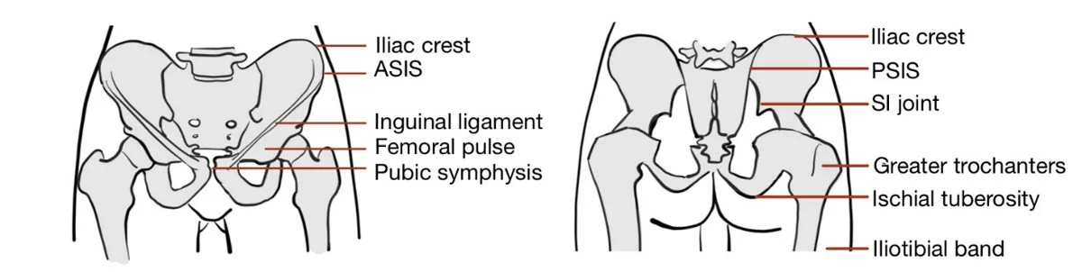
- Approach: Important landmarks include the Pubic Symphysis, the ASIS, AIIS and the Iliac Crests.
- Commenting on: TESTCA – Tenderness, Effusions, Swelling, Temperature, Crepitus, & Atrophy
- Palpation Order: Easiest to palpate in an ordered sequence, usually from anterior to posterior
- Anterior structures – Along a line from Pubic Symphysis to ASIS: With patient supine, begin with the medial anterior Pubic Symphysis (if indicated and with patient consent) → Inguinal Ligament → Femoral Pulse → Anterior Inferior Iliac Spine (AIIS) → Anterior Superior Iliac Spine (ASIS) → Iliac Crest
- Note: Pubic symphysis angle acute in males, vs. obtuse in females
- Posterior structures – Beginning at the Iliac Crests:
- Ask patient to change position to to lateral decubitus position to palpate the posterior Iliac Crest (bony prominence) → Dimples of Venus at Posterior Superior Iliac Spine (PSIS, symmetrical sagittal indents located at S2) → SI Joint (a palpable depression superior to PSIS)
- Ask patient to flex leg to 90 degrees to palpate the Ischial Tuberosity (lateral to PSIS)
- Ask patient to return leg to neutral position to palpate Greater Trochanters (Bony prominence of proximal lateral femur) and Iliotibial Band
- Anterior structures – Along a line from Pubic Symphysis to ASIS: With patient supine, begin with the medial anterior Pubic Symphysis (if indicated and with patient consent) → Inguinal Ligament → Femoral Pulse → Anterior Inferior Iliac Spine (AIIS) → Anterior Superior Iliac Spine (ASIS) → Iliac Crest
Why Check: Palpation may be helpful to assess pain source. Ask patient to localize pain, palpate accordingly.
- Tenderness at Ischial tuberosity may indicate Ischio-gluteal bursitis
- Tenderness at Greater Trochanter may indicate Trochanteric bursitis
- Tenderness at distal 1/3rd of Iliotibial Band can indicate Iliotibial band syndrome
- Tenderness at inguinal ligament mildly increases probability of Hip Osteoarthritis (LR+ 2.4, LR- 0.53)
Range of Motion (ROM)
AFMC level Pre-clerkship 3 · Clerkship 4
- Approach: Perform Active ROM, and then Passive ROM if Active ROM is limited (assess end-feel i.e. Bony End-Feel i.e. Bony pathology vs. Soft End-Feel i.e. Soft-tissue pathology)
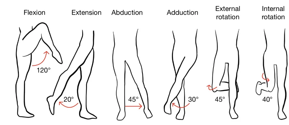
- Flexion (120°): Knee to Chest
- Extension (20°): Have patient lying prone. If patient lying on side, stabilize hemipelvis with one hand
- Abduction (45°): Moving leg laterally away from midline
- Adduction (30°): With other leg abducted out of the way, adduct leg medially as far as possible without having to lift it over other leg (which will flex the hip being tested)
- Internal Rotation (35-40°): Flex hip and knee to 90°, stabilize knee with one hand. Bring foot outwards with femur as axis of rotation – Internal rotation usually 1st movement limited in hip joint pathology
- External Rotation (45°): Same position as Internal Rotation, except patient brings lower leg inwards
Why Check: Assessing passive range of motion if active range of motion is limited helps distinguish between ROM limitations caused by pain, tendon rupture, etc. and those caused by structural limitation. Asymmetry with passive range of motion may indicate arthritis (bony end-feel), joint effusion (e.g. septic arthritis with soft end-feel), or internal derangements such as Femoroacetabular impingement or fracture (bony end-feel). Specifically examining for osteoarthritis, the likelihood of hip osteoarthritis increases mildly with decreased range of movement (LR+ 2.9, LR- 0.34), decreased hip extension (LR+ 2.4, LR- 0.35), decreased hip abduction (LR+ 1.6, LR -0.26), decreased hip adduction (LR+ 4.2, LR- 0.25), decreased hip internal rotation (LR+ 3.2, LR- 0.43) and decreased hip external rotation (LR+ 2.1, LR- 0.37). The likelihood of hip osteoarthritis increases moderately with groin pain on hip abduction or adduction (LR+ 5.7, LR- 0.71), and increases mildly with hip pain on active hip abduction (LR+ 1.5, LR- 0.43-0.59) and active hip internal rotation (LR+ 1.4, LR- 0.31-0.45)
Power Assessment
AFMC level Pre-clerkship 3 · Clerkship 4
- Approach: Resisted Isometric Testing i.e. patient resists force applied by clinician.
- Sitting Up Approach:
- Flexion Strength: Examiner braces hip, pushes down on quadriceps; patient resists motion.
- Extension Strength: Examiner braces hip, pushes up on hamstrings; patient resists motion.
- Abduction: Examiner braces hip, pushes on lateral aspect of thigh; patient resists motion.
- Adduction: Examiner braces hip and pushes out on medial aspect of thigh; patient resists motion.
- Supine Approach: Similar to above, with slight setup differences.
- Flexion, Extension: With patient lying supine, have the patient’s hip and knees flexed to 90°.
- Abduction and Adduction: With patient lying supine, have the patient’s hips and knees extended to be flat on exam table, and slightly abducted.
- Reporting: For each muscle group tested, assess per the Medical Research Council Power Grading. If it is all 5/5, you can say “Power was 5/5 bilaterally in both upper and lower limbs”, and report specific deficits. NOTE: This scale is detailed in Chapter 31 – Screening Neurological Examination
Special Tests:
Thomas Test
Say: I will support the low back while asking you to use both hands to pull the knee towards the chest to assess for a hip contracture.
Do:
- Patient supine. Clinician places one hand under lumbar spine. Patient places hands on knee and pulls knee towards their chest.
- Observe if contralateral thigh rises off exam table. Repeat on other side.
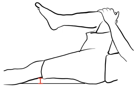
Report: I note no movement of the contralateral thigh off the exam table – Thomas Test is negative.
Why Check: Positive when contralateral thigh rises above bed, indicating Contralateral Hip Contracture. To maintain posture, patients with a contralateral hip contracture may compensate by arching the lumbar spine (↑ lordosis). The Thomas Test aims to reverse lumbar lordosis to make hip contracture more apparent on testing.
Ober Test
Say: I will move the leg backwards to assess for a tight Iliotibial band. Please turn on your side to face away from me, and keep the legs relaxed.
Do:
- Patient in decubitus position upper leg extended backwards and knee bent. Clinician braces upper leg and extends it backwards.
- Release the upper leg. Assess whether the knee touches the exam table.
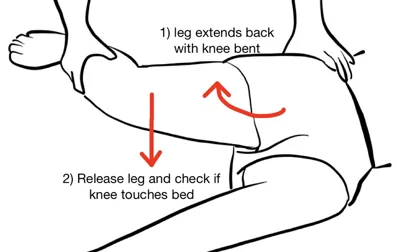
Report: The knee of upper leg touched exam table on release – Ober Test is negative.
Why Check: Positive when upper leg’s knee does not touch exam table, indicating a Tight Iliotibial Band.
FABER Test (same as in Back Exam – Chapter 29)
Say: I will rest the foot on the opposite knee and apply pressure to the inside of the knee to look for signs of Hip or Sacroiliac Joint pain when moving the hip. Please let me know if you feel pain, and if so, where.
Do:
- Patient lying supine, Flex, Abduct, and Externally Rotate one hip (placing heel on opposite knee, making a figure-of-4).
- Push down on knee; press on opposite hemipelvis to stabilize.
- Observe for pain in lower back or buttock
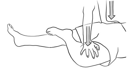
Assess: No pain elicited with FABER test.
Why Check: FABER (Flexion, Abduction, External Rotation) test with inability to achieve FABER position or eliciting pain in the anterior/lateral of the hip or groin pain suggests a Hip Joint Pathology (can cause pain in healthy patients due to stretch); pain elicited in lower back/gluteal region suggests a Sacroiliac Joint Pathology.
Part 3: Select Special Tests – Not Part of Screening Hip Exam But Seen in Practice:
Barlow & Ortolani Maneuvers (Neonate)
AFMC level Pre-clerkship 1 · Clerkship 3
Also seen in Ch 17 – Newborn Exam – MF3
Say: I will check the baby’s hip joints for stability, which we check for every newborn.
Do: Infant supine on examination table with diaper off.
- Barlow Maneuver: Stabilize infant’s pelvis with one hand and grasp abducted and flexed thigh with other hand. Hip flexed to 90 degrees. Adduct the hip with an additional posterior force on the anterior hip. A positive test is felt as a clunk.
- Ortolani Maneuver: If a clunk is felt on Barlow Maneuver, Abduct the hip with an additional anterior force on the posterior thigh. A positive test is the reduction of the dislocation (may also be felt as a clunk). Repeat with contralateral hip.
Report: The hip joints are stable on each side – Barlow and Ortolani screen is negative for Developmental Dysplasia of the Hip, which is reassuring.
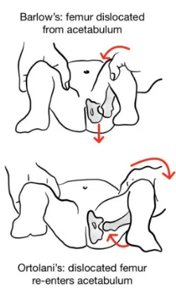
Why Check: Part of Newborn Screening Exam that requires referral to a pediatric orthopedic surgeon and correction. Risk factors include ligamentous laxity, female, positive FMHx, breech presentation, primiparous parent, oligohydramnios & postnatal infant position. Sensitivity of test is lowered in an agitated, crying infant.
FADIR Test
Say: I will flex the knee and hip, then rotate the hip inwards to check for Femoral Acetabular Impingement.
Do:
- Patient supine and both knees extended/relaxed. Passively move hip to 90° flexion and knee to 90° flexion.
- Clinician adducts and internally rotates the hip. Ask patient if it reproduces their hip pain symptoms.
Report: FADIR is negative as it did not reproduce the patient’s pain.
Why Check: The FADIR test is used to assess for Femoroacetabular Impingement (FAI). A negative FADIR test moderately decreases the probability of FAI (LR+ 1.1, LR- 0.4). There are three kinds of FAI: Cam (abnormal femur); Pincer (over-coverage of femoral head by acetabulum); and Mixed (combination of Cam + Pincer). Note that FAI may present as groin pain as well.
Leg Length Discrepancy
Say: I will use a measuring tape to measure the length of each leg to look for signs of true or apparent leg length discrepancies. Please keep the legs straight.
Do:
- Palpate for ASIS and medial malleolus on each side, and measure distance between them –true leg length.
- Palpate the Umbilicus and medial malleolus, and measure distance between them – apparent leg length.
Report: No true or apparent leg length discrepancies detected.
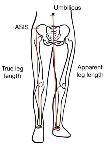
Why Check: A true leg length discrepancy occurs when discrepancy arises below level of hip (e.g. Genu Valgus or Genu Varus). In contrast, an apparent leg length discrepancy is where the true leg lengths are equal, but the apparent leg lengths are different, e.g. when discrepancy arises at level of hip – for example, Trauma (acquired), Pelvic Tilt, Adduction/Abduction deformities of hips, or Congenital Dysplasia of hip.
Helpful Videos to Review:
- Video 1: Hip Exam – McMaster MSK Examination Series (Link: HIP EXAMINATION - McMASTER UNIVERSITY)
- Video 2 and 3: Bates’ Visual Guide to the Physical Exam – Anatomy Review and Examining the Hips (Link: https://www.medportal.ca/ug/educational-resources/bates/lists/bates/musculoskeletal)
- Video 4: Dr. Janice Harvey’s Joint Assessment Made Easy – Hip Assessment (Link: https://www.medportal.ca/ug/educational-resources/joint-assessment-made-easy)
Part 4: Relevant Likelihood Ratios – from Rational Clinical Examination JAMA series
Clinical Question 1: Does this infant delivered with Breech Presentation have Developmental Dysplasia of the Hip?2
| Procedure | Sensitivity | Specificity |
|---|---|---|
| Clinical Examination (Ortolani & Barlow maneuvers) | 0.667 | 0.958 |
Clinical Question 2: Does This Patient have Hip Osteoarthritis?3
| Procedure | LR+ (95% CI) | LR- (95% CI) | |
|---|---|---|---|
| History | General Features – Family history of arthritis | 2.1 (1.2-3.6) (about +15%) | 0.79 (0.67-0.93) (about −15%) |
| General Features – History of knee osteoarthritis | 2.1 (1.1-3.8) (about +15%) | 0.80 (0.68-0.95) (about −15%) | |
| General Features – Age > 60 years | 1.5 (1.3-1.7) (about +15%) | 0.11 (0.02-0.78) (about −30%) | |
| General Features – Age > 50 years | 1.3 (1.1-1.5) (about +15%) | 0.32 (0.16-0.62) (about −30%) | |
| General Features – Morning stiffness < 60 minutes | 1.5-1.7 (about +15%) | 0.22-0.65 (about −30%) | |
| Location of Pain – Worst pain in medial thigh | 7.8 (1.7-37) (about +30%) | 0.89 (0.77-1.0) (no change: 95% CI includes 1) | |
| Location of Pain – Worst pain in buttock | 0.38-2.0 (about −30%) | 0.38-0.76 (about −30%) | |
| Activity Effect on Pain – Climbing stairs or walking down slopes worsens pain | 2.1 (1.6-2.8) (about +15%) | 0.47 (0.35-0.63) (about −30%) | |
| Activity Effect on Pain – Pain on initial steps after rest | 1.2 (1.1-1.4) (about +15%) | 0.31 (0.08-1.2) (no change: 95% CI includes 1) | |
| Activity Effect on Pain – Pain on walking | 1.1-1.2 (about +15%) | 0.25-0.58 (about −30%) | |
| Activity Effect on Pain – Pain relieved by sitting | 1.4 (0.2-1.6) (no change: 95% CI includes 1) | 0.24 (0.06-0.92) (about −30%) | |
| Physical | General Findings – Abductor Weakness | 4.5 (2.4-8.4) (about +15%) | 0.62 (0.43-0.88) (about −15%) |
| General Findings – Limp | 1.5 (1.2-1.9) (about +15%) | 0.35 (0.20-0.61) (about −30%) | |
| Palpation – Inguinal ligament tenderness | 2.4 (1.6-3.8) (about +15%) | 0.53 (0.33-0.87) (about −15%) | |
| Palpation – Tensor fascia lata tenderness | 2.0 (1.1-3.4) (about +15%) | 0.75 (0.54-1.1) (no change: 95% CI includes 1) | |
| Pain on movement and provocation tests – Squat causing posterior pain | 6.1 (1.3-29) (about +30%) | 0.79 (0.62-1.0) (no change: 95% CI includes 1) | |
| Pain on movement and provocation tests – Groin pain on abduction or adduction | 5.7 (1.6-20) (about +30%) | 0.71 (0.52-0.97) (about −15%) | |
| Pain on movement and provocation tests – Scour test | 2.4 (1.4-4.3) (about +15%) | 0.51 (0.29-0.90) (about −15%) | |
| Pain on movement and provocation tests – Pain on hip abduction | 1.5 (about +15%) | 0.43-0.59 (about −30%) | |
| Pain on movement and provocation tests – Pain on hip internal rotation | 1.4 (about +15%) | 0.31-0.45 (about −30%) | |
| Range of Motion – Restricted movement in 3 planes | 4.4 (2.4-8.3) (about +15%) | N/A | |
| Range of Motion – Restricted movement in 2 planes | 1.5 (0.90-2.6) (no change: 95% CI includes 1) | N/A | |
| Range of Motion – Restricted movement in 1 plane | 1.3 (0.85-2.0) (no change: 95% CI includes 1) | N/A | |
| Range of Motion – Restricted movement in 0 planes | 0.91 (0.78-1.1) (no change: 95% CI includes 1) | N/A | |
| Range of Motion – Decreased hip adduction | 4.2 (3.0-6.0) (about +15%) | 0.25 (0.11-0.54) (about −30%) | |
| Range of Motion – Decreased hip internal rotation | 3.2 (1.7-6.0) (about +15%) | 0.43 (0.31-0.60) (about −30%) | |
| Range of Motion – Decreased range of movement | 2.9 (2.1-4.0) (about +15%) | 0.34 (0.25-0.47) (about −30%) | |
| Range of Motion – Decreased hip extension | 2.4 (1.8-3.2) (about +15%) | 0.35 (0.17-0.71) (about −30%) | |
| Range of Motion – Decreased hip external rotation | 2.1 (1.6-2.8) (about +15%) | 0.37 (0.19-0.76) (about −30%) | |
| Range of Motion – Decreased hip abduction | 1.6 (1.3-2.1) (about +15%) | 0.26 (0.09-0.77) (about −30%) |
Clinical Question 3: Does This Patient have Femoral Acetabular Impingement (FAI)?4
| Procedure | Sensitivity | Specificity | LR+ | LR- |
|---|---|---|---|---|
| FADIR Test | 0.96 | 0.11 | 1.1 (about +15%) | 0.4 (about −30%) |
Clinical Question 4: Does This Adult Patient Have Septic Arthritis?5
Note: This is for any joint (e.g. hip, knee, ankle, etc.) – included here for reference.
| Type | Finding | Sensitivity | Specificity | LR+ (95% CI) | LR- (95% CI) |
|---|---|---|---|---|---|
| Risk Factor | Hip or Knee Prosthesis and Skin Infection | 24 | 98 | 15.0 (8.1-28.0) (about +45%) | 0.77 (0.64-0.93) (about −15%) |
| Recent Joint Surgery | 24 | 96 | 6.9 (3.8-12.0) (about +30%) | 0.78 (0.64-0.94) (about −15%) | |
| Age > 80 years old | 19 | 95 | 3.5 (1.8-7.0) (about +15%) | 0.86 (0.73-1.00) (no change: 95% CI includes 1) | |
| Hip or Knee Prosthesis | 35 | 89 | 3.1 (2.0-4.9) (about +15%) | 0.73 (0.57-0.93) (about −15%) | |
| Skin Infection | 32 | 88 | 2.8 (1.7-4.5) (about +15%) | 0.76 (0.60-0.96) (about −15%) | |
| Diabetes Mellitus | 12 | 96 | 2.7 (1.0-6.9) (no change: 95% CI includes 1) | 0.93 (0.83-1.00) (no change: 95% CI includes 1) | |
| Rheumatoid Arthritis | 68 | 73 | 2.5 (2.0-3.1) (about +15%) | 0.45 (0.32-0.72) (about −30%) | |
| HIV-1 Infection | 79 | 50 | 1.7 (1.0-2.8) (no change: 95% CI includes 1) | 0.47 (0.25-0.90) (about −30%) | |
| Symptoms | Joint pain | 85 | N/A | N/A | N/A |
| History of joint edema | 78 | N/A | N/A | N/A | |
| Fever | 57 | N/A | N/A | N/A | |
| Sweats | 27 | N/A | N/A | N/A | |
| Rigors | 19 | N/A | N/A | N/A | |
| Physical Exam | Fever | 46 | 31 | 0.67 (0.43-1.00) (no change: 95% CI includes 1) | 1.7 (1.0-3.0) (no change: 95% CI includes 1) |
| Serum Lab Values | CRP > 100 mg/L | 77 | 53 | 1.6 (1.1-2.5) (about +15%) | 0.44 (0.24-0.82) (about −30%) |
| Peripheral WBC Count > 10.0 x 10⁹/L | 90 | 36 | 1.4 (1.1-1.8) (about +15%) | 0.28 (0.07-1.10) (no change: 95% CI includes 1) | |
| ESR > 30mm/hour | 95 | 29 | 1.3 (1.1-1.8) (about +15%) | 0.17 (0.20-1.30) (no change: 95% CI includes 1) | |
| Synovial Fluid Studies | WBCs > 100 x 10⁹/L | 29 | 99 | 28.0 (12.0-66.0) (about +45%) | 0.71 (0.64-0.79) (about −15%) |
| WBCs > 50 x 10⁹/L | 62 | 92 | 7.7 (5.7-11.0) (about +30%) | 0.42 (0.34-0.51) (about −30%) | |
| WBCs > 25 x 10⁹/L | 77 | 73 | 2.9 (2.5-3.4) (about +15%) | 0.32 (0.23-0.43) (about −30%) | |
| Polymorphonuclear Cells ≥ 90% | 73 | 79 | 3.4 (2.8-4.2) (about +15%) | 0.34 (0.25-0.47) (about −30%) |
References
-
Orthopaedics. Choosing Wisely Canada. Accessed July 22, 2022. https://choosingwiselycanada.org/recommendation/orthopaedics/ ↩
-
Sulaiman AR, et al. DDH screening using Ortolani and Barlow testing on breech delivered neonates? Malays Orthop J. 2011;5(3):13-16. ↩
-
Metcalfe D, et al. Does this patient have hip osteoarthritis? JAMA. 2019;322(23):2323-2333. ↩
-
Ranawat A, et al. Foot progression angle walking test: a dynamic diagnostic assessment for FAI and hip instability. Orthop J Sports Med. 2017;5(1):2325967116679641. ↩
-
Margaretten ME, et al. Does this adult patient have septic arthritis? JAMA. 2007;297(13):1478-1488. ↩