Medical Foundations 2
Chapter 10Genitourinary (GU) History and Renal Examination
Part 1: Focused History – Based on Chief Complaint (Begin with Open-Ended Questions)
HPI, Assoc. Symptoms
Urinary Changes
- FUND (When): Commonly seen in urinary tract infections (UTI)
- Frequency: Do you feel the need to urinate many times during day and/or night?
- Urgency: Do you feel a sudden overwhelming urge to urinate? Is it difficult to control? Do you sometimes not make it to the bathroom in time or wet your pants?
- Nocturia: Does the need to urinate wake you up at night? Do you go a lot at night?
- Dysuria: Is there a painful or burning sensation with urination?
- WISE (Stream): Commonly seen in urinary outlet obstruction (e.g. Benign Prostatic Hypertrophy, BPH, in older male patients – explored in Ch 19 – Sensitive Pelvic Exam – MF3)
- Weak: Is the urine stream weak?
- Intermittent: Does the stream stop and go? Is there dribbling after?
- Straining: Do you have to strain for urine to come out?
- Incomplete emptying: Do you feel like you have not completely emptied your bladder after you urinate?
- Also ask about constipation (can cause urinary retention)
- ABCCO (Qualitative):
- Amount: Has the amount of urine changed (i.e. more or less)?
- Oliguria/Anuria: acute kidney injury, hypovolemia, shock, obstruction
- Polyuria: diabetes mellitus, diabetes insipidus, primary polydipsia
- Blood: Do you see any blood in the urine?
- Painless gross hematuria painless hematuria is bladder cancer until proven otherwise. Other causes are usually painful: infections, stones, trauma
- Hematuria, blood at urethral meatus, inability to void, pelvic trauma/fracture, and high-riding prostate on DRE → urethral injury. Avoid DREs, can worsen trauma
- Colour: Frothy (proteinuria)? Dark (glomerulonephritis)?
- Catheter: Recent foley catheter (each day with a catheter is a 10-15% chance of UTI. 75% of hospital-acquired UTIs are catheter-related).
- Odour: Foul odour? (Common in UTIs)
- Amount: Has the amount of urine changed (i.e. more or less)?
- Note: Urine samples are usually collected mid-stream. When collecting a sample, ask the patient to urinate a little bit, stop, then collect the sample.
- Can’t Miss Diagnosis – Acute Urinary Retention: sudden onset painful inability to void, suprapubic pain/discomfort palpable bladder, restlessness/distress. Etiologies include urinary outlet obstruction (see below), neurogenic (see below – rule out neurogenic causes e.g. spinal cord compression), medications (see below) or trauma (e.g. to pelvis, urethra, or penis)
Flank Pain
- Location: Unilateral vs. bilateral?
- Onset: Acute vs. chronic?
- Palliating/Provoking: Does anything make the pain better or worse?
- Quality: Is the pain sharp or dull?
- Radiation: Does the pain travel anywhere?
- Severity: How bad is it on a scale of 1-10
- Timing: Does it come and go? Or is it consistent?
- Constitutional symptoms: Fevers, night sweats, chills, unintentional weight loss, malaise?
- Potential causes: pyelonephritis, nephrolithiasis, pancreatitis, bowel obstruction, abdominal aortic aneurysm (AAA), bowel perforation, biliary colic
- Nephrolithiasis typically presents with progressively worsening, unilateral, colicky pain associated with hematuria, nausea/vomiting, dysuria and urinary urgency. Upper ureteral or kidney pelvic obstruction cause flank pain, where lower ureteral obstruction radiates to the lower abdomen/groin. The location of pain may change as the stone migrates.
- Can’t miss diagnosis – Pyelonephritis: presents with severe unilateral/bilateral flank pain, dysuria/recent UTI, constitutional symptoms (e.g. fevers, chills, rigors, malaise/fatigue), and sometimes nausea/vomiting and abdo/pelvic pain.
Obstetrical and Menstrual History
(Explored further in Ch 19 – Sensitive Pelvic Exams – MF3)
- GTPAL: G = Gravida (Gestations including current pregnancy); T = Term Births (carried to 37 weeks and delivered); P = Preterm Births (carried 20-36 weeks and delivered); A = Abortions (Elective or Spontaneous < 20 weeks); L = Living Children (Live births)
- Delivery Method: C Section; Vaginal → Assisted/Instrumental (Vacuum or Forceps), Spontaneous, or Augmented (Oxytocin)
- Menstrual History: Menarche (age at first period)? When was your last menstrual period? Are cycles regular or irregular? How long is one cycle? How often do you change your pad/tampon in a day? High flow? Recent changes in flow?
Acute Female Pelvic Pain
(Pain < 3 months duration)
- Gastrointestinal causes further explored in Ch 13 – Abdo Exam – MF3
- Gynecologic causes further explored in Ch 19 – Sensitive Pelvic Exams – MF3
- Obstetric causes further explored in Ch 20 – Obstetric Visit Notes – MF3
- Note: While introduced here for reference, pelvic pain is explored in detail in Chapter 19 – Sensitive Pelvic Exams – MF3
- “Is there a chance you could be pregnant?” → impacts differential diagnosis
- Location: Unilateral vs. bilateral? Diffuse vs. focal pain?
- Bilateral: Pelvic Inflammatory Disease (PID)
- Unilateral: Ruptured ovarian cyst, ovarian torsion, Ectopic pregnancy; Mittelschmerz; nephrolithiasis, pyelonephritis, inguinal hernia
- Specifically Left-sided: Diverticulitis; Specifically Right-sided: Appendicitis
- Diffuse: Ectopic pregnancy; ovarian torsion; Endometriosis; Adenomyosis; Leiomyomas
- Onset: Acute vs. chronic?
- Onset during or shortly after menses: Pelvic inflammatory disease
- Onset with sexual intercourse or after strenuous physical exercise: Ruptured ovarian cyst
- Palliating/Provoking: Does anything make the pain better or worse?
- Dyspareunia (pain with intercourse): Pelvic inflammatory disease, endometriosis
- Worse with jarring movements: Pelvic inflammatory disease
- Dysmenorrhea (painful menses): Endometriosis, adenomyosis, leiomyomas, mittelschmerz
- Quality: Is the pain sharp or dull?
- Radiation: Does the pain travel anywhere?
- Severity: How bad is it on a scale of 1-10
- Timing: Does it come and go? Or is it consistent?
- Associated Symptoms: Nausea/Vomiting
- Constitutional symptoms: Fevers, night sweats, chills, unintentional weight loss, malaise?
- Purulent Endocervical/Vaginal Discharge: Gonorrhea; Chlamydia; Bacterial Vaginosis; Trichomonas; Vulvovaginal; Candidiasis; Pelvic Inflammatory Disease
- Can’t Miss Gynecologic Diagnoses of Pelvic Pain – Not known to be pregnant:
- Ectopic pregnancy; Ovarian torsion; Ruptured Ovarian Cyst; Pelvic Inflammatory Disease
- Non-Obstetric/Gynecological Can’t Miss-Diagnoses of Acute Female Pelvic Pain:
- Gastrointestinal: Appendicitis; Diverticulitis; Bowel obstruction; Inguinal Hernia
- Renal: Cystitis; Urethritis; Nephrolithiasis; Pyelonephritis; Acute Urinary Retention
- Common Gynecologic Causes of Female Pelvic Pain:
- Endometriosis; Adenomyosis; Uterine leiomyomas; Mittelschmerz
- Can’t Miss Obstetric Diagnoses of Pelvic Pain – Pregnant:
- Ectopic Pregnancy; Early Pregnancy Loss; Placental Abruption; Uterine Rupture; Endometritis
Acute Male Pelvic Pain
- Gastrointestinal causes further explored in Ch 13 – Abdo Exam – MF3
- Male pelvic pain further explored in Ch 19 – Sensitive Pelvic Exams – MF3
- Note: Included here for reference; pelvic pain explored in Ch 19 – Sensitive Pelvic Exams – MF3
- Location: Unilateral vs. bilateral scrotal pain?
- Focal pain (e.g. epididymitis) vs. diffuse pain (e.g. testicular torsion)?
- Onset: Acute vs. chronic?
- Palliating/Provoking: Does anything make the pain better or worse?
- Quality: Is the pain sharp or dull?
- Radiation: Does the pain travel anywhere?
- Severity: How bad is it on a scale of 1-10
- Timing: Does it come and go? Or is it consistent?
- Associated Symptoms: Nausea/vomiting; Cloudy urine (e.g. in acute bacterial prostatitis)?
- Constitutional symptoms: Fevers, night sweats, chills, unintentional weight loss, malaise?
- FUND Urinary Symptoms: Frequency, Urgency, Nocturia, Dysuria (pain with urination)
- Can’t Miss Diagnoses:
- Testicular torsion; Epididymitis; Trauma; Acute Bacterial Prostatitis
- Renal Causes: Cystitis, Urethritis, Nephrolithiasis, Pyelonephritis, Acute Urinary Retention
- Gastrointestinal Causes: Appendicitis; Diverticulitis; Bowel obstruction; Inguinal Hernia
Past Medical History (PMHx)
- Chronic Kidney Disease (CKD): Diabetes and hypertension are two most common causes of CKD
- Polycystic Kidney Disease: Includes autosomal dominant and autosomal recessive types.
- Foley catheter placement: each day with catheter is 10-15% chance of UTI; accounts for 75% of hospital acquired UTIs
- Urinary Tract Infection (UTIs): previous infections increase risk for future infections
- Acute Urinary Retention:
- Urinary outlet obstruction causes:
- Male patients: Benign prostatic hypertrophy (BPH), prostate cancer, phimosis or paraphimosis, acute bacterial prostatitis → Explored in Ch 19 – Sensitive Pelvic Exams – MF3
- Female patients: Pelvic organ prolapse such as cystocele, or pelvic masses in female patients → Explored in Ch 19 – Sensitive Pelvic Exams – MF3
- Other causes → Constipation, bladder cancer, urethral stricture or urolithiasis
- Neurogenic causes: e.g. spinal cord compression, stroke, diabetic neuropathy, Guillain-Barre syndrome – with urinary retention preceding overflow incontinence
- Urinary outlet obstruction causes:
- Shock/hypovolemia/hypotension: cause of pre-renal acute kidney injury (AKI) or acute tubular necrosis (ATN)
- Diagnostic imaging with contrast: can cause contrast-induced renal AKI
- Benign, neurogenic bladder, urethral caliculi, tumours: can cause post-renal AKI
- Nephrolithiasis: increased risk for repeat stones – helpful if patient can capture stone to send for analysis (impacts dietary management and risk factor reduction to reduce risk of stone recurrence)
- Increased risk of Calcium Stones (non-infectious stone, radio-opaque on imaging): Hypertension; Hyperparathyroidism; Malabsorption (e.g. Crohn’s, bariatric surgery, Pancreatic insufficiency, bowel resection – re. hyper-oxaluria for calcium oxalate stones)
- Increased risk of Uric Acid Stones (non-infectious stone, radiolucent on imaging): Gout; Hypertension; Diabetes; Obesity; Tumor Lysis; Myeloproliferative disorders
- Increased risk of Struvite Stones (infectious stone, poorly radio-opaque on imaging): Urinary Tract Infections
- Increased risk of Ammonium Urate Stones (Infectious stone, radiolucent on imaging): Urinary Tract Infections, Malabsorption, Hypokalemia, Malnutrition
Relevant clinical scores/calculators:
- Cockcroft-Gault Equation for Creatinine clearance (CrCl): Estimates kidney function based on demographic info and Cr, used for drug dosing
- Fractional Excretion of Sodium (FENa): Used to determine if AKI is pre-renal, renal or post-renal based on serum and urine Na and Cr
- Fractional Excretion of Urea (FEUrea): For patients on diuretics (impacting FENa), used to determine if AKI is pre-renal, renal or post-renal based on serum and urine Urea or Bun and Cr
- Free water deficit in hypernatremia (NOTE: hyper-Na is usually an issue of water rather than sodium)
- Sodium deficit in hyponatremia (NOTE: hypo-Na is usually an issue of water rather than sodium)
- Winter’s formula for metabolic acidosis: Calculates expected pCO2 compensation in metabolic acidosis
- Urine anion gap: Detects urine acidosis for evaluation of non-anion gap metabolic acidosis
- AKIN/RIFLE/KDIGO: Different criteria defining AKI (AKIN); (RIFLE); (KDIGO)
- Kidney Failure Risk Calculator – Predicts 5-year risk of progression to kidney failure in patients with chronic kidney disease (CKD)
Past Surgical History (PS Hx)
- Recent Angiography → risk of contrast-induced renal acute kidney injury (AKI)
- Prostate: Transurethral resection of the prostate (TURP) – e.g. for Benign Prostatic Hypertrophy or Prostate Cancer
- Recent Transrectal Prostate Biopsy → increases risk for acute bacterial prostatitis
- Nephrolithiasis: Stones <5mm typically pass on own or with medical expulsive therapy (see Medications below)
- Stone 10-20 mm → Shockwave Lithotripsy (for proximal stones) vs. Ureteroscopy (for distal stones)
- Stone >20 mm → Percutaneous nephrolithotomy (for larger/complex stones); Laparoscopic stone removal (rare)
- Renal Diseases:
- Post-Renal Causes:
- Nephrostomy tube (connection from skin to renal pelvis to divert from distal obstruction)
- Ureteral stent (stent placed in lumen of ureter to maintain patency of ureter)
- Urostomy (e.g. Ileal conduit – for patient with cystectomy i.e. removed bladder, segment of ileum exteriorized to abdominal wall as a stoma, with ureters draining into the ileal conduit)
- Foley catheter (transurethral tube); Suprapubic catheter (connection from skin to bladder)
- End-Stage Renal Disease: Hemodialysis; Peritoneal dialysis; Kidney transplant
- Post-Renal Causes:
Family Medical History (FMHx)
- Diabetes Mellitus: Common cause of chronic kidney disease
- Polycystic Kidney Disease: May be autosomal dominant or autosomal recessive type
- Renal Calculi/Nephrolithiasis
- Malignancy: Renal Cell Cancer, Bladder Cancer, Prostate Cancer
Social Hx
Sexual History
(Explored further in Ch 18 – Detailed Sexual Histories – MF3)
General Sexual History (seen in Chapter 1 – General Medical History – MF1)
- Normalizing Opening Statements2: Is it okay if I ask you a few questions about your sexual health? Some of questions are very personal. I ask everyone these questions, and I consider them to be very important. You do not have to answer any questions you don’t want to.
- Confidentiality Statement: Now and for the rest of our visits, this info will be kept confidential. I might take notes and put those in your chart here, but everything will be stored securely.
- General Sexual History34:
- Are you currently sexually active? (Are you having sex?) If not, have you ever been sexually active?
- How would your partners identify themselves in terms of gender?
- What kind of sex do you have (or have you had in the past): Vaginal sex (penetration of vagina)? Anal sex (penetration of anus)? Oral sex (mouth on penis/vagina/anus)? Manual stimulation of penis/vagina/anus?
- Are you the insertive or receptive partner or both? (Are you a top or a bottom?)
- How many different people have you had sex with in the past 2 months? In the past year?
- Note: Patients may interpret this question as irrelevant or invasive – inquire only when necessary, e.g. for STBBI testing or partner notification per public health reporting.
- Do you have any sexual concerns you would like to discuss?
- Note: A detailed sexual history may be added to a general sexual history, depending on patient care needs. To discuss sexual health in an effective and patient-centred way, it is important to ensure a safe, private and respectful environment. The 5P’s Model of Harm Reduction4 (Practices; Partners; Protection from Sexually Transmitted and Blood Borne Infections = STBBIs, Past History of STBBIs; and Pregnancy) is introduced in Ch 18 – Detailed Sexual Histories – MF3
Specific Questions to ask on Genitourinary History if concerns identified:
- Any abnormal discharge from the vagina/penis? Post-coital bleeding (after intercourse)?
- Any itchiness or pain in the genital area at rest? What about during urination?
- Any pain during/after intercourse (dyspareunia)? Pain while urinating (dysuria)?
- Do you notice any new lesions/spots/marks/moles/ulcers in the genital area?
- Have you ever had a Sexually Transmitted and Blood Borne Infections (STBBI)? – previous STBBI infections increase risk for future STBBI infections
Substance Use History
- Smoking (important risk factor for bladder cancer)
- Pack years = packs/day x years smoking: Use of cigarettes? E-cigarettes? Vapes?
- Have you thought about quitting within the next 6 months? How long ago did you quit smoking?
- The benefits of smoking cessation begin with the last cigarette.
- 5As approach taking a smoking Hx (Explored in Chapter 38 – Motivational Interviewing – IF)
- Pro-Comp Connection: Regarding patient’s position, do not jump to thinking in terms of “compliance” with recommendations for smoking cessation.
- Alcohol Use: How often do you drink alcohol (Drinks/Week)? What is the size of 1 drink? What kinds of drinks? → Risk factor for Gout → Uric Acid stone formation
- Recreational Drug Use: Do you now use, or have you used any of the following substances: Cannabis? Prescription drugs for non-medical purposes? Street drugs (cocaine, heroin, fentanyl, MDMA, etc.)?
Dietary History
(Dietary history further explored in Ch 38 – Prescribing Lifestyle Interventions in Medicine – IF)
- Dietary Recommendations to prevent nephrolithiasis recurrence
- Fluid: Recommend 2-2.5 litres/day (enough to produce 2 litres of Urine per day)
- Sodium: Recommend sodium restriction <2300mg per day
- Encourage fruits and vegetable intake (except high oxalates in calcium stones)
- Calcium Stones:
- Calcium: Recommend 800-100mg per day dietary calcium (calcium supplements may not be helpful or ↑ risk of calcium oxalate stone recurrence)
- Avoid High Oxalate Foods: Spinach, Rhubarb, Potatoes, Peanuts, Cashews, Almonds
- Avoid supplemental vitamin C, sucrose and fructose intake
Medications, Allergies
Anti-hypertensive Medications
- Angiotensin converting enzyme inhibitors (ACEi): end in “-pril” – e.g. Perindopril, Ramipril
- May be renal protective effect (decreases intraglomerular pressure: Angiotensin II preferentially constricts efferent arteriole; glomerular proliferation)
- Pts with renal artery stenosis may have a bump in Cr for a few days after starting
- Avoid ACEi in patients with bilateral renal artery stenosis
- Angiotensin Receptor Blockers (ARB): end in “-sartan” – e.g. Candesartan, Irbesartan
- Beta-blockers (BB): end in “-lol” – e.g. Bisoprolol, Metoprolol, Carvedilol, Atenolol
- Calcium Channel Blockers (CCB)
- Dihydropyridines: end in “-pine” – e.g. Amlodipine, Nifedipine
- Non-dihydropyridines: Diltiazem, Verapamil
- Diuretics
- Loop diuretics: Furosemide (Lasix), Bumetanide
- Thiazide-like diuretics: Hydrochlorothiazide, Chlorthalidone, Metolazone, Indapamide
- Mineralocorticoid receptor antagonists (MRA): Spironolactone, Eplerenone
- Angiotensin Receptor-Neprilysin Inhibitor (ARNI): Sacubitril/Valsartan (Entresto)
NSAIDs
- Ibuprofen, naproxen, celecoxib, indomethacin, high-dose acetylsalicylic acid (ASA), ketorolac
- Can cause pre-renal AKI (vasoconstricts afferent arteriole) or acute interstitial nephritis (AIN)
Other Nephrotoxic drugs
- Ask about recent new medications or changes to medications
- Acute tubular necrosis (ATN): Aminoglycosides, vancomycin, cisplatin, radiocontrast media
- Acute interstitial nephritis: NSAIDs, Antibiotics (particularly cephalosporin, penicillin’s, sulfa drugs e.g. trimethoprim-sulfamethoxazole, fluoroquinolones, rifampin), Allopurinol, Proton pump inhibitors, 5-Aminosalicylates, Anti-epileptic drugs (e.g. Phenytoin)
- During an Acute Kidney Injury, hold all nephrotoxic medications, as well as most medications that are renally cleared until the AKI resolves
Nephrolithiasis
- Medical Expulsive Therapy (for stones 5-10 mm; <5 mm expected to pass on own):
- Alpha Blocker (e.g. Tamsulosin – most common)
- Calcium Channel blocker (e.g. Nifedipine - less common)
- Medications to Prevent Nephrolithiasis Recurrence:
- Calcium Stones:
- Thiazide diuretics (e.g. Chlorthalidone, Hydrochlorothiazide, Indapamide – ↓ urine Ca)
- Urine alkalinization (e.g. Potassium citrate or Potassium bicarbonate – ↑urine citrate)
- Xanthine Oxidase inhibitors (e.g. Allopurinol, Febuxostat
- Uric Acid Stones:
- Urine alkalinization (e.g. Potassium citrate or Potassium bicarbonate – ↑urine citrate)
- Xanthine Oxidase inhibitors (e.g. Allopurinol, Febuxostat) – off label use
- Calcium Stones:
Medications with risk of Acute Urinary Retention
- Anticholinergics: e.g. Atropine, Scopolamine, Glycopyrrolate, Oxybutynin
- Antidepressants: e.g. Tricyclic Antidepressants (Amitriptyline, Nortriptyline); Doxepin
- Anti-Parkinsonian Medications: e.g. amantadine, levodopa, bromocriptine, benztropine
- Antipsychotics: e.g. Haloperidol, Chlorpromazine, Prochlorperazine
- Antihistamines: e.g. diphenhydramine, Hydroxyzine
- Antihypertensives: e.g. Nifedipine, Hydralazine
- Muscle Relaxants: e.g. Diazepam, Baclofen, Cyclobenzaprine
- Opioid Analgesics: e.g. Morphine, Hydromorphone, Oxycodone
Part 2: Pertinent Notes about the Physical Exam
- Note about Renal Issues: In general, you will need to order bloodwork and urinalysis to differentiate these issues. There are very few specific tests for the Renal system; instead, use your discretion and clinical judgment, based on your differential diagnosis (see Appendix 3 – Differential Diagnoses Practice – MF1) to guide what pieces from other examinations you should incorporate
- Key idea for Renal Histology: For the kidneys, every day is garbage day – the kidneys will export its contents at the glomeruli in nephrons, and then specific transporters will bring specific contents back. It is a future proof system, one that enables us to introduce novel drugs into our systems that the kidneys at a rate they could never evolve to accommodate, while still being capable of being cleared by the renal system.
Part 3: Specific Maneuvers in the Physical Exam – Say, Setup, Do, Report
Notes on Vitals
Vitals – Always do HR, BP, RR, Temp
In This Exam
AFMC level Pre-clerkship 4 · Clerkship 4
- Do Orthostatic vitals re volume status, a critical piece of regulation of volume status is the renal system
Other Examinations to Perform (not exhaustive)
Respiratory
See Chapter 5 – Resp Exam (MF1)– should perform a scanning Respiratory physical exam, particularly listening to the lungs at the bases
Why Check: Kidneys, lungs and heart are all interconnected with respect to the hematological system and maintaining homeostasis of its contents.
Cardiovascular
See Chapter 6 – Cardiac Examination (MF1) – should perform a scanning Cardiac physical examination, particularly auscultating the heart for murmurs and extra heart sounds, and assessing JVP
Why Check: Kidneys, lungs and heart are all interconnected with respect to the hematological system and maintaining homeostasis of its contents.
Volume Status
See Chapter 9 – Volume Status Exam (MF2) – should perform a more complete Volume Status examination if you suspect renal issues
Why Check: The renal system is particularly important for maintaining volume homeostasis; consider how variable our fluid intake can be, and how well the renal system and supporting endocrine signalling paths, etc. can accommodate for this flexibility in behavior.
Abdominal
Explored in Ch 13 – Abdo Exam (MF3) – should perform a scanning Abdominal physical exam: specifically, look for signs of liver disease and splenomegaly, listen to bowel sounds, and palpate the liver and spleen
Why Check: Liver is vital in clearance (and many other functions), and so is strongly interconnected with kidneys. It is important to ask about bowel history when asking about urinary history.
Palpation
Ballottement of Kidneys
AFMC level Pre-clerkship 2 · Clerkship 4
Say: I will palpate the abdomen and the back for the kidneys. Could you hold the arms across the chest? (avoids tensing up abdominal muscles)
Do: Standing at right side of patient (normally the one that is palpable is right side, if at all, slightly inferior vs left kidney). Place your right hand anteriorly, and left hand posteriorly in the iliac region (just below 12th rib, more lateral than midline). Ask patient to breathe in deeply (this causes diaphragm expansion to push kidneys down). Use pancaking maneuvers to attempt ballottement of kidneys. Can be normal to be able to do ballottement of kidneys in children or thin adults.
Report: Kidneys are non-palpable.
Why Check: If palpable, would raise suspicion of kidney enlargement or excursion.
Costovertebral Angle (CVA) Tenderness
AFMC level Pre-clerkship 2 · Clerkship 4
Say: I will palpate the back to assess for pain from the kidneys. Please tell me if you are in discomfort.
Do: Stand behind the patient and place a hand flat on the patients back at the level of the CVA and use a fist to tap with some force at costovertebral angle. An appropriate force exerted causes the patient’s head to bob forward. Observe for a severe pain response. Stop the exam if the patient is in distress.
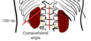
Report: No costovertebral angle tenderness is observed.
Why Check: Sign of inflammation in the area of the kidney, such as pyelonephritis, ureterolithiasis, or perinephric abscess. Positive renal tenderness mildly increases the likelihood of suspected ureterolithiasis (LR+ 3.6), while absence of renal tenderness moderately decreases the likelihood of suspected ureterolithiasis (LR- 0.2). When present with fevers, nausea/vomiting, malaise, and/or UTI signs, positive CVA tenderness may be more concerning for suspected pyelonephritis.
Bladder Palpation
(Also seen in Ch 41 – Adult Palliative Care Assessment – IF)
Say: I will palpate the lower abdomen for a distended bladder. Could you please lie flat on the exam table?
Setup: Expose lower abdomen to be able to palpate/landmark symphysis pubis.
Do: Palpate over lower abdomen – check if bladder palpable above symphysis pubis
Report: I detect an enlarged bladder on palpation – would recommend a bladder scan.
Why Check: Urinary retention can cause post-obstructive acute kidney injury, with inability to pass urine, suprapubic pain, and delirium. While a Bladder Scan (ultrasound) is best to assess bladder volume, a palpable bladder has an LR+ of 1.9, LR- of 0.3 for detecting ≥400mL in bladder5 to determine who would benefit from a bladder scan and intervention e.g. with catheterization if appropriate. Causes include urinary outlet obstruction (e.g. benign prostatic hypertrophy, pelvic organ prolapse, constipation), neurogenic causes (e.g. spinal cord compression, stroke) and medication causes (e.g. anticholinergics, opioids)
Auscultation for Bruits
Abdominal Bruits Exam
AFMC level Pre-clerkship 3 · Clerkship 4
(Further explored in Ch 13– Abdo Exam – MF3)
Say: I will use the bell of my stethoscope to auscultate key arteries for any signs of turbulent blood flow.
Do: With patient in supine position, auscultate epigastric, R/L renal, R/L iliac and R/L femoral arteries at the following locations:
- Abdominal Aorta: Slightly left and superior to umbilicus
- Right and Left Renal: 5cm superior and 5cm lateral to umbilicus
- Right and Left Iliac: 5cm inferior and 5cm lateral to umbilicus
- Right and Left Femoral: Midway between ASIS and Symphysis pubis
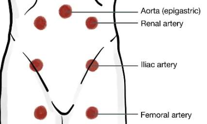
If you detect a renal bruit (“whoosh” sound), clarify if it is systolic or systolic-diastolic (e.g. via timing of carotid upstroke on palpation). Also clarify if it unilateral vs. bilateral.
Report: I note no bruits on auscultation.
Why Check: In a normal vessel, a pulse can be palpated but not auscultated. Bruits occur due to turbulent flow within a vessel, where the pitch and radiation are a function of flow and direction of the turbulent stream. Bruits can be innocent (e.g. in young, normotensive and asymptomatic patients), typically described as short, faint and mid-systolic. A systolic-diastolic abdominal bruit greatly increases the likelihood of renovascular hypertension (LR+ 39, LR- 0.62) (e.g. renal artery stenosis), a cause of secondary hypertension.
Helpful Stethopedia Videos to Review
- Video 1 – Renal and Volume Status Exam OSCE Style (Link: https://videopress.com/v/ej80D4WR)
Part 4: Relevant Likelihood Ratios – from Rational Clinical Examination JAMA series
Clinical Question 1: Does this woman have an Uncomplicated UTI (Acute)?6
| Procedure | LR+ (95% CI) | LR- (95% CI) |
|---|---|---|
| Dipstick Result | 4.2 (about +15%) | 0.3 (about −30%) |
| Dysuria | 1.5 (1.2-2.0) (about +15%) | 0.5 (0.3-0.7) (about −15%) |
| Frequency | 1.8 (1.1-3.0) (about +15%) | 0.5 (0.4-1.0) (no change: 95% CI includes 1) |
| Vaginal Discharge | 0.3 (0.1-0.9) (about −30%) | 3.1 (1.0-9.3) (no change: 95% CI includes 1) |
| Vaginal Irritation | 0.2 (0.1-0.9) (about −30%) | 2.7 (0.9-8.5) (no change: 95% CI includes 1) |
Clinical Question 2: Does this child have a UTI?7
Table 1: Non-Verbal Children
| Procedure | LR+ (95% CI) | LR- (95% CI) |
|---|---|---|
| Suprapubic Tenderness | 4.4 (1.6-12.4) (about +15%) | 0.96 (0.90-1.01) (no change: 95% CI includes 1) |
| Un-circumcised male infants | 2.8 (1.9-4.3) (about +15%) | 0.33 (0.18-0.63) (about −30%) |
| Jaundice | 2.1 (0.3-17.4) (no change: 95% CI includes 1) | 0.98 (0.91-1.06) (no change: 95% CI includes 1) |
| Ill Appearance (infants 3-24 mo.) | 1.9 (1.5-2.4) (about +15%) | 0.68 (0.53-0.88) (about −15%) |
| No other source for fever on Px | 1.4 (1.1-1.8) (about +15%) | 0.69 (0.55-0.80) (about −15%) |
| Ill Appearance (infants <3mo) | 0.59 (0.22-1.59) 1.1 (0.9-1.3) (no change: 95% CI includes 1) | 1.03 (0.99-1.08) 0.95 (0.84-1.08) (no change: 95% CI includes 1) |
Table 2: Verbal Children
| Procedure | LR+ (95% CI) | LR- (95% CI) |
|---|---|---|
| Abdominal Pain | 6.3 (2.5-16.0) (about +30%) | 0.80 (0.65-0.99) (about −15%) |
| New Onset Urinary Incontinence (Day/Night) | 4.6 (2.8-7.6) (about +15%) | 0.79 (0.69-0.90) (about −15%) |
| Back Pain | 3.6 (2.1-6.1) (about +15%) | 0.84 (0.75-0.95) (about −15%) |
| Frequency | 2.8 (2.0-4.0) (about +15%) | 0.72 (0.60-0.86) (about −15%) |
| Dysuria | 2.4 (1.8-3.1) (about +15%) | 0.65 (0.51-0.81) (about −15%) |
| Dysuria/Frequency | 2.2 (1.1-4.3) (about +15%) | 0.71 (0.45-1.13) (no change: 95% CI includes 1) |
| Offensive Urine Odor | 0.93 (0.34-2.51) 0.82 (0.34-1.96) (no change: 95% CI includes 1) | 1.01 (0.95-1.07) 1.2 (0.6-2.4) (no change: 95% CI includes 1) |
Clinical Question 3: Does this patient have Ureterolithiasis?8
| Procedure | LR+ (95% CI) | LR- (95% CI) |
|---|---|---|
| Microscopic hematuria | 73.1 (41.7-128.1) (about +45%) | 0.3 (0.2-0.4) (about −30%) |
| Loin tenderness | 27.7 (10.7-71.9) (about +45%) | 0.9 (0.8-0.9) (about −15%) |
| Renal tenderness | 3.6 (3.1-4.1) (about +15%) | 0.2 (0.1-0.3) (about −30%) |
Clinical Question 4: Is Listening for Abdominal Bruits Useful in the Evaluation of Hypertension?9
| Finding | LR+ (95% CI) | LR- (95% CI) |
|---|---|---|
| Systolic-Diastolic Bruits | 39 (10-145) (about +45%) | 0.62 (0.49-0.73) (about −15%) |
| Systolic Bruit | 4.3 (2.3-8.0) (about +15%) | 0.52 (0.34-0.78) (about −15%) |
| History of Atherosclerotic Disease | 2.2 (1.8-2.8) (about +15%) | 0.52 (0.40-0.66) (about −15%) |
References
-
Recommendations. Choosing Wisely Canada. Accessed August 1, 2022. https://choosingwiselycanada.org/recommendations/#list ↩
-
Centers for Disease Control and Prevention. Accessed March 11, 2019. https://www.cdc.gov/std/treatment/sexualhistory.pdf ↩
-
National LGBT Health Education Center. Taking Routine Histories of Sexual Health: A Systems Wide Approach for Health Centers. 2015. ↩
-
Canadian Public Health Association. Discussing Sexual Health, Substance Use and STBBIs. 2017. ↩ ↩2
-
Weatherall M, Harwood M. The accuracy of clinical assessment of bladder volume. Arch Phys Med Rehabil. 2002;83:1300-1302. ↩
-
Does this woman have an acute uncomplicated urinary tract infection – update. In: Rational Clinical Examination Series. Chapter 51. 2009:689. ↩
-
Shaikh N, Morone N, Lopez J. Does this child have a urinary tract infection? JAMA. 2007;298:2895-2904. ↩
-
McGee S. Evidence Based Physical Diagnosis. Saunders; 2012:830. ↩
-
Is listening for abdominal bruits useful in the evaluation of hypertension? – update. In: Rational Clinical Examination Series. Chapter 3. 2009:35. ↩