Medical Foundations 2
Chapter 12Dermatological Examination and Longitudinal Dermatology Curriculum Overview
Overview of Pre-Clerkship Clinical Skills Dermatology Curriculum at McMaster University
- Dermatology clinical skills is structured as a longitudinal learning path with integrated American Academy of Dermatology Curriculum Modules (https://www.aad.org/member/education/residents/bdc). Students are assigned 4 modules per MF to support asynchronous student learning of dermatology in pre-clerkship. The modules integrate findings for patients with skin of colour within case-based vignettes and includes assessment quizzes. A total of 20 of the 30 AAD Basic Curriculum modules are required to be completed in pre-clerkship. Students may complete the other 10 modules on an optional basis.
- For more information on the Dermatology Clinical Skills Longitudinal Integrated Learning Path, visit (https://www.medportal.ca/ug/mf/1/integrated-dermatology-curriculum) – Requires Medportal Login.
- The McMaster Clinical Skills Guide is not intended to be a comprehensive guide to dermatology. It is intended to help establish a foundation of dermatological clinical skills and lesion/rash recognition to build on through the longitudinal learning path supported by American Academy of Dermatology modules, select Clinical Skills Active Learning LGS series in dermatology, and through clinical learning experiences.
Helpful Resources
- Required Preparation – American Academy of Dermatology (AAD) Basic Curriculum e-modules (https://www.aad.org/member/education/residents/bdc): The AAD has a 2-week Basic Dermatology Curriculum, with 30 online modules designed around core principles of dermatology that all physicians should know. Each module is listed as less than 30 minutes in length. The curriculum also integrates findings in patients with skin of colour through case examples.
- McMaster Dermatology Pocket Guide (https://ecampusontario.pressbooks.pub/macdermpocketguide/): This pocket guide developed by McMaster medical students, dermatology fellows and faculty is created to serve as a concise, photo-rich reference for common skin conditions students will encounter in clerkship particularly in Internal Medicine and Family Medicine. It is intended as a practical tool to support your learning on the wards and in clinic. The dermatology pocket guide is developed separately from the McMaster Clinical Skills Guide and the McMaster UGME program.
- DermNet NZ (www.dermnetnz.org): A free resource to support informed, evidence-based decisions on how to care for skin conditions. DermNet generally encourages use of watermarked images for the purpose of education within a Creative Commons licence. All images included in this chapter are credited to DermNet, presented unmodified, with links to each image available in the References section. Please note that the McMaster Clinical Skills Guide is not affiliated with DermNet and does not endorse the McMaster Clinical Skills Guide or use of DermNet images.
- VisualDx (https://library.mcmaster.ca/databases/visualdx): Diagnostic decision support system designed by clinicians to aid medical professionals in diagnosis of visually identifiable diseases at the point of care. This includes a helpful dermatology section with a dedicated portion on dermatology for skin of colour. It also includes helpful images for findings seen on inspection for other clinical exams.
- British Academy of Dermatology (BAD) Resources for Medical Students (https://www.bad.org.uk/education-training/medical-students/): Helpful free-to-access educational reference for medical students, including a Handbook and the BAD’s Medical Student App, available on iOS and Google Play.
Helpful Resources focused on Inclusive Dermatology Clinical Skills
- Required Preparation – Mind the Gap (https://www.blackandbrownskin.co.uk/mindthegap): Mind the Gap is a clinical handbook developed in the UK to illustrate signs and symptoms in black and brown skin. It is a helpful resource for medical students, including common dermatologic findings for patients with systemic diseases.
- American Academy of Dermatology (AAD) Skin of Colour Curriculum: The AAD Skin of Colour Curriculum is intended to serve as a complete in-patient and out-patient curriculum for the diagnosis and effective treatment of diseases and conditions in patients with skin of colour (https://edhub.ama-assn.org/aad-education/provider-referrer/13778). The Skin of Color Curriculum features five guides that include pediatric, inpatient, outpatient, surgical, and cultural competence. The primary audience for this curriculum is dermatology residents, faculty, and medical students on a dermatology rotation.
- University of New Mexico Inclusive Dermatology Photo Gallery (https://hsc.unm.edu/medicine/departments/dermatology/inclusive-dermatology/gallery.html): This resource includes high-quality images of common and can’t-miss dermatologic findings for patients with skin of colour.
Part 1: Focused History – Based on Chief Complaint (Begin with Open-Ended Questions)
HPI & Associated Symptoms
All dermatological exams
The TEST mnemonic can be used for any dermatological history to ensure that the basics are covered after seeing every patient:
- Timing: When did this start? When was the first episode?
- Evolution: Has it changed in any way (size, shape, texture, colour)?
- Symptoms: Is there any burning, itching, pain, or bleeding?
- Treatments: What topicals and systemics have they tried? Did they work? Were there adverse reactions to previous treatments?
Rash
- When did it start? When was the first episode?
- Where on the body did it start? How has it spread (pattern of spread)?
- Any injury or trauma to the site?
- Any discolouration? Have the individual lesions changed in appearance (evolved)?
- Any provoking factors or triggers that make it worse?
- Does it itch, burn, or hurt?
- Pain out of proportion with localized numbness over lesion (re: necrotizing fasciitis)?
- Any bleeding or discharge? Colour of discharge?
- Tried any systemic or topical agents which helped in lesions’ appearance or symptoms?
- Can’t Miss Diagnoses:
- New rash in patient with chronic immunosuppression (e.g. solid organ transplant patients, HIV infection, long-term glucocorticoid use)
- Differential Diagnosis includes HIV; Kaposi’s Sarcoma; Disseminated Herpes Zoster; Shingles; Meningococcemia; Steven Johnson Syndrome (SJS); Toxic Epidermal Necrolysis (TEN); Syphilis; Erythema Multiforme Major; Thrombotic thrombocytopenic purpura (TTP); Necrotizing fasciitis; Disseminated fungal infection.
- New rash in patient with recent travel
- Dengue fever: Mosquito-borne illness; many areas of outbreaks in the world (Americas, Africa, Middle East, Asia, Pacific Islands – about 4 billion people live in areas with a risk of Dengue)
- Typhus: Includes epidemic typhus (Louse-borne illness caused by Rickettsia prowazekii); scrub typhus (mite-borne illness caused by Orientia tsutsugamushi); and murine typhus (Flea-borne illness caused by Rickettsia typhi)
- Lyme Disease: Tick-borne illness caused by Borellia burgdorferi; risk with travel to endemic areas, transmission most likely from May to October.
- Rocky Mountain Spotted Fever (RMSF): Tick-borne illness caused by Rickettsie rickettsii; mainly northern climate in spring, later in southern climates.
- Zika Virus: Mosquito-borne viral illness transmitted primarily by Aedes mosquitoes; may also be a sexually transmitted infection (e.g. vaginal, anal, oral sex), or via maternal-fetal transmission. Widespread in tropical and subtropical regions, including parts of Africa, the Americas, Asia, and the Pacific. Symptoms are usually mild but can include fever, rash, joint pain, and conjunctivitis. For pregnant patients with Zika virus exposure, there is association with congenital microcephaly.
- Leptospirosis: Bacterial illness transmitted through contact with water or soil contaminated with urine from infected animals; common in tropical areas, particularly after heavy rainfall or flooding. Symptoms include fever, headache, muscle aches, and jaundice.
- Schistosomiasis: Parasitic infection acquired through contact with contaminated freshwater bodies infested with Schistosoma parasites; prevalent in parts of Africa, South America, the Caribbean, and Southeast Asia. Symptoms vary but may include fever, abdominal pain, and skin rash.
- New rash in patient with chronic immunosuppression (e.g. solid organ transplant patients, HIV infection, long-term glucocorticoid use)
Growth
- When did you first notice the lesion was present?
- Has the lesion changed since you first noticed it? – Changed in size, shape, or colour?
- Does the lesion itch? Does it bleed?
Burns
- How and when did the burn occur?
- Was it a thermal burn (e.g. heat) or non-thermal burn (radiation, chemical, electrical)?
- Is it painful? – Absence of pain raises concern for full thickness burn.
- Is, or was there any blistering? – Suggests superficial or partial thickness burn.
- When was your last tetanus shot? – Burns have increased risk of C. tetani proliferation.
- What treatments have you tried?
- Shock symptoms: Any light-headedness, difficulty breathing, loss of consciousness, etc.? – due to large losses of fluid and proteins with burn injuries.
Constitutional Symptoms
- Any fevers, night sweats, chills, hot flashes, weight loss? (re: infection or malignancy)
- Altered mental status?
Past Medical History (PMHx)
- Full Medical History - Any chronic medical conditions which affect the skin?
- Past Skin Cancer Diagnoses: Basal Cell and Squamous Cell Carcinoma are most common.
- Skin Cancer risk factors: Chronic immunosuppression; previous radiation therapy; chemical exposure (e.g. arsenic), UV exposure (including tanning beds), skin pigmentation differences (e.g. Basal Cell Carcinoma most common in patients with lighter skin tones; Squamous Cell Carcinoma most common in patients with darker skin tones)
- Atopic Triad
- (1) Asthma
- (2) Allergies (environmental allergies e.g. ragweed, hay fever, tree, pollen)
- (3) Eczema (e.g. red itchy rashes on back of knees or inside of elbows)
- Diabetes Mellitus: Increases risk of cellulitis and foot ulcers (↑ risk of osteomyelitis)
- Peripheral Vascular Disease: Impaired wound/ulcer healing – for Peripheral Arterial Disease, look for Critical Limb Ischemia (≥2 weeks of ischemic rest pain, non-healing wound/ulcer, or gangrene in 1 or more legs attributable to objectively proven arterial occlusive disease) as a Can’t-Miss Diagnosis.
Past Surgical History (PS Hx)
- Melanoma: When was your surgery? Do you recall Breslow tumor depth? Any ulceration? Were lymph nodes removed?
- Breslow Tumor Depth informs tumor staging: T1 = ≤ 1mm; T2 = 1-2mm; T3 = 2-4mm; T4 = >4mm.
- Ulceration informs tumor staging: For given staging, ulceration =Stage b (e.g. T3b - >4mm thickness with ulceration)
- Lymph Nodes (LN) inform tumor staging: N0 = no LN involved; N1 = 1 LN; N2 = 2-3 LN; N3 = 4+ LN.
- Past Diagnostic Dermatology Interventions: Any punch biopsies, shave biopsies, or excisional biopsies? Mohs surgery?
- Punch biopsy: Cone-shaped core biopsy widest at skin surface and narrowest at biopsy base using punch biopsy tool.
- Shave biopsy: Biopsy nearly parallel to skin surface, extending to epidermis only (superficial shave biopsy) or to limited superficial dermis (deep shave biopsy) using scalpel blade – do NOT use if concern for melanoma.
- Excisional biopsy: Allows for histopathologic examination of entire lesion –helpful for biopsy of suspected melanoma.
- Mohs surgery: Specialized surgical technique to remove locally invasive, high-risk skin cancers.
Family Medical History (FMHx)
- Any family history of similar skin conditions? Any family members with biopsy-confirmed melanoma?
Social Hx
- Occupational History: Occupational sun UV exposure or history of extensive sun exposure.
- Alcohol: A common trigger for flushing in patients with rosacea, while alcohol associated hepatic dysfunction can result in spider angiomas, palmar erythema, and pruritis
- Smoking: Negatively impacts wound healing, accelerates skin aging process, and is a risk factor for psoriasis, hidradenitis suppurativa, systemic lupus erythematous, and skin cancers
- Environmental Exposures: Exposure to environmental pollutants, toxins, or allergens can contribute to conditions like contact dermatitis, allergic reactions, or chemical burns. Previous tanning bed use, history of extensive UV exposure, or history of severe sunburns significantly increases risk of skin cancer.
- Stress: Psychological stress can exacerbate existing skin conditions like eczema, psoriasis, or acne, and may also trigger conditions such as alopecia areata or urticaria.
Medications Hx & Allergies
- Drug Allergies: Be sure to ask what the allergic reaction looked like - patients may incorrectly name side effects as drug allergies (e.g. acute nausea/vomiting) which doesn’t qualify as a drug reaction; or they might not remember the details.
- Red Flag – Any new medication changes? – Consider Drug Eruptions (Drug-Induced Skin Reaction), including:
- Immediate Reaction (<1 hr after start of medication) – e.g. Urticaria, Angioedema, and Anaphylaxis
- Delayed Reaction (>6 hrs after med start) – e.g. Exanthematous eruptions, fixed drug eruption, DRESS (Drug Reaction with Eosinophilia and Systemic Symptoms), Steven Johnson Syndrome (SJS)/ Toxic Epidermal Necrolysis (TEN)
- SJS/TEN High Risk Medications – 5As + I:
- Allopurinol – also high risk for DRESS
- Anti-epileptics (e.g. phenytoin, lamotrigine, phenobarbital, carbamazepine) – also high risk for DRESS
- Antibiotics (sulfamethoxazole, sulfonamides including sulfasalazine – a Disease-Modifying Anti-Rheumatic Drug) – also high risk for DRESS
- Anti-inflammatories (especially Oxicam NSAIDs)
- Anti-retroviral therapy (nevirapine) – also high risk for DRESS
- Immune checkpoint inhibitors (pembrolizumab, nivolumab)
- Additional high-risk medications for DRESS: Vancomycin, Minocycline, Anti-Tuberculosis Medications (e.g. rifampicin, ethambutol, isoniazid, pyrazinamide)
- Thiazide Diuretics (e.g. hydrochlorothiazide) – photosensitizing drug with ↑ risk of damage from UV exposure.
- Chronic Dermatological Condition Treatments:
- Barrier Creams, Emollients and Moisturizers: e.g. in atopic dermatitis, skin hydration and emollients are key to management, and also helpful adjunct in psoriasis; use of gentle skin cleansers in acne vulgaris and rosacea
- Topical Steroids – Principle: use lowest potency steroid for shortest duration to achieve clinical benefit. 7 groups according to potency (Group 1 – Super High Potency → Group 4 – Medium Potency → Group 7 – Least Potent)
- Caution: If dermatophyte present, topical steroids could exacerbate cutaneous fungal infections.
- Topical steroids may exacerbate some inflammatory conditions (e.g rosacea and perioral dermatitis)
- Topical calcineurin inhibitors (e.g. tacrolimus, pimecrolimus) – e.g. used in mild-moderate atopic dermatitis and in treatment of psoriasis in sensitive areas (facial, intertriginous)
- Topical Antibiotics: e.g. Topical metronidazole in rosacea; Topical clindamycin in mild acne vulgaris.
- Additional Topicals in mild Acne Vulgaris: Retinoids (e.g. tretinoin, adapalene), salicylic acid, benzoyl peroxide
- Oral Antibiotics: Tetracyclines (e.g. tetracycline, doxycycline, minocycline) in moderate-severe rosacea or acne vulgaris
- Anti-Androgenic Hormonal Therapies: e.g. Combined Oral Estrogen-Progestogen Contraceptives (OCP), Spironolactone in moderate-severe acne vulgaris in female patients.
- Isotretinoin: Treatment for severe nodulocystic acne, or acne with scarring (significant risks including teratogenicity, increased liver enzymes, dyslipidemia, etc. – requires close monitoring with dose titration until treatment response)
- Biologic Agents (e.g. dupilumab – a IL-4/13 inhibitor in moderate-severe atopic dermatitis; TNF-α inhibitors e.g. etanercept, infliximab, adalimumab in psoriasis)
- Disease-Modifying Anti-Rheumatic Drug (DMARD): e.g. methotrexate, cyclosporine. Used in psoriasis, scleroderma, etc.
- Phototherapy: UVA and/or UVB used to treat various conditions (e.g. psoriasis; atopic dermatitis to control pruritus)
Part 2: Pertinent Notes about the Physical Exam
Process of Total Body Skin Examination
- Setup: You should have a ruler to accurately measure lesion sizes; a penlight to help distinguish flat and raised lesions, and solid vs. fluid-filled lesions; and a tool to magnify lesions for closer inspection
- Draping: In this examination, draping is particularly important, with sequential exposure of examined areas after obtaining consent. For dermatological exam in sensitive areas, consider the role of a chaperone. Recommend a Head-to-Toe exam.
- Modification for Patients who are Transgender:
- Transmasculine Patients (FTM): Patients on masculinizing hormone therapy may have oilier skin, more acne, increased facial and body hair, or decreased scalp hair – important to normalize these changes.
- Transfeminine Patients (MTF): Patients on feminizing hormone therapy may have softening of skin/decreased skin oiliness, less acne, thinning/slowed growth of facial and body hair, or arrested progression of scalp hair loss – important to normalize these changes.
Part 3: Approach to Lesions1
Primary Lesions – Directly Associated with Disease Process
| Descriptor | Small | Large |
|---|---|---|
| Raised | Papule (<1cm) | Plaque (>1cm) |
| Not Raised | Macule (<1cm) | Patch (>1cm) |
| Solid | Nodule (1-2cm) | Mass/Tumor (>2cm) |
| Fluid-Filled | Vesicle (<1cm) | Bulla (>1cm) |
Additional Primary Lesions
- Pustule (<1cm): Vesicle filled with pus.
- Wheal: Firm, edematous, transient, circumscribed papule/plaque due to infiltration of dermis with fluid, often with erythematous borders & pale center (e.g. urticaria)
Secondary Lesions – Evolution of Primary Lesion or Modification from Patient’s Activities
Breaks
- Erosions: Focal loss of epidermis, surface break (non-scarring)
- Excoriation: Erosion of epidermis, skin abrasions (usually caused by scratching)
- Ulcers: Focal loss of epidermis + dermis, deeper break (scarring)
- Fissure: Epidermis, linear/wedge shaped sharply defined tear
Additional Secondary Lesions
- Scale: Visible fragments of stratum corneum (outermost epidermis layer) shed from skin
- Crust: Liquid exudate (serum/pus) dried on surface of skin – “scab”
- Lichenification: Area of epidermal thickening with accentuated skin lines due to scratching
- Petechiae: Round, pinpoint, flat purplish spot due to intradermal or subdermal hemorrhage, <5mm diameter
- Purpura: Blue or purple in colour; secondary to hemorrhage in skin; >5mm diameter
- Scar: excess collagen production after injury, secondary to injury or surgery
Approach to Lesions Based on Morphology
Skin Cancer
Description:
- Generally macules or papules, sometimes nodules or patches
- Caused by UV exposure
Examples:
- Basal Cell Carcinoma (BCC)
- Squamous Cell Carcinoma (SCC)
- Merkel Cell Carcinoma (MCC)
- Melanoma
Papulosquamous Disease
Description: WERS Mnemonic:
- Well-defined
- Elevated
- Red
- Scales
Examples:
- Psoriasis (plaque, guttate, pustular, erythrodermic inverse)
- Lichen planus (7Ps: Papule, Purple, Pruritic, Planar, Polygonal, Periphery, Penis) – found on ventral wrists, anterior shins, trunk.
- Pityriasis rosea (1st phase herald patch, 2nd phase Christmas tree pattern following Langer’s lines of skin tension)
- Dermatophyte infection (scales at leading edge, central clearing)
- KOH exam to rule out dermatophyte cause - “all that scales must be scraped”
- Secondary syphilis (palm and sole involvement, lymphadenopathy)
Dermatitis (Eczema)
Description:
- Inflammation of the skin
- Poorly demarcated erythematous patches or plaques
Examples:
- Asteatotic Dermatitis (“winter itch” common in older adults)
- Atopic Dermatitis
- Contact Dermatitis (irritant versus contact mechanisms)
- Dyshidrotic Dermatitis
- Nummular Dermatitis
- Seborrheic Dermatitis
- Stasis Dermatitis
- Perioral Dermatitis
Vesiculobullous Diseases
Description: Vesicular and/or bullous (fluid-filled) morphologies
Examples:
- SJS and TEN - a spectrum of disease
- SJS < 10% BSA
- TEN >30% BSA, blistering eruptions with mucosal membrane involvement
- Bullous Pemphigoid
- PemphigoiD = Dee
- Tense blistering
- Negative Nikolsky sign
- Pemphigus Vulgaris
- VulgariS = Superficial blistering disease
- Positive Nikolsky sign
- Mucosal membrane involvement
- Dermatitis Herpetiformis (associated with Celiac disease)
Approach to Can’t Miss Diagnoses
Petechial/Purpuric
Red lesions caused by breaks in capillaries → bleeding into skin. Non-blanchable. May or may not be raised/palpable. May appear brown on darker skin tones.
- Petechiae <5mm
- Purpura >5mm
- Febrile/Toxic Appearing
- Palpable: Meningococcemia, Disseminated gonococcal infection (e.g. IgA Vasculitis), Endocarditis, Rocky Mountain Spotted Fever (RMSF)
- Non-palpable: Disseminated Intravascular Coagulation (DIC), Thrombotic thrombocytopenic purpura (TTP), Purpura fulminans
- Afebrile/ Non-Toxic Appearing
- Palpable: Autoimmune vasculitis (e.g. in SLE, RA, Sjogren’s Syndrome, Systemic Vasculitides)
- Non-palpable: Idiopathic thrombocytopenic purpura (ITP)
Erythematous
Red skin secondary to capillary congestion. Assess whether Nikolsky Sign is present (skin sloughs off, leaving red raw base). May appear ashen gray or dark brown on darker skin tones.
- Febrile
- Nikolsky +: Steven Johnson Syndrome (SJS)/Toxic epidermal necrolysis (TEN), Staphylococcal scalded skin infection (SSSS - pediatrics)
- Nikolsky -: Toxic Shock Syndrome (TSS; mucous membranes), Kawasaki Disease (pediatrics), Scarlet Fever (“sandpaper rash”)
- Afebrile
- Nikolsky +: SJS/TEN
- Nikolsky -: Anaphylaxis, Cellulitis
Maculo-papular
Seen in viral illnesses, bacterial infections, drug reactions and immune-mediated syndromes. Very common with a wide DDx.
- Macules – Flat, red circular
- Papules – Solid, raised
- Febrile
- Central (chest, abdo, back): Viral exanthems, Lyme disease (Erythema migrans)
- Peripheral (extremities)
- Target lesions: Early SJS/TEN (atypical target lesions), erythema multiforme
- No target lesions: Meningococcemia, RMSF, Syphilis, Lyme disease (Erythema migrans)
- Afebrile
- Central: Drug reaction, Pityriasis (herald patch)
- Peripheral
- Flexors: Eczema, Scabies
- Extensors: Psoriasis
Vesiculo-bullous
Fluid-filled lesions with involvement of the dermal-epidermal junction.
- Vesicles <1cm
- Bullae >1cm
- Pustule – pus-filled
- Febrile
- Diffuse: Varicella, Smallpox, Disseminated gonococcal infection, Purpura fulminans, DIC
- Localized: Necrotizing fasciitis; Hand-foot-and-mouth disease
- Afebrile
- Diffuse: Bullous pemphigoid, Pemphigus vulgaris
- Localized: Contact dermatitis, Herpes zoster, Dyshidrotic eczema, Burns
Part 4: Skin Cancers
Skin Cancer In Darker Skin Tones2
Skin cancer is one of the most common malignancies in Canada and has traditionally been associated with lighter skin, as it is much more common in Caucasians. Despite this, skin cancer does present in people with darker skin, and often early signs can get missed. This oversight can lead to a delayed diagnosis, with skin cancer typically being detected at more advanced and aggressive stages in people of colour compared to their lighter-skinned counterparts. The following information can help us understand the clinical picture of three common skin cancers in those with lighter vs darker skin.
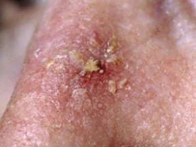
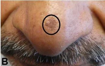
Actinic keratosis (AK) – a premalignant Squamous Cell Carcinoma lesion
Actinic Keratoses arise from keratinocyte cells in the epidermis. They are part of a disease spectrum from photo-damaged skin → Actinic Keratosis → Squamous Cell Carcinoma. Not all AKs transform or progress to Squamous Cell Carcinomas (SCC) – annual risk of lesion transformation from AK to SCC is about 8%.
| Characteristic | Lighter Skin | Darker Skin |
|---|---|---|
| Risk Factors | UV exposure, increased age, male sex, fair skinned individuals, immunosuppression, occupational sun exposure. | UV exposure, increased age, immunosuppression, genetic factors, occupational sun exposure. |
| Typical Clinical Presentation | Ill demarcated, extremely flat, yellow to brown scale, erythematous papule with a subtle, adherent scale that feels like sandpaper. May be tender to touch. | Ill demarcated, extremely flat, reddish-brown papule with a subtle, adherent scale that feels like sandpaper. May be tender to touch. |
| Anatomic Distribution | Head, neck, extensor forearms, dorsum of hands, and back region. | Head, neck, extensor forearms, dorsum of hands, legs, and groin. AKs can also appear in less sun-exposed areas in darker skin tones. |
Basal Cell Carcinoma (BCC)
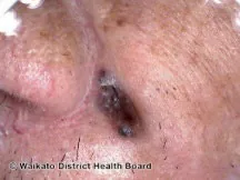
Basal Cell Carcinomas arise from the basal layer of the epidermis. BCC is the most common skin cancer in those with lighter skin. It is also more common in those with skin of colour that are lighter in complexion. While the clinical features are similar between the two groups, it is important to recognize that the risk factors and pigmentation of the lesion can vary significantly.
| Characteristic | Lighter Skin | Darker Skin |
|---|---|---|
| Risk Factors | Sun exposure (including occupational), fair skin, previous radiation therapy, genetic disorders. | Sun exposure (including occupational), scars, ulcers, previous radiation therapy, genetic disorders. |
| Typical Clinical Presentation | Erythematous, pearly papule with central erosion and telangiectasias. | Pigmented BCC with a black, pearly appearance (pigmented BCC often leads to incorrect diagnoses of seborrheic keratoses). |
| Anatomic Distribution | Head and neck regions | Head and neck regions |
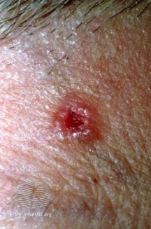
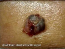
Squamous Cell Carcinoma (SCC)
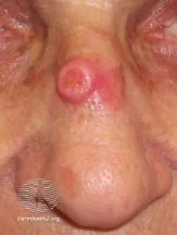
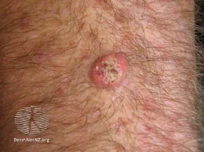
Squamous cell carcinomas arise from keratinocyte cells in the epidermis. Squamous Cell Carcinomas are not as common as Basal Cell Carcinomas, only comprising 20% of all skin cancers. However, it is the most common skin cancer for those with darker complexions. The risk factors for SCC vary significantly as people with darker skin tend to be predisposed to SCC if they have chronic or severe scars. The anatomic distribution significantly differs as well. SCC can present in a more advanced and even metastatic disease state in those with darker skin due to missed signs in earlier stages.
| Characteristic | Lighter Skin | Darker Skin |
|---|---|---|
| Risk Factors | Sun exposure, fair skin, immunosuppression, HPV infection, scarring, occupational sun exposure. | Chronic and/or severe scarring/inflammation from burns, leg ulcers, radiation, lupus, immunosuppression, HPV infection, occupational sun exposure. |
| Typical Clinical Presentation | Hyperkeratotic superficial lesion arising from an indurated, rounded, elevated red to violaceous base. | Sore that will not heal (bleeds or develops a crust). |
| Anatomic Distribution | Sun exposed areas (head, neck, hands) | Skin infrequently exposed to sun (legs) |
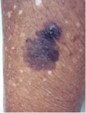
Melanoma
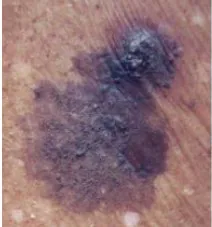
Melanomas arise from melanocytes in the basal layer of the epidermis. Melanomas are the third most common skin cancer in all skin groups, though it is also the deadliest due to high risk of metastasis. Therefore, it is important to recognize the differences in presentation and anatomic distribution, with a focus on screening and early detection for patients at increased risk. While all skin tones present with rapidly spreading, dark macules/patches, acral melanoma on the palms, soles and subungual (under nail) can be more common in those with darker skin.
| Characteristic | Lighter Skin | Darker Skin |
|---|---|---|
| Risk Factors | Sun exposure, fair skin, family history, increased number of moles (nevi), dysplastic nevi, occupation (e.g. farmer, construction worker, etc.). | Sun exposure, family history, occupational sun exposure (e.g. farmer, construction worker). |
| Typical Clinical Presentation | Dark, rapidly spreading macules or patches arising from pigmented nevi. | Dark, rapidly spreading macules or patches arising from pigmented nevi; Rapidly changing pigmented band on nail (Hutchinson’s sign). |
| Anatomic Distribution | Often on trunk or lower legs | Often on palmar, plantar and subungual areas |
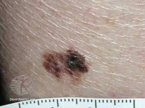
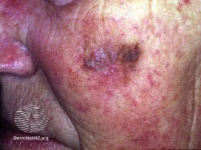
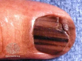
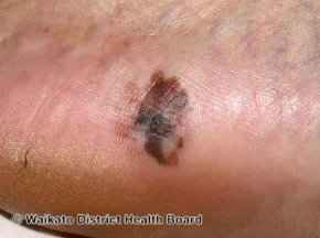
While the gold standard is excisional biopsy to examine histopathology, the first step used clinically in the physical examination is the ABCDE mnemonic. A patient with a lesion suspicious for melanoma should be urgently referred to dermatology.
| Descriptor | Melanocytic Nevi (Mole - Benign) | Melanoma (Malignancy) |
|---|---|---|
| Asymmetrical Shape | Symmetrical (one half matches the other) | May be asymmetrical (one half unlike the other) |
| Border Irregularity | Regular border (even, distinct border) | May have irregular borders (uneven, indistinct border) |
| Colour Variation | Uniform colour | May have multiple shades of red, blue, black, gray, or white |
| Diameter >6mm | <6 mm | May be >6mm |
| Evolution over time | Stable over time | May evolve over time (change in size, shape, colour) Melanomas growing in vertical axis (Breslow Depth of tumor) tend to have worse prognosis. |
It is important to be able to differentiate a mole (melanocytic nevus) from a melanoma, as follows:
Nevi
Benign circumscribed overgrowth of cell lines normally found in the skin.
- A mole is melanocytic nevus
Melanoma
A malignant tumor of melanocytes.
- Most common in areas of intermittent sun exposure – e.g. the back or the legs.
- Risk factors: light skin, atypical/dysplastic nevi, blistering sunburn history, and Family history
- Ugly Duckling sign: Most patients with multiple nevi have a “signature nevus” where nevi tend to have similar appearances. The Ugly Duckling sign refers to a pigmented lesion that is obviously different from others for a given patient.
Notes on Screening for Skin Cancer1213
- Screening Exams:
- Full Body Skin Exam – a focused inspection of entire surface of skin, particularly areas of sun-exposure by trained healthcare provider or dermatologist – Note: inclusion of genital exam here should be a discussion balancing patient preference and risk of patient discomfort against risk of genital melanoma (0.2-0.8% of all melanomas)12
- Skin Self-Exam – patient performs self-exam at home, where another person (e.g. partner, family member, friend) may help especially with hard to see areas (e.g. back, scalp) – helpful patient-instruction video (98 seconds)13
Very High Risk (≥10X risk of skin cancer vs. general population)
Risk Factors:
- On immunosuppressive therapy after organ transplant
- Personal history of skin cancer
- ≥2 first-degree relatives with melanoma
- More than 100 nevi in total or 5+ atypical nevi
- Received more than 250 treatments with psoralen-ultraviolet A (PUVA) radiation for psoriasis.
- Received photodynamic therapy for cancer as a child
Recommendation (Cancer Care Ont.)14:
- Offered total body skin exam by dermatologist or trained healthcare provider on yearly basis.
- Counseled about skin self-exam and skin cancer prevention.
- Monitor site of radiation therapy (for childhood cancer survivors)
High Risk (5X risk of skin cancer vs. general population)
Risk Factors:
- First degree relative with melanoma
- Many (50-100) nevi
- One or more atypical (dysplastic) nevi
- Naturally red or blond hair
- Blue or green eyes
- A tendency to freckle
- Skin that burns easily and tans poorly or not at all
- Others: Outdoor occupation, childhood spent at latitude <35° (Canada’s lowest latitude 41°), use of tanning beds during teens and 20s, radiation therapy as an adult.
Recommendation (Cancer Care Ont.)14:
- Counseled about skin self-exam (particularly focused on site of therapeutic radiation if pertinent) and skin cancer prevention.
- Should be seen once a year by healthcare provider trained in screening for skin cancers.
Average or Low Risk (Same as general population)
Risk Factors:
- None of the risk factors identified above
Recommendation (Cancer Care Ont.)14:
- No evidence for or against screening
- Routine total body skin exam is not recommended here.
- Routine counseling on skin self-exam is not recommended here.
Part 5: Skin and Soft Tissue Infections (SSTI)
- Common etiologies of SSTI: Trauma, wounds, breaks in skin, recent surgery, foreign bodies.
- Risk factors for Skin and Soft Tissue Infections: Older age; cardiopulmonary disease; hepatorenal disease; obesity; asplenia; Immunocompromise (e.g. patients on chemotherapy, patients with HIV); peripheral vascular disease; peripheral neuropathy; lymphedema; IV or subcutaneous drug use; human/animal bites; water exposure (salt or freshwater)
- Risk factors for Methicillin-Resistant Staphylococcus aureus (MRSA) Infections:
- Healthcare associated risk factors: Recent hospitalization; residence in long-term care; recent surgery; hemodialysis.
- Additional risk factors: HIV infection; Injection drug use; prior antibiotic use
- MRSA outbreak factors: Incarceration; military service; sharing sports equipment; sharing needles/razors/sharp objects.
Approach to Skin and Soft Tissue Infections (SSTI)
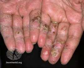
Impetigo15
Definition: Infection of the epidermis.
Clinical Features:
- Commonly in young children
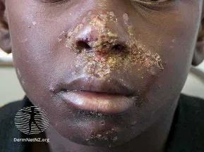
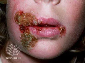
- Non-Bullous: Begins as a single erythematous macule evolving into pustules or vesicles that rupture with serous contents drying into “honey-crusted” lesions.
- Minimal or no surrounding erythema.
- No systemic symptoms (e.g. fever, chills).
- Bullous: Quickly appearing superficial, small or thin roofed bullae spontaneously rupturing and oozing yellow fluid with scaley rim (collarette).
- May have systemic symptoms (fever, malaise, lymphadenopathy).
Locations: Non-Bullous: Commonly on face (perioral) arms, legs
Bullous: Usually on face, trunk, extremities, buttocks, perineal.
Pathogens: GAS, S. aureus
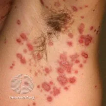
Example Images:
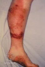
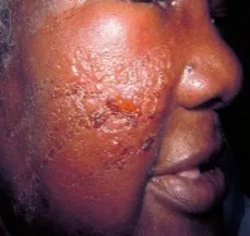
Erysipelas16
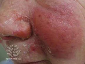
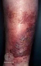
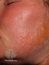
Definition: Infection of the upper dermis and cutaneous lymphatics.
Clinical Features:
- Sharp raised edge
- Well demarcated
- Warm plaque
- Erythematous
- Systemic symptoms indicate serious infection (spreads via lymphatics)
Locations: Face (e.g. butterfly distribution on cheeks across nasal bridge), legs
Pathogens: GAS (beta-hemolytic streptococci)
Example Images:
Cellulitis1718
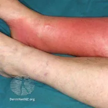
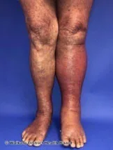
Definition: Infection of the deep dermis and subcutaneous fat.
Clinical Features:
- Flat
- Poorly demarcated
- Warm
- Erythematous
- Tender
- May have vesicles
- Not uniformly raised
- First sign of illness is often systemic symptoms (e.g. fever, chills, rigors – re. bacteremia)
- May be complicated by lymphadenopathy or abscess formation
Locations: Legs; can occur by itself, or a complication of underlying skin condition or wound.
Pathogens: GAS, S. aureus (most common), H. influenzae (Periorbital), Pasturella multocida (cat/dog bite)
Example Images:
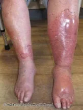
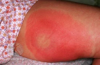
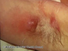
Cutaneous Abscess19
Definition: Cavity filled with pus (pyoderma or sepsis – with leukocytes, dead tissue and bacteria).
Clinical Features:
- Warm
- Erythematous
- Tender
- Swollen lump
- May be complicated by lymphadenopathy and systemic symptoms (e.g. fever), with risk of progression to septicemia
- Incision and Drainage is core part of treatment here
Locations: May occur anywhere on skin; Common under arms, base of spine (pilonidal disease), genitals, or anus.
Pathogens: S. aureus including MRSA most common.
Example Images:
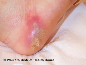
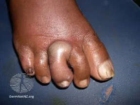
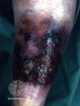
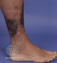
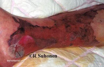
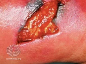
Necrotizing Fasciitis20
Definition: Rapidly progressing limb/life threatening infection of subcutaneous fat, deep fascia and muscle, with extensive necrosis of involved layers within hours.
Clinical Features:
- Diffuse erythema
- Extreme tenderness (pain out of proportion)
- Edema
- Induration
- Crepitus (due to gas in tissues)
- Bullae
- Purple skin discolouration
- Paresthesia (with destruction of peripheral nerves)
- Systemic symptoms (fever, chills, altered mental status, sepsis)
Locations:
- Most common site lower leg. Other sites include upper limb, perineum, buttocks, truck, head/neck. May progress from pre-existing SSTI.
- Fournier Gangrene: Necrotizing Fasciitis of external genitalia.
Pathogens:
- Type 1: Polymicrobial (e.g. mix of aerobic, anerobic pathogens)
- Type 2: Monomicrobial (e.g. Hemolytic GAS or S. aureus including MRSA)
- Type 3: Gas gangrene e.g. due to Clostridium
Example Images:
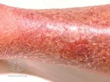
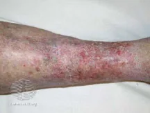
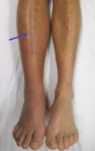
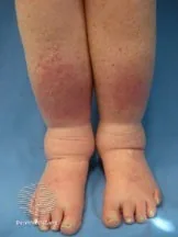
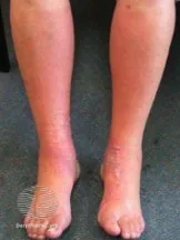
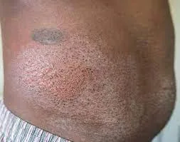
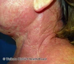
Common Skin and Soft Tissue Infection Mimics (Differential Diagnosis)
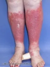
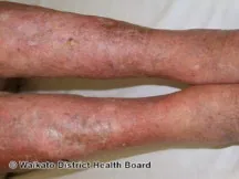
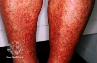
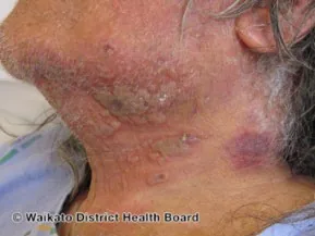
Part 6: Burns
Types of Burns
Burn Severity is determined by surface area and depth/degree of burn.
- Thermal: Injury related to contact temperature, exposure duration, thickness of skin (e.g. flames, hot liquids/solids, steam)
- Cold injury (e.g. frostbite due to intracellular ice crystal formation and microvascular occlusion causing tissue anoxia)
- Non-Thermal:
- Chemical
- Alkali burns (causes liquefaction necrosis of tissue) are generally more serious than acid burns (causes coagulation necrosis of tissue) because alkali materials able to penetrate deeper tissues
- If dry powder (chemical residue) is still present on skin, brush it away before irrigating with water
- Electrical
- e.g. high voltage >1000 volts; low voltage <1000 volts; arc flash burns; lightning strikes
- Electrical energy transforms into heat as current passes through poorly conducting body tissues
- Serious deep tissue injuries may present with less obvious superficial findings (e.g. underlying muscle necrosis)
- Radiation
- e.g. radiofrequency energy, ionizing radiation – including sunburn, therapeutic radiation, nuclear industry injury
- Injury related to dose, time of exposure, and type of particle exposure.
- Chemical
Effects of Burns
Damage to the skin leads to loss of the skin’s functions (more serious consequences in higher surface area or depth/degree):
| Function | Result | Treatment |
|---|---|---|
| Fluid regulation | Large losses of body water and proteins → hypovolemic | Aggressive fluid resuscitation (as per Rule of 9s and Parkland Formula) |
| Prevent infection | Loss of mechanical barrier (intrinsic immunity) → high risk of infection | Antimicrobial dressings, consider systemic antibiotics, tetanus prophylaxis |
| Thermoregulation | Loss of insulation → hypothermic | Keep warm |
Burns can also cause hemolysis and muscle damage (with hemoglobinuria or myoglobinuria leading to secondary acute tubular necrosis), Disseminated Intravascular Coagulation (DIC), Acute Respiratory Distress Syndrome (ARDS), cardiac arrhythmias (re. electrical burns), and sepsis/systemic inflammatory response syndrome (SIRS)/shock.
Three zones of burns (Jackson’s Burn Model)
| Zone | Location | Description | Viability |
|---|---|---|---|
| Zone of Hyperemia | Outermost | Inflammation leading to vasodilation and increased blood flow/perfusion | Completely viable - Cells recover within 7 days (unless severe sepsis or prolonged hypoperfusion) |
| Zone of Stasis | Middle | Decreased perfusion and early ischemia. | Progressive necrosis, may be viable – goal of resuscitation to increase tissue perfusion in zone of stasis |
| Zone of Coagulation | Innermost | Lack of blood flow and irreversible necrosis. Extent of determined by duration of contact and amount of heat transfer. | Irreversible necrosis → Non-viable |
Depth of Burns
1st Degree: Superficial
- Layers: Epidermis – sparing dermal-epidermal junction
- Clinical Features:
- Appearance: Erythema, edema, dry skin, no vesicles/bullae
- Pain: Localized
- Sensation: Intact
- Blanchable: Blanches with pressure
- Prognosis: Recovery in 3-6 days, no scarring
2nd Degree (2a): Superficial partial thickness
- Layers: + upper (papillary) dermis
- Clinical Features:
- Appearance: Erythema, edema, vesicle/bullae, hair follicles present
- Pain: Hypersensitivity (e.g. to touch, temperature, air current)
- Sensation: Intact
- Blanchable: Blanching may be sluggish
- Prognosis: Recovery in 1-3 weeks, minimal to no scarring
2nd Degree (2b): Deep partial thickness
- Layers: + deep (papillary and reticular) dermis
- Clinical Features:
- Appearance: Vesicles/bullae (fragile), Variable colour (white, mottled pink, red, patchy), some hair follicles, softer than 3rd degree
- Pain: May or may not be painful (to pressure only)
- Sensation: Dull; Painful to pressure only
- Blanchable: No
- Prognosis: 3+ weeks, scarring, recommend grafting. May progress to full-thickness burns in few days post-injury if untreated.
3rd Degree: Full thickness
- Layers: + subcutaneous tissue
- Clinical Features:
- Appearance: Necrosis with eschar (leather-like skin, waxy, white, grey or charred). Dried, inelastic.
- Pain: No (surrounding areas may be painful)
- Sensation: None
- Blanchable: No
- Prognosis: Requires debridement and grafting or flap
4th Degree: Deep full thickness
- Layers: + deeper structures (fascia, muscle, bone)
- Clinical Features:
- Appearance: Necrosis with eschar (leather-like skin, waxy, white, grey, or charred), dried, inelastic, thrombosed veins
- Pain: No (surrounding areas may be painful)
- Sensation: None
- Blanchable: No
- Prognosis: Requires debridement and amputation or flap
Total Body Surface Area (TBSA)
To calculate burn surface area, can use the Rule of 9s in adults with 2nd/3rd degree burns (unreliable in patients <10y). Note that underestimation of burn size may lead to under-resuscitation, while overestimation of burn size may lead to over-resuscitation.
| Location | Total Surface Area | Anterior | Posterior |
|---|---|---|---|
| Head | 9% | 4.5% (0.5*9) | 4.5% (0.5*9) |
| Trunk | 36% (4*9) | 18% (2*9) | 18% (2*9) |
| Arm (each) | 9% | 4.5% (0.5*9) | 4.5% (0.5*9) |
| Thighs (each) | 9% | 4.5% (0.5*9) | 4.5% (0.5*9) |
| Lower legs/feet (each) | 9% | 4.5% (0.5*9) | 4.5% (0.5*9) |
| Genitals | 1% | - | - |
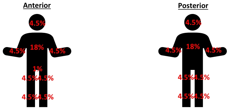
| Anterior | Posterior |
|---|---|
| 4.5% | 4.5% |
| 4.5% 18% 4.5% | 4.5% 18% 4.5% |
| 1% | |
| 4.5% 4.5% | 4.5% 4.5% |
| 4.5% 4.5% | 4.5% 4.5% |
Alternative Methods
- The Lund-Browder Chart can be used to estimate burn surface area in all patients, including children <10y. The chart has percentages that vary across age groups in the head (↓ from infancy to adulthood) and thighs/legs (↑ from infancy to adulthood), while the percentages remain constant across age groups everywhere else in the body.
- To estimate TBSA for small or patchy burn, compare burn to patient’s palm, which represents ~1% (includes palm, fingers)
Initial Volume Resuscitation
The amount of fluid (Ringer’s Lactate) to be provided for volume resuscitation can be calculated by the Parkland Formula.
Volume = 4 mL / kg × %TBSA × body weight (kg)
- The first half of volume is administered in first 8 hours (front-loaded resuscitation due to ↑ capillary permeability, protein leak, edema, loss of plasma volume in early post-burn period). The second half is administered in the following 16 hours.
- For superficial (1st degree burns), this is not included in the Total Body Surface Area (TBSA) used to calculate volume resuscitation requirements – not life-threatening.
Red Flags of Burns
Inhalation injury
Findings:
- Burns in a confined space.
- Burns to face, neck, upper torso.
- Singed eyebrows and no nasal vibrissae (hair)
- Carbon deposits (soot), acute inflammatory change in oropharynx
- Carbonaceous sputum
- Stridor, Dysphonia, dysphagia, drooling
- Coughing, wheezing, dyspnea
- Explosion with burns to head and torso
Can’t Miss Diagnosis and Management Implications:
- Can cause Acute Respiratory Distress Syndrome (ARDS), Carbon Monoxide poisoning, or laryngeal edema with eventual airway obstruction (often difficult to intubate AND VENTILATE once laryngeal edema has progressed).
- Consider early intubation due to projected clinical course of laryngeal edema and loss of airway.
- Clinical manifestations of inhalation injury may be subtle– need to maintain a high index of suspicion.
Barotrauma or other associated trauma
Findings:
- Explosion
Carbon Monoxide (CO) Poisoning
Findings:
- Headache, dizziness, or history of loss of consciousness at scene
- Burn in an enclosed space
Can’t Miss Diagnosis and Management Implications:
- t½ on room air: 250 minutes
- t½ on 100% FiO2: 40 minutes → give high-flow oxygen via non-rebreather mask if suspect CO exposure
Non-accidental trauma
Findings:
- Burn wound inconsistent with history
Acute limb ischemia or compartment syndrome
Findings:
- Circumferential burns of extremities, chest, or abdomen
- ↑ intrabdominal pressure: Distended, painful abdomen, jugular venous distension, hypotension, tachycardia); Intrabdominal organ dysfunction (oliguria, dyspnea)
- 6 P’s: pain (out of proportion, with passive muscle stretch); late findings – pallor, poikilothermia, paresthesia, paralysis, pulselessness
- Compartment pressure > 30mmHg
Can’t Miss Diagnosis and Management Implications:
- Burn compartment syndrome typically develops over >6 hours to point of requiring fasciotomy
- May require escharotomy.
- Fasciotomy may be needed to restore circulation e.g. associated skeletal trauma, crush injury, high-voltage electrical injury, burns involving tissue beneath fascia.
Respiratory distress/compromise
Findings:
- Burn eschar restricting chest wall expansion (e.g. circumferential burn, full thickness burns of anterior and lateral chest wall leading to severe chest wall motion
Can’t Miss Diagnosis and Management Implications:
- May require chest wall escharotomy
Electrical burns
Findings:
- Cardiac arrhythmias (e.g. asystole, ventricular fibrillation, sinus tachycardia, heart blocks)
- Rhabdomyolysis (re. muscle contraction with electricity) – including dark urine (due to myoglobinuria)
- Electrical burns: Due to different rates of heat loss from superficial vs. deep tissues, relatively normal overlying skin may coexist with deep muscle necrosis – the body surface area on inspection may not reflect true injury
Can’t Miss Diagnosis and Management Implications:
- Fasciotomy may be needed re. deeper tissue injuries
- Fluid and electrolyte management (re. acute kidney injury e.g. from rhabdomyolysis)
- Management of cardiac arrhythmias (e.g. Advanced Cardiac Life Support (ACLS) protocols)
Burn Centre Referral Criteria22
- Partial thickness burns > 10% TBSA
- Any Full thickness (3rd degree) burns
- Burns involving face, hands, feet, genitalia, perineum or major joints; as well as evidence of Inhalation Injury
- Non-Thermal Burns: Electrical burns (including lightning injury), Chemical burns.
- Burn injury in patients with comorbidities that could complicate management, prolong recovery, or affect mortality.
- Burn injuries in patients who will require special social, emotional, or rehabilitative intervention.
- Patient with burns and concomitant trauma (e.g. fractures) where burn injury poses greatest risk of morbidity or mortality. If trauma poses greater immediate risk, patient may be initially stabilized in trauma centre before burn unit transfer.
- Burned children in hospitals without qualified personnel or equipment for the care of children.
Part 7: Approach to Dermatitis (Eczematous Dermatoses)
Dermatitis is a very common group of itchy, inflammatory conditions characterized by epidermal changes. Presentation can vary based on the type of eczema, the population, and the acuity. In general, eczema presents with poorly demarcated erythematous patches or plaques, pruritis and occasionally pain. Acute eczema presents with papules or vesicles; subacute eczema with crusting, scaling or excoriations; and chronic eczema with Lichenification, xerosis or fissuring.
Types of Dermatitis (Eczematous Dermatoses – Non-Exhaustive)
Note: Other types of eczematous dermatoses exist than those described below.
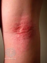
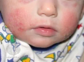
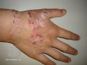
Atopic Dermatitis (Eczema)24
Clinical Features: Presents as subacute and chronic eczematous rash. Pruritis (hallmark of atopic dermatitis) → scratching → inflammation, Lichenification, excoriations. Chronic or relapsing history (re. flares of eczema). Common triggers include irritants (e.g. soaps, detergents), skin infections, contact allergens, foods/inhalant triggers, stress. May appear dark brown, purple, or gray on darker skin tones.
Common Locations:
Infants: face/head, extensor surfaces.
Children: face, neck, extensor surfaces, flexor surfaces; skin creases (e.g. antecubital/popliteal fossae)
Adult: flexor surfaces; face/neck less frequent
*Spares groin, axillary regions
Notes: Affects about 20% of children and 3% if adults. 60% develop before 1 year old, 90% by 5 years old.
Positive family history. Atopic dermatitis forms part of Atopic triad – including asthma and allergic rhinitis.
Example Images:
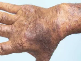
Contact Dermatitis (Eczema)2526
Clinical Features: Presents as erythematous papulovesicular rash, eruption, swelling, pruritis.
Acute insult – quick, well-demarcated.
Cumulative insult – slow, poorly-demarcated. May appear dark brown, purple, or gray on darker skin tones.
Common Locations:
Irritant: hands; face (e.g. volatile irritants, cosmetics)
Allergic: areas exposed to allergen
Notes: May be irritant (e.g. soaps, detergents, organic solvents) or allergic (less common – due to immune sensitization to an allergen e.g. nickel, hair dye, rubber, perfumes).
Example Images:
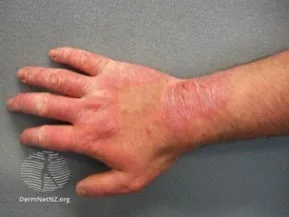
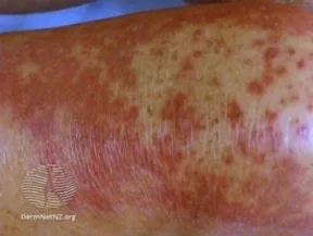
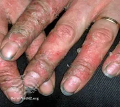
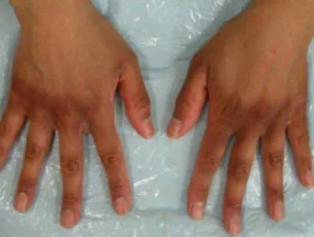
Dyshidrotis Dermatitis (Acute palmoplantar eczema)27
Clinical Features: Presents as a sudden, symmetric eruption of papulovesicular rash (“tapioca pudding” appearance). Very pruritic → plaques → painful tearing and chronic Lichenification. May be episodic, commonly recurs in warmer months.
Common Locations: Palms, later and dorsal aspect of fingers, or on soles.
Notes: May be complicated by secondary infection (e.g. Staphylococcus aureus).
Example Images:
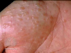
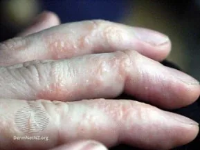
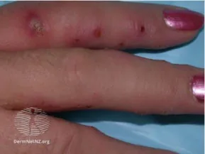
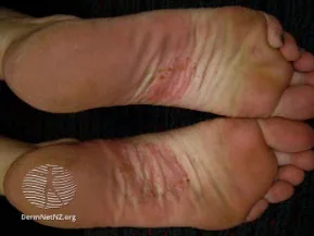
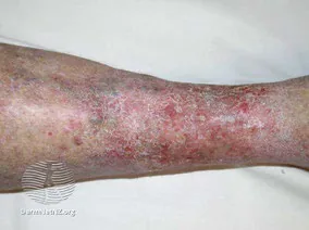
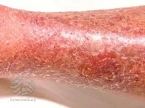
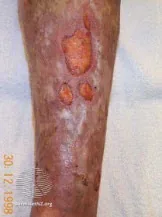
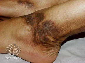
Stasis Dermatitis (Venous Eczema)28
Clinical Features: Presents as erythematous, scaly plaques in lower legs, ± hemosiderin (brown) deposits, woody fibrosis, atrophy blanche, lipodermatosclerosis, or varicose veins. Pruritus is variable, but if present results from Lichenification from chronic scratching/rubbing.
Common Locations: Lower legs (medial malleolus, most frequent, may extend up to knee), usually bilaterally
Notes: Late manifestation of chronic venous disease. May mimic cellulitis or DVT. May present with secondary lymphedema, varicosities, secondary cellulitis, or ulceration.
Example Images:
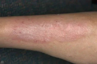
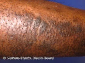
Lichen Simplex Chronicus29
Clinical Features: Pruritic, well-demarcated, erythematous patches and plaques of lichenified skin ± excoriations. “Itch-scratch cycle”: Itch → scratching → Lichenification
Common Locations: Neck, scalp, lower extremities, wrists, extensor forearms, genitals
Notes: If generalized pruritis, rule out systemic cause.
Example Images:
Part 8: Other Important Immune-Mediated Rashes
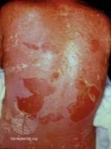
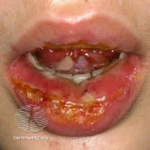
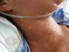
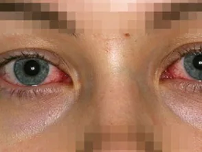
Steven Johnson-Syndrome (SJS)/Toxic Epidermal Necrolysis (TEN)30
Clinical Features:
- Prodrome with fever, flu-like symptoms, malaise, myalgia, arthralgia.
- 1-3 days later, develop mucocutaneous lesions.
- Rash – Targetoid lesions ± painful macules/purpura ± diffuse erythema
- Nikolsky sign – sloughing off of the epidermal layer of the skin with gentle lateral pressure.
- Can present with shock.
- Secondary infection may occur.
- Anemia, lymphocytopenia, neutropenia may occur.
- SCORTEN Score (TEN prognosis, indication for ICU/specialized burn unit)
Common Locations: Neck, scalp, lower extremities, wrists, extensor forearms, genitals
Notes:
- Rare condition leading to epidermal detachment. Associated with high mortality and complications rate. SJS and TEN are the same entity, but are differ based on involved surface area:
- <10% body surface area = SJS
- 10-30% body surface area = SJS/TEN overlap
- >30% body surface area = TEN
- Triggers: Drugs (Esp. Allopurinol, Aromatic antiseizure medications, lamotrigine, sulfonamide antibiotics, sulfasalazine, oxicam NSAIDs), infections (e.g. Mycoplasma pneumonaie, CMV, herpes), acute graft-versus-host disease, idiopathic.
- Risk factors: HIV, active cancer, certain HLA phenotypes.
Example Images:
Psoriasis31
Clinical Features:
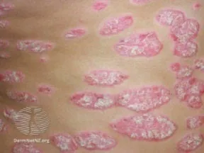
- Well-demarcated erythematous skin lesions with silver-white scaly plaques, pink and/or red papules in lighter skin tones and purple papules in darker skin tones
- Pruritis
- Nail involvement – nail pitting, brittleness, onycholysis, or oil drop sign/salmon spot
- Auspitz Sign – Pinpoint bleeding when scales of cutaneous lesions are scraped off.
Common Locations:
- Extensor surfaces (e.g. scalp, gluteal cleft, extensor surface of elbows and knees in plaque psoriasis)
- Trunk and proximal extremities in Guttate psoriasis
- Generalized/Widespread in Erythrodermic or Pustular psoriasis.
- Intertriginous areas (inguinal, perineal, genital, intergluteal, axillary, inframammary) in Inverse psoriasis
- Nails
- Psoriatic arthritis:
- Asymmetrical oligo-arthritis
- Involvement of DIPs (vs. spared in Rheumatoid Arthritis), PIPs, dactylitis.
- Spondyloarthropathy (sacroiliitis, spondylitis)
Notes:
- Chronic inflammatory condition with disease course involving intervals of symptom-free periods and relapse.
- Types of psoriasis:
- Plaque (symmetrically distributed cutaneous plaques)
- Guttate (abrupt appearance of multiple small, psoriatic papules and plaques)
- Erythrodermic (uncommon, generalized erythema and scaling of most of body surface area)
- Pustular (uncommon, can be life-threatening – sudden onset widespread erythema, scaling and sheets of superficial pustules)
- Inverse/Intertriginous (Well-demarcated, smooth, shiny plaques with absent or minimal scale)
- Psoriatic arthritis (in up to 30% of psoriasis patients)
Example Images:
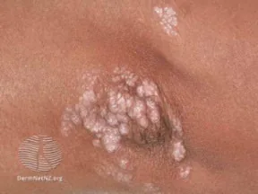
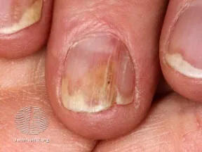
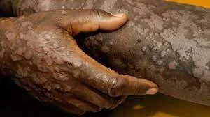
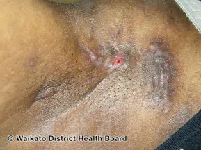
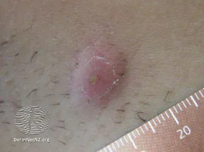
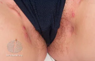
Hidradenitis Suppurativa (HS)32
Clinical Features:
- Nodules and Abscesses: Typically begins with the formation of painful, deep-seated nodules beneath the skin. These nodules can gradually increase in size and become filled with pus, forming abscesses.
- Sinus Tracts: Chronic inflammation and repeated abscess formation can result in the development of sinus tracts or tunnels beneath the skin. These sinus tracts may connect multiple nodules and abscesses, forming a network of interconnected channels.
- Scarring and Fibrosis: As lesions heal, they often leave behind thick, fibrotic tissue and hypertrophic scars.
Common Locations:
- Axillae
- Groin
- Buttocks
- Intertriginous areas (e.g., beneath breasts): These regions are characterized by friction, heat, and sweat accumulation, creating an environment conducive to the development and exacerbation of HS lesions.
- Apocrine glands: Perianal and perineal regions
Notes:
- Chronic inflammatory skin condition characterized by recurrent, painful nodules, abscesses, and sinus tracts.
- Risk factors: More common in women.
- Triggers: Exacerbated by factors such as hormonal changes, obesity, and smoking.
Example Images:
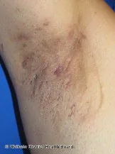
Identifying Toxic Epidermal Necrolysis (TEN) or Steven Johnson Syndrome (SJS)
Nikolsky Sign
Say: I will now inspect the skin for signs of skin separation with gentle pressure.
Do: Identify the lesion. With sterile gloves on, apply gentle lateral pressure on skin. Observe whether this dislodges upper layers of epidermis from lower layers, causing skin to slough off.
Report: Nikolsky sign is negative.
Why Check: The Nikolsky sign describes the ability to extend the area of superficial sloughing by applying gentle lateral pressure on the surface of the skin at an apparently uninvolved site with normal-appearing skin. It is particularly important in identifying patients with Steven Johnson Syndrome (SJS) and Toxic Epidermal Necrolysis (TEN) - explained below. A positive Nikolsky sign is also seen in pemphigus vulgaris, bullous pemphigoid, and staphylococcal scaled skin syndrome.
Part 9: Approach to Acne (Acne Vulgaris)3334
Acne vulgaris is an inflammatory disease of the pilosebaceous unit of the skin. It is a common diagnosis affecting approximately 9% of patients, and 85% of those aged 12-24 years. It is marked by comedone formation (dilated hair follicles filed with keratin squamae, bacteria and sebum), papules, pustules, nodules, and cysts. Secondary signs include scars, erythema and hyperpigmentation. Its presentation varies across individuals, influenced by factors such as age, hormone levels, and genetic predisposition. Acne severity can range from mild to severe, impacting both physical appearance and psychological well-being.
Types of Acne (Acne Dermatoses – Non-Exhaustive)
Note: Other types of acne dermatoses exist than those described below.
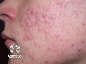
Acne Vulgaris (common)
Clinical Features:
- Comedonal Acne: Characterized by Superficial lesions composed of open comedones (“blackheads”, follicle partially blocked) and closed comedones (“whiteheads”, follicle completely blocked).
- Inflammatory Acne: Characterized by papules and pustules.
- Nodulocystic Acne: Severe from of acne characterized by deeper lesions comprised of nodules and cysts affecting the face and upper trunk that typically resolve with associated scarring.
- Secondary lesions in acne vulgaris encompass excoriations, erythematous macules, and pigmented macules primarily affecting individuals with darker skin tones.
- Moderate to severe acne may present with scarring, particularly atrophic scaring, including ice-pick scars, rolling scars, and box-car scars.
Common Locations:
- Commonly found in areas with sebaceous glands (predilection sites: face, shoulders, upper chest, and back)
Notes:
- Individual acne lesions usually last less than 2 weeks. Deeper papules and nodules may persist for months.
- Many acne patients also have oily skin (seborrhoea).
- Triggers for acne flares include: Polycystic Ovarian Syndrome (PCOS), Drugs (e.g. Steroids), Occlusive cosmetic use, high environmental humidity, and diets high in dairy products and high glycemic foods.
- For each patient, describe severity including presence of scarring to inform appropriate management:
- Mild: Comedones with few inflammatory lesions
- Moderate: Comedones with marked number of inflammatory lesions
- Severe: Extensive inflammatory lesions with diffuse scarring.
- Cystic or scarring acne should be treated aggressively to prevent permanent sequelae (e.g. scarring, post-inflammatory hyperpigmentation)
Example Images35:
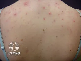
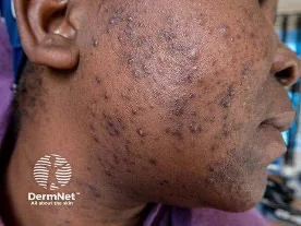
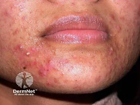
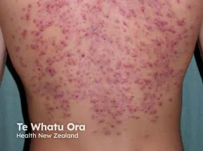
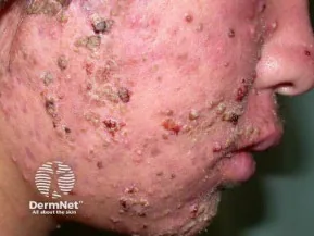
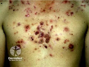
Acne Fulminans: Severe form of nodulocystic acne (rare)
Clinical Features: Acute onset of large, painful nodules, plaques, and ulcers with hemorrhagic crusts. Also may be accompanied with systemic symptoms, including fever, myalgia, weight loss, arthralgia, and arthritis. In rare instances, patients may present with hepatosplenomegaly, erythema nodosum, or septic osteomyelitis.
Common Locations: Most commonly seen on the chest, back, and face
Notes:
- Acne fulminans has been associated with increased androgens (male hormones), autoimmune complex disease, genetic predisposition, and hypersensitivity reaction to surface bacteria (Cutibacteria acnes)
- Acne Fulminans Precipitants: Testosterone and anabolic steroids; oral isotretinoin (used in treatment of severe nodulocystic acne)
Example Images36:
Part 10: Approach to Rosacea3738
Rosacea is a chronic inflammatory skin disorder that primarily affects the central face area (e.g. cheeks, nose, chin, and forehead) and the eyes. Its onset typically initiates between the ages of 30 and 60 years and is estimated to affect 5% of adults. Rosacea's manifestations vary among individuals and may be influenced by factors such as age, gender, ethnicity, and environmental triggers like sun exposure. Diagnostic features include persistent centro-facial erythema and phymatous changes (skin thickening with irregular contours, most commonly affecting the nose), with cycles of exacerbation and remission due to trigger factors. Common trigger factors include extremes of temperature, sunlight, alcohol, spicy foods, exercise, acute psychologic stressors, menopausal hot flashes, and medications (e.g. steroid creams, vasodilator medications).
Exacerbation features which occur in the centro-facial distribution include flushing/transient erythema, inflammatory papules/pustules and telangiectasias (visible blood vessels, excluding nasal alar area where telangiectasias are common in the general adult population). Patients may endorse burning, stinging or dry sensations of the skin. More than half of patients with rosacea have ocular features including dryness, foreign body sensation, photophobia, conjunctivitis, blepharitis, and in rare cases, keratitis can lead to long-term vision loss. Comedones are not present in rosacea.
Types of Rosacea (Rosacea Dermatoses – Non-Exhaustive)
Note: Other types of rosacea dermatoses exist than those described. Patients often present with morphological characteristics of more than one rosacea subtype and may complain of increased sensitivity of the facial skin with burning, stinging and itch.
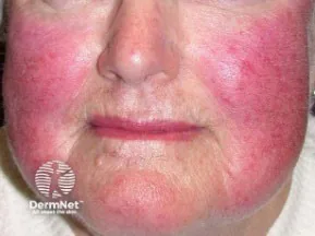
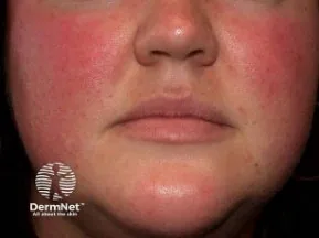
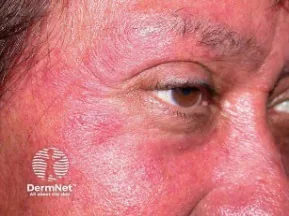
Erythematotelangiectatic rosacea
Clinical Features:
- Transient facial flushing with persistent centrofacial erythema.
- Telangiectasias are common but not required for diagnosis.
Common Locations:
- Persistent central facial erythema (cheeks, nose, chin, forehead). Erythema may also involve peripheral face, ears, neck and upper chest, but periocular skin is typically spared.
Example Images39:
Papulopustular rosacea
Clinical Features:
- Persistent central facial erythema.
- Transient, central facial papules and/or pustules.
Common Locations:
- Persistent central facial erythema (cheeks, nose, chin, forehead).
Example Images39:
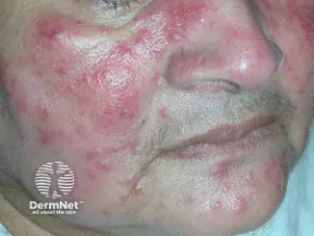
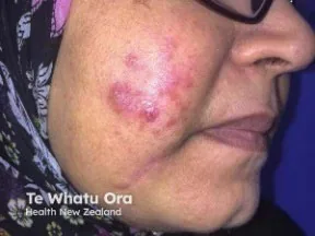
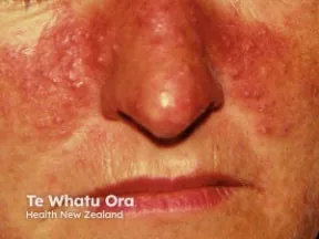
Phymatous rosacea
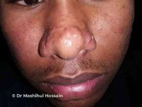
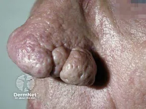
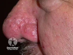
Clinical Features:
- Thickening of skin with irregular surface nodularities and hyperplasia of sebaceous glands.
- Rhinophyma: enlarged, bulbous nose (almost exclusively in male patients)
Common Locations:
- Central facial area (nose, cheeks, chin, forehead) and ears
Example Images39:
Ocular rosacea
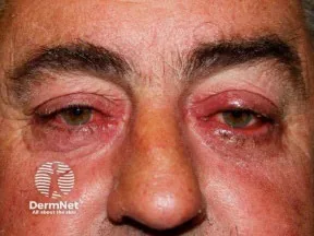
Clinical Features:
- Conjunctival hyperemia (redness of conjunctivae), blepharitis (inflammation of eyelid margin), styes (hordeolum externum), chalazion, dry eyes, keratitis (corneal inflammation –emergency re. vision loss risk).
- Patients may present with foreign body sensation in the eye, burning or stinging, dryness, itching, ocular photosensitivity, and blurred vision.
Common Locations:
- Eyes, periocular region
Notes: Seen in association with other subtypes of rosacea.
Example Images39:
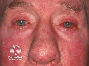
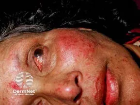
Part 11: Relevant Likelihood Ratios – from Rational Clinical Examination JAMA series
Clinical Question 1: Does this patient have a Mole or Melanoma?40
- Note: Here, “E” in ABCDE checklist is Elevation, not Evolution. A Positive test result is defined as 1+ of ABCDE criteria met.
| Procedure | Sensitivity | Specificity | LR+ (95% CI) | LR- (95% CI) |
|---|---|---|---|---|
| Dermatologists use of ABCDE Checklist | 0.92 (82-96) | N/A | N/A | N/A |
| Chart review of Dermatologists’ Examinations | 1.0 (0.54-1.0) | 0.984 (0.95-0.99) | 62 (19-170) (about +45%) | 0 (0-0.5) (about −45%) |
References
-
emdocs.net. Accessed June 23, 2022. http://www.emdocs.net/9009-2/ ↩
-
Bradford P. Skin cancer in skin of color. Dermatol Nurs. 2009;21(4):170-178. ↩
-
Actinic keratosis. dermnetnz.org. Accessed May 16, 2023. https://dermnetnz.org/topics/actinic-keratosis ↩
-
Abudu B, et al. Pigmented actinic keratosis: case report and review of an uncommon actinic keratosis variant that can mimic melanoma. Cureus. 2019;11(5):e4721. ↩
-
Basal cell carcinoma. dermnetnz.org. Accessed June 26, 2022. https://dermnetnz.org/topics/basal-cell-carcinoma ↩
-
Basal cell carcinoma in skin of colour. dermnetnz.org. Accessed June 26, 2022. https://dermnetnz.org/topics/basal-cell-carcinoma-in-skin-of-colour ↩
-
Squamous cell carcinoma on the face images. dermnetnz.org. Accessed June 26, 2022. https://dermnetnz.org/topics/squamous-cell-carcinoma-on-the-face-images ↩
-
Squamous cell carcinoma of limbs images. dermnetnz.org. Accessed June 26, 2022. https://dermnetnz.org/topics/squamous-cell-carcinoma-of-limbs-images ↩
-
67-year-old African American woman presents lesion right leg. cancernetwork.com. Accessed June 26, 2022. https://www.cancernetwork.com/view/67-year-old-african-american-woman-presents-lesion-right-leg ↩
-
Melanoma. dermnetnz.org. Accessed June 26, 2022. https://dermnetnz.org/topics/melanoma ↩
-
Melanoma in skin of colour. dermnetnz.org. Accessed June 26, 2022. https://dermnetnz.org/topics/melanoma-in-skin-of-colour ↩
-
Houston NAM. Patient preferences during skin cancer screening examination. JAMA Dermatol. 2016;152(9):1052-1054. ↩ ↩2
-
American Academy of Dermatology. Accessed June 26, 2022. https://www.aad.org/public/diseases/skin-cancer/find/check-skin ↩ ↩2
-
From L, et al. Screening for Skin Cancer: A Quality Initiative of the Program in Evidence-Based Care (PEBC). CCO; 2015. ↩ ↩2 ↩3
-
Impetigo. dermnetnz.org. Accessed June 26, 2022. https://dermnetnz.org/topics/impetigo ↩
-
Erysipelas. dermnetnz.org. Accessed June 26, 2022. https://dermnetnz.org/topics/erysipelas ↩
-
Cellulitis. dermnetnz.org. Accessed June 26, 2022. https://dermnetnz.org/topics/cellulitis ↩
-
Cellulitis mimics. dermnetnz.org. Accessed June 26, 2022. https://dermnetnz.org/topics/cellulitis-mimics ↩ ↩2 ↩3 ↩4 ↩5 ↩6 ↩7 ↩8
-
Cutaneous abscess. dermnetnz.org. Accessed June 26, 2022. https://dermnetnz.org/topics/cutaneous-abscess ↩
-
Necrotising fasciitis. dermnetnz.org. Accessed June 26, 2022. https://dermnetnz.org/topics/necrotising-fasciitis ↩
-
File:Deep vein thrombosis of the right leg.jpg. Wikimedia Commons. Accessed June 26, 2022. https://commons.wikimedia.org/wiki/File:Deep_vein_thrombosis_of_the_right_leg.jpg ↩
-
Burn center referral criteria. ameriburn.org. Accessed June 26, 2022. https://ameriburn.org/wp-content/uploads/2017/05/burncenterreferralcriteria.pdf ↩
-
Recommendations. Choosing Wisely Canada. Accessed August 1, 2022. https://choosingwiselycanada.org/recommendations/#list ↩
-
Atopic dermatitis. dermnetnz.org. Accessed June 26, 2022. https://dermnetnz.org/topics/atopic-dermatitis ↩
-
Contact dermatitis. dermnetnz.org. Accessed June 26, 2022. https://dermnetnz.org/topics/contact-dermatitis ↩
-
Irritant contact dermatitis. dermnetnz.org. Accessed June 26, 2022. https://dermnetnz.org/topics/irritant-contact-dermatitis ↩
-
Vesicular hand dermatitis. dermnetnz.org. Accessed June 26, 2022. https://dermnetnz.org/topics/vesicular-hand-dermatitis ↩
-
Venous eczema. dermnetnz.org. Accessed June 26, 2022. https://dermnetnz.org/topics/venous-eczema ↩
-
Lichen simplex. dermnetnz.org. Accessed June 26, 2022. https://dermnetnz.org/topics/lichen-simplex ↩
-
Stevens-Johnson syndrome/toxic epidermal necrolysis. dermnetnz.org. Accessed June 26, 2022. https://dermnetnz.org/topics/stevens-johnson-syndrome-toxic-epidermal-necrolysis ↩
-
Psoriasis. dermnetnz.org. Accessed June 26, 2022. https://dermnetnz.org/topics/psoriasis ↩
-
Hidradenitis suppurativa. dermnetnz.org. Accessed May 24, 2024. https://dermnetnz.org/topics/hidradenitis-suppurativa ↩
-
Eichenfield DZ, Sprague J, Eichenfield LF. Management of acne vulgaris: a reivew. JAMA. 2021;326(20):2055-2067. ↩
-
Cipriano SD, et al. American Academy of Dermatology Basic Dermatology Curriculum module: acne and rosacea. American Academy of Dermatology. Updated 2023. Accessed June 24, 2024. https://www.aad.org/member/education/residents/bdc ↩
-
Acne. dermnetnz.org. Accessed June 23, 2024. https://dermnetnz.org/topics/acne ↩
-
Acne fulminans. dermnetnz.org. Accessed June 23, 2024. https://dermnetnz.org/topics/acne-fulminans ↩
-
Rainer BM, Kang S, Chien AL. Rosacea: epidemiology, pathogenesis, and treatment. Dermatoendocrinol. 2017;9(1):e1361574. ↩
-
Oge LK, Muncie HL, Phillips-Savoy AR. Rosacea: diagnosis and treatment. Am Fam Physician. 2015;92(3):187-196. ↩
-
Rosacea. dermnetnz.org. Accessed June 23, 2024. https://dermnetnz.org/topics/rosacea ↩ ↩2 ↩3 ↩4
-
Whited JD, Grichnik JM. Does this patient have a mole or a melanoma? JAMA. 1998;279(9):696-701. ↩
