Medical Foundations 1
Chapter 8Peripheral Vascular Examination
Part 1: Focused History – Based on Chief Complaint (Begin with Open-Ended Questions)
HPI, Assoc. Symptoms
Claudication
- Onset: When did the leg pain/cramping start? What were you doing when it started? Was it gradual onset or acute onset?
- Palliating/Provoking:
- Is leg pain/cramping worse on exertion? How far can you walk before pain occurs? Has the distance you can walk before the pain occurs diminished over time?
- Is the pain relieved by rest <10 mins?
- Is pain position-dependent in lower legs/feet?
- Peripheral arterial disease: Pain worse on elevation, relieved by dependency
- Peripheral venous disease: Pain worse with prolonged standing or seated with feet dependent; improved with limb elevation and walking
- Any pain at rest (e.g. wakes you from sleep? – RED FLAG SYMPTOM)
- Quality: Burning?
- Region: Unilateral vs. Bilateral?
- Diffuse lower extremity pain (may indicate acute onset ischemia)
- Specific locations:
- Buttock/hip (suggests aortoiliac disease)
- Thigh (suggests common femoral artery disease)
- Calf (most common– suggests superficial femoral artery disease or popliteal artery disease)
- Foot (suggests tibial or peroneal artery disease)
- Severity: How would you rate this on a scale of 1-10? Does the pain cause you to stop walking?
- Timing: Does the pain come and go? When you feel the pain, how long does it last?
- Definitions
- Intermittent Claudication – Fatigue, discomfort, cramping or pain of vascular origin in lower extremity muscles, consistently induced by exercise and relieved by rest within 10 minutes.
- Rest pain – Pain at rest, occurring with disease progression. Worsens with reclining, improves with hanging feet off bed or standing.
- Critical Limb Ischemia (≥2 weeks) – Limb-threatening arterial occlusion, characterized by ischemic rest pain, non-healing wound/ulcers, or gangrene in 1 or both legs.
- Acute Limb Ischemia (<2 weeks) – Acute severe hypoperfusion of a limb due to arterial occlusion; causes include arterial thrombus, embolism or dissection. Characterized by 6 Ps (Pain, pallor, pulselessness, paralysis, paresthesia, poikilothermia i.e. cold). Limb threatening ischemia particularly concerning if there is sensory or motor loss.
- Associated symptoms:
- Arterial disease – Noticed coldness, numbness or pallor in legs or feet? Hair loss and shiny skin? Ulcers on pressure points of feet? Injuries poorly healing or non-healing in legs or feet?
- Venous disease – Noticed warmness, blue/purple/brownish discolouration in legs or feet? Ulcers on tibial plateau or medial malleolus? Flakey skin? Peripheral edema?
Past Medical History (PMHx)
- Peripheral Arterial Disease Risk Factors: Age; Smoking; Diabetes; Hypertension; Dyslipidemia; Coronary Artery Disease
- Peripheral Venous Disease Risks: Age; Smoking; ↑ BMI; Hx lower extremity trauma; prior venous thrombosis; pregnancy
- Venous Thromboembolism Risk, Including Deep Vein Thrombosis (DVT) and Pulmonary Embolism (PE):
- Virchow’s Triad – (1) Stasis of blood flow (e.g. immobilization, venous insufficiency), (2) endothelial injury (e.g. Hypertension, Atherosclerosis) & (3) hypercoagulability (e.g. smoking, coagulation disorders, malignancy)
- Risk Factors (as seen in Well’s Score for DVT): Active cancer, bedridden >3 days, major surgery within 12 weeks, paralysis/paresis/plaster immobilization of lower extremity, previous Deep Veinous Thrombosis.
Relevant clinical scores:
- Well’s Score for DVT (Risk of deep venous thrombosis based on clinical criteria)
- Well’s Score for PE (Risk of pulmonary embolism based on clinical criteria)
- YEARS Algorithm for PE (Helps rule out PE, validated in pregnant patients with clinically suspected PE)
- PERC Rule (Clinically ruling out pulmonary embolism in low-risk patients)
- HAS-BLED Score for Major Bleeding Risk – bleeding risk for patients on anti-coagulation
Past Surgical History (PS Hx)
- Risk factor for venous thromboembolism: Major surgery within 12 weeks (re. Virchow’s Triad)
- Peripheral Arterial Disease:
- Revascularization (e.g. for claudication; or as a limb salvage procedure in chronic limb-threatening ischemia): e.g. stent-grafting, angioplasty, bypass (intent for restoration of inflow)
- Amputation: e.g. unsalvageable extremity due to critical limb ischemia, severe osteomyelitis infection, etc.
- Foot amputation (ambulation preserving potential): e.g. digit, trans-metatarsal, mid-/hind-foot amputation
- Others: Below-knee amputation (BKA), knee disarticulation, above knee amputation (AKA), hip disarticulation
- Peripheral Venous Disease:
- Symptomatic Superficial Venous Insufficiency: e.g. saphenous vein stripping, minimally invasive ablation)
- Venous Thromboembolism (most managed with anticoagulation alone): e.g. thrombectomy; IVC Filter
- Wound management: e.g. Operative wound debridement, negative pressure wound therapy, etc.
Family Medical History (FMHx)
- Coagulation disorders (e.g. Factor V Leiden mutation, Protein C or Protein S deficiency, Antithrombin deficiency)
- Peripheral venous disease
Social Hx
Smoking
- Important risk factor for peripheral artery disease; smoking cessation key component of treatment
- Pack years = packs/day x years smoking; Have you thought about quitting within the next 6 months?
- Cigarettes? Marijuana? E-cigarettes/vapes?
- 5As approach taking a smoking Hx (Explored in Chapter 38 – Motivational Interviewing – IF)
- Pro-Comp Connection: Regarding patient’s position, do not jump to thinking in terms of “compliance” with recommendations for smoking cessation.
Alcohol
- How often do you drink alcohol (Drinks/Week)? What is the size of 1 drink? What kinds of drinks?
Travel
- Any recent long travels? (Prolonged immobilization ↑ risk venous thromboembolism re: Virchow’s triad)
Pregnancy
- Estrogen increases risk of Venous Thromboembolism (e.g. DVT, PE)
Occupation
- Impact of claudication symptoms on functioning at work and quality of life
- Impact of prolonged standing at work (e.g. for peripheral venous disease) and quality of life
Exercise
- Structured exercise training programs important component of management of intermittent claudication
- Programs may be supervised or unsupervised; and high-intensity or low-intensity
- General: Exercise 45-60 minutes at least 3 times/week for minimum 12 weeks, at exercise level at sufficient intensity to elicit claudication symptoms.
- Explored further in Chapter 38 – Prescribing Lifestyle Interventions in Medicine – Integration Foundation
Medications Hx, Allergies
Statin Use
Statins can cause myalgias/cramps; muscle damage → muscle weakness; DDx for claudication.
Anti-platelet, Anti-coagulant Medications
(Note: Explored further in MF2)
- Anti-platelets: primary hemostasis – e.g. ASA (Aspirin), Clopidogrel (Plavix), Ticagrelor
- Anti-coagulants: secondary hemostasis
- Heparins: Unfractionated Heparin (UFH), Low molecular weight heparin (LMWH)
- Vitamin K antagonists: Warfarin/Coumadin
- Direct Oral Anticoagulants (DOAC): Dabigatran, Apixaban, Rivaroxaban, Edoxaban
Oral Contraceptives/ Hormone replacement Therapy
- Estrogen increases risk of Venous Thromboembolism
- Combined oral contraceptive pills (COC) ↑ risk vs. Progestin-only contraceptive pills (POP)
- ↑ risk for COC with >35mcg ethinyl estradiol formulations
Wound Management
- Barrier creams to protect adjacent skin; wound dressings (frequency of dressing changes, involvement of wound care specialists, home care involvement, etc.)
Claudication Pain Management
Compression Stockings
- Essential component of chronic venous disease management e.g. static compression therapy (bandages, stockings) – ensure no coexistent moderate-severe peripheral arterial disease before starting compression stockings re. risk of ↑ limb ischemia.
Part 2: Pertinent Notes about the Physical Exam
- OSCE Tip: Always start with the vitals in an OSCE situation with physical exam component
- Chapter Connections: Given that poorly controlled Diabetes Mellitus can lead to Peripheral Vascular Disease (PVD) complications, the Peripheral Vascular examination (Chapter 8) is closely tied to the Diabetes Exam (Ch 14 – MF3)
Part 3: Specific Maneuvers in the Physical Exam – Say, Setup, Do, Report
Notes on Vitals
Vitals – Always do HR, BP, RR, Temp
In This Exam
AFMC level Pre-clerkship 4 · Clerkship 4
Standard Vitals measurements
Inspection
Varicose Veins
AFMC level Pre-clerkship 1 · Clerkship 2
Say: I will inspect for varicose veins (appropriately drape patient).
Do: Look for varicosities (swollen veins visible under skin – caused by surface vein damage, loss of valve integrity facilitating pooling) – typically examined with patient standing
Report: I note no varicosities present on inspection bilaterally
Why Check: Sign of peripheral venous disease. Varicosities can be supported with compression stockings.
Arterial vs Venous Disease
AFMC level Pre-clerkship 1 · Clerkship 2
| ACRONYM – CHUSE-T | Peripheral Arterial Disease | Peripheral Venous Disease |
|---|---|---|
| Colour | Pallor | Brown, Blue, Purple |
| Hair | Loss | No Change |
| Ulceration | Pressure points – soles, ventral surface of foot Tend to be dry lesions | Tibial plateau, Medial Malleolus Tend to be wet lesions (e.g. wound drainage) |
| Skin Changes | Shiny | Flaky |
| Edema | N/A | Peripheral Edema |
| Temperature | Cold | Warm |
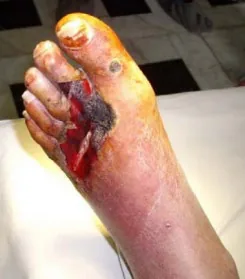
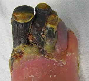
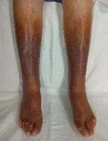
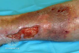
Why Check: Differentiates peripheral arterial vs. peripheral venous disease. Note that in peripheral arterial disease, critical limb ischemia is defined by chronic (≥2 weeks) ischemic rest pain, nonhealing wound/ulcer, or gangrene in 1 or both legs attributable to proven arterial occlusive disease. Gangrene can be divided into:
- Dry gangrene: Presents with hard, dry texture; often a clear border of viable and black, necrotic tissue. This can be seen in photo above, with dry gangrene of the first 3 digits of the right foot.
- Wet gangrene: Moist, swollen appearance with blistering – a surgical emergency.
Auscultation: BEFORE PALPATION OF PULSES
Bruits
AFMC level Pre-clerkship 1 · Clerkship 2
Say: I will use the bell of my stethoscope to auscultate key arteries for any signs of turbulent blood flow.
Do: With patient in supine position, auscultate Carotid → Epigastric → Abdominal aorta → Renal → Iliac → Femoral Arteries for bruits.
- R/L Carotid: Medial to sternocleidomastoid muscle, roughly at level of cricoid cartilage
- Abdominal Aorta: Slightly left and superior to umbilicus
- Right and Left Renal: 5cm superior and 5cm lateral to umbilicus
- Right and Left Iliac: 5cm inferior and 5cm lateral to umbilicus
- Right and Left Femoral: Midway between ASIS and Symphysis pubis
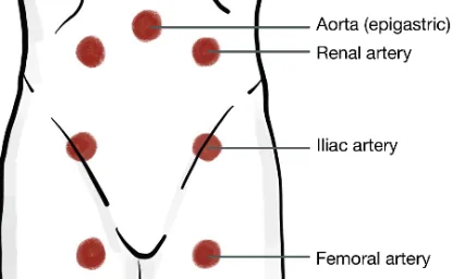
Report: I note no bruits on auscultation.
Why Check: Bruits may be indicative of obstructive arterial diseases e.g. atherosclerotic processes, or an aortic dissection (aortic bruit). An abdominal bruit in a patient with hypertension raises concern for Renal Artery Stenosis as a secondary cause of hypertension. Also explored in Chapter 13 – Abdominal Exam – MF3.
Palpation
Temperature to Touch
AFMC level Pre-clerkship 1 · Clerkship 2
Say: I will palpate the top of each foot to check temperature on each side.
Do: Feel the dorsum of each foot with back of hand – assess whether it is cold to touch, or warm to touch. Migrate proximally on each side and compare temperature between each.
Report: I note no temperature deviation at the dorsal surface of the feet.
Why Check: Cold feet is a sign of arterial deficiency; Warmer feet is a sign of venous deficiency
Note: Kneecap (patella) can be physiologically slightly cooler than rest of body
Capillary Refill Time
AFMC level Pre-clerkship 4 · Clerkship 4
(Also seen in Ch 9 – Volume Status Exam – MF2)
Say: I will palpate for circulation in the fingers and toes. Can you raise one arm above the level of the heart?
Do: Press down on the nail bed until it turns colourless. If unable to see nail bed (e.g. with nail polish present), can press on pad of finger/toe as a backup. Record time when colour returns to the nail.
Normal Values: 2s for children and adult men, 3s for women, 4s for elderly; evidence suggests >5s for moderate-severe peripheral arterial disease.
Report: I note normal capillary refill time.
Why Check: Volume status sign, and also used to assess moderate-severe peripheral arterial disease.
Edema
AFMC level Pre-clerkship 4 · Clerkship 4
(Also seen in Ch 6 – Cardiac Exam – MF1)
Say: I will palpate for leg swelling (Sacral edema if patient immobile). Please tell me if you are in discomfort.
Do: Press firmly with pads of your thumbs on the dorsal surface of patient’s leg, just above the ankle at bony prominence. Hold for 10s, then release. Look for pitting edema – skin does not bounce back immediately (i.e. pits). How high does it go on the leg? Is it pitting (transudate) or non-pitting (exudate)? Look at patient for signs of discomfort and stop exam signs of distress.
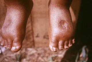
Pedal Edema6
Grading: 0 = none; 1+ = 2mm depression; 2+ = 4mm depression; 3+ = 6mm depression; 4+ = 8mm depression
Report: I note (grade) pitting edema bilaterally, up to the level of (location e.g. ankles).
Why Check: Specific to the Peripheral Vascular Exam, pitting edema can be a sign of peripheral venous disease. Edema is excess interstitial fluid accumulation causing swelling, particularly in gravity dependent areas (e.g. feet). Normally, this fluid returns through lymphatic drainage. Pitting edema = transudate (non-protein filled) – sign of increased hydrostatic pressure (e.g. congestive heart failure, decreased venous return) or decreased oncotic pressure (e.g. nephrotic syndrome, hypoalbuminemia). Non-pitting edema = exudate (protein) filled, or an issue with lymphatic drainage (e.g. lymphedema).
Calf Circumference
Say: I will now measure calf circumference in each leg, looking for symmetry.
Do: Using a measuring tape, measure the largest circumference bilaterally. Assess whether difference > 3cm.
Report: Calf circumference difference is less than 3 cm at the largest circumference.
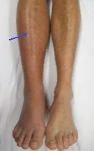
DVT of Right leg7
Why Check: Difference in calf circumference > 3cm is a clinical sign of DVT (see Well’s DVT Criteria below)
Peripheral Pulses
AFMC level Pre-clerkship 1 · Clerkship 2
Say: I will palpate the peripheral pulses in the arms, lower legs and feet.
Do: Palpate for pulses bilaterally. You can palpate the most distal pulse in each limb first. If non-palpable, move more proximally until pulses are palpable. Pulses should be symmetrical, a regular rate and rhythm with no delay between them.
Where to Palpate – Lower Extremities:
- Dorsalis Pedis: Dorsal side of foot, just lateral to extensor tendon of big toe
- Posterior Tibial: Posterior and inferior to medial malleolus = bony prominence on side of ankle
- Popliteal: Behind knees, use 2 hands
Where to Palpate – Upper Extremities:
- Radial: Along radius bone just proximal to wrist on palmar side of hand
- Brachial: Medial to biceps tendon and 2-3cm above antecubital fossa
Grading: 0 = Non-palpable; 1 = Barely palpable; 2 = Easily Palpable (Normal); 3 = Bounding; 4 = Aneurysmal.
Report: I palpated the dorsalis pedis and radial arteries as most distal pulse in each extremity. They were grade 2, symmetrical bilaterally with no delay, and regular rate and rhythm.
Why Check: Assess extent of arterial insufficiency (distal pulses lost prior to more proximal pulses).
6P’s of Acute Limb Ischemia: Pulseless, Pallor, Pain, Paresthesia (Pins/Needles), Polar (Cold) & Paralysis.
Modification for Patients with Obesity: Use a handheld Doppler Ultrasound to locate pulses if difficult to appreciate by palpation. In this population, handheld Doppler may be more sensitive than palpation.
Central Pulses
AFMC level Pre-clerkship 1 · Clerkship 2
Say: I will palpate the central pulses in the neck, arms and abdominal region.
Do: Palpate pulses and note any differences bilaterally. You should observe symmetry in amplitude, contour and upstroke.
Where to Palpate – Central Pulses:
- Carotid: Medial to sternocleidomastoid muscle, roughly at level of cricoid cartilage
- Abdominal Aorta: Slightly left and superior to umbilicus
- Femoral: Midway between ASIS and Symphysis pubis
Grading: 0 = Non-palpable; 1 = Barely palpable; 2 = Easily Palpable (Normal); 3 = Bounding; 4 = Aneurysmal.
Report: I note symmetry in amplitude, contour and upstroke at the arterial pulses examined.
Why Check: Assess signs of obstructive arterial pathologies. Note that Renal and Iliac Arteries cannot be palpated and are assessed through auscultation alone.
Modification for Patients with Obesity: Use a handheld Doppler Ultrasound to locate pulses if difficult to appreciate by palpation. In this population, handheld Doppler may be more sensitive than palpation.
Special Tests
Brodie-Trendelenburg Test
Say: I will assess each leg’s venous drainage for valve competency.
Do: Patient in supine position. Raise the patient’s leg, placing a tourniquet around the upper thigh (Normal: occluding the veins). Have patient stand up with tourniquet on. Look for venous system to fill from below the superficial saphenous system within 35-60 seconds.
Report: Brodie-Trendelenburg Test showed a venous refilling time within normal limits.
Why Check: If re-filling occurs faster than 35 seconds, evidence of valvular incompetence.
Buerger’s Test
AFMC level Pre-clerkship 1 · Clerkship 2
Say: I will hold each leg up, then lower the leg to inspect for colour changes.
Do: With patient supine, elevate leg for 30-60s and look for pallor on elevation. Normally, the leg should not become pale after 60s. Then, have patient sit with legs down for 15s, to assess delayed rubor on dependency (erythema). Normally, colour should return to the leg within 10 seconds (in a darker-skinned patient, look for time to venous filling <15 seconds).
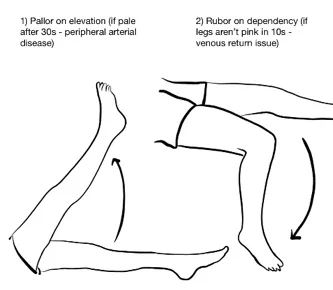
Report: I note no pallor on elevation or rubor on dependency.
Why Check: Pallor on Elevation → Peripheral Arterial Disease. The angle at which pallor begins correlates with the severity of arterial disease (e.g. pallor at <20° indicates severe ischemia). Rubor on Dependency → >15s colour return correlates with arterial disease; extreme red/pink colour suggests venous disease.
Allen’s Test
Say: I will assess each hand for the collateral circulation between the ulnar and radial arteries. Please raise the arm to the level of the heart and clench a tight fist for 30 seconds.
Do: After the 30s of patient making a fist, apply pressure over ulnar and radial arteries to occlude them. Have patient open hand while still occluding the arteries – the hand should be blanched. Release one artery. See whether the colour returns to whole hand in 5 seconds. Repeat for the other artery in the same hand. Repeat on each hand.
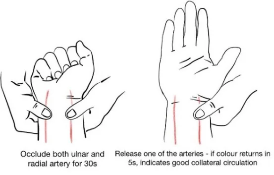
Report: Allen’s Test shows normal collateral circulation.
Why Check: Confirms adequate collateral circulation (anastomosis) to hand. Typically done in advance of arterial line placement, arterial blood gas sampling, or catheterization.
Ankle Brachial Index (ABI)
AFMC level Pre-clerkship 1 · Clerkship 2
Say: Using a Doppler Ultrasound (U/S), I will assess blood pressure differences between the Ankle and Arm.
Setup: Requires Doppler U/S Equipment. Lay patient supine.
Do:
Brachial Systolic BP: Place the manual cuff 2-3cm above crease of the elbow (as you would for normal BP measurement – see Chapter 4 – Vital Signs – MF1). Use the Doppler as your stethoscope by placing the U/S probe in the antecubital fossa. Inflate the cuff to 20 mmHg above expected systolic BP (where Doppler signal disappears). Deflate cuff until the Doppler signal has re-appeared. This is sBP for arm. Repeat for the patient’s other arm.
Ankle Systolic BP: Place the manual BP cuff just proximal to the ankle malleoli. Place the U/S probe on the dorsum of the foot to find dorsalis pedis pulse, or posterior to the medial malleolus for posterior tibial pulse. Use the higher sBP between dorsalis pedis and posterior tibial pulses.
Calculating ABI: Calculate the ratio between the highest sBP from either arm and the highest sBP of the foot of interest. (e.g Right ABI = Highest systolic BP in R Foot / Highest brachial systolic BP in either arm.)
Grading:
| ABI | Interpretation |
|---|---|
| >1.4 | Abnormal: Non-compressible vessels (e.g. hardening from PAD/calcification |
| 1.0 – 1.4 | Normal (sBP higher in leg than arm) |
| 0.9 – 0.99 | Borderline peripheral arterial disease |
| 0.5 – 0.89 | Abnormal: Mild-moderate peripheral arterial disease |
| <0.5 | Abnormal: Severe peripheral arterial disease |
Report: I note a normal ratio of Ankle BP to Brachial BP of 1.0.
Why Check: The Ankle-Brachial Index is a more accurate test for assessing the severity of Peripheral Arterial Disease. It is not assessed on the OSCE exam (requiring proficiency in use of doppler ultrasound), but you may encounter it in clinical practice. For patients with suspected peripheral arterial disease (PAD) or critical limb ischemia (CLI), the ABI is a key investigation to send patients for to guide next steps in management – where a key differentiation between PAD and CLI guiding urgency of investigation and management is the presence of rest pain, non-healing wounds, and/or gangrene8.
Helpful Stethopedia Videos to Review
- Video 1 – Peripheral Vascular Exam OSCE Style (Link: https://videopress.com/v/4flIWwRG https://videopress.com/v/ZMrJXn)
- Video 2 – Peripheral Vascular Exam Special Tests (Link: https://videopress.com/v/nrpSxbFC)
Part 4: Relevant Likelihood Ratios – from Rational Clinical Examination JAMA series
Clinical Question 1: Does this Patient have a DVT (Deep Vein Thrombosis)?9
Diagnosing a DVT depends on a 2-step process; (1) a clinically validated tool called the Wells Score, and (2) a D-dimer test.
Step 1: Clinical Prediction Rule for DVT-Wells Score3
| Clinical Variable | Score |
|---|---|
| Active Cancer (treatment ongoing or within previous 6 months, or palliative) | 1 |
| Paralysis, paresis, or recent plaster immobilization of the lower extremities | 1 |
| Recently bedridden for 3 days or more, or major surgery within previous 12 weeks requiring general or regional anesthesia | 1 |
| Localized tenderness along the distribution of the deep venous system | 1 |
| Entire leg swelling | 1 |
| Calf swelling at least 3 cm larger than the asymptomatic leg (measured 10 cm below the tibial tuberosity) | 1 |
| Pitting edema confined to the symptomatic leg | 1 |
| Collateral superficial veins (non-varicose) | 1 |
| Previously documented DVT | 1 |
| Alternative diagnosis at least as likely as DVT | -2 |
| After Adding Total Score, Find Score 3 or Higher → High Probability | DVT Probability 53% (44-61) |
| After Adding Total Score, Find Score of 1-2 → Moderate Probability | DVT Probability 17% (13-23) |
| After Adding Total Score, Find Score of Less than 1 → Low Probability | DVT Probability 5.0% (4-8) |
Step 2: D-Dimer Result Paired with Clinical Prediction Rule9
| Test | Lab Result | High Probability (Wells) | Mod Probability (Wells) | Low Probability (Wells) |
|---|---|---|---|---|
| High Sensitivity D-Dimer | Positive | 63% | 25% | 11% |
| High Sensitivity D-Dimer | Negative | 8.6% | 1% | 0.5% |
| Moderate Sensitivity D-Dimer | Positive | 67% | 34% | 17% |
| Moderate Sensitivity D-Dimer | Negative | 19% | 4.4% | 0.9% |
Clinical Question 2: Does the clinical examination predict Lower Extremity Peripheral Arterial Disease?10
Note: Screening denotes diagnosis of PAD in asymptomatic patients; Cap Refill has little clinical utility among asymptomatic patients with diabetes mellitus in screening for PAD.
| Study Type | Severity (By ABI) | Symptom or Sign | LR+ (95% CI) | LR – (95% CI) |
|---|---|---|---|---|
| Claudication – Screening | Any disease | “Definite”/ ”Probable” Claudication | 3.30 (2.30-4.80) (about +15%) | N/A |
| No claudication | N/A | 0.57 (0.43-0.76) (about −15%) | ||
| Moderate-Severe disease | No claudication | N/A | 0.89 (0.78-1.00) (no change: 95% CI includes 1) | |
| Skin Changes – Screening | Moderate-Severe disease | Hair, temperature, colour or atrophic change | 1.50 (1.20-1.70) (about +15%) | 0.81 (0.72-0.92) (about −15%) |
| Skin Changes – Symptomatic | Any disease | Cooler to touch | 5.90 (4.10-8.60) (about +30%) | 0.92 (0.89-0.95) (about −15%) |
| Wounds or sores | 5.90 (2.60-13.40) (about +30%) | 0.98 (0.97-1.00) (no change: 95% CI includes 1) | ||
| Discoloration | 2.80 (2.40-3.30) (about +15%) | 0.74 (0.69-0.79) (about −15%) | ||
| Bruits – Screening | Any disease | Femoral bruit | 4.80 (2.40-9.50) (about +15%) | 0.83 (0.73-0.95) (about −15%) |
| Bruits – Symptomatic | Any disease | Femoral bruit | 5.70 (4.70-7.00) (about +30%) | 0.74 (0.70-0.78) (about −15%) |
| At least 1 bruit (iliac, femoral, popliteal) | 5.60 (4.70-6.70) (about +30%) | 0.39 (0.34-0.45) (about −30%) | ||
| Pulse Palpation – Screening | Any disease | Any palpable pulse abnormality | 3.10 (1.40-6.60) (about +15%) | 0.48 (0.22-1.04) (no change: 95% CI includes 1) |
| Moderate-Severe disease | Any palpable pulse abnormality | 3.00 (2.30-3.90) (about +15%) | 0.44 (0.30-0.66) (about −30%) | |
| Pulse Palpation – Symptomatic | Any disease | Any palpable pulse abnormality | 4.70 (2.20-9.90) (about +15%) | 0.38 (0.23-0.64) (about −30%) |
| Capillary Refill – Screening | Severe disease | Cap Refill >5 seconds | 1.90 (1.20-3.20) (about +15%) | 0.80 (0.70-1.00) (no change: 95% CI includes 1) |
References
-
Recommendations. Choosing Wisely Canada. Accessed August 1, 2022. https://choosingwiselycanada.org/recommendations/#list ↩ ↩2 ↩3
-
Kalinchenko S, Zemlyanoy A, Gooren LJ. Improvement of the diabetic foot upon testosterone administration to hypogonadal men with peripheral arterial disease. Report of three cases. Cardiovasc Diabetol. 2009;8:19. ↩
-
File:GangreneFoot.JPG. Wikimedia Commons. Accessed February 21, 2023. https://commons.wikimedia.org/wiki/File:GangreneFoot.JPG ↩ ↩2
-
File:Chronicvenousinsufficiency.jpg. Wikimedia Commons. Accessed February 21, 2023. https://commons.wikimedia.org/wiki/File:Chronicvenousinsufficiency.jpg ↩
-
Stasis ulcer. dermnetnz.org. Accessed February 21, 2023. https://dermnetnz.org/topics/stasis-ulcer ↩
-
Centers for Disease Control and Prevention. Accessed February 21, 2023. https://phil.cdc.gov/Details.aspx?pid=16536 ↩
-
File:Deep vein thrombosis of the right leg.jpg. Wikimedia Commons. Accessed February 21, 2023. https://commons.wikimedia.org/wiki/File:Deep_vein_thrombosis_of_the_right_leg.jpg ↩
-
Gerhard-Herman MD, et al. AHA/ACC guideline on the management of patients with lower extremity peripheral artery disease. Circulation. 2016;135(12):e686-e725. ↩
-
Does this patient have a deep vein thrombosis – update. In: Rational Clinical Examination Series. Chapter 18B. 2009:246. ↩ ↩2
-
Khan, et al. Does the clinical examination predict lower extremity peripheral arterial disease? Rational clinical examination review. JAMA. 2006;295(5):536-546. ↩


