Medical Foundations 3
Chapter 13Abdominal Examination
Part 1: Focused History – Based on Chief Complaint (Begin with Open-Ended Questions)
HPI, Assoc. Sx
Abdominal Pain
- Location: Is the pain well-localized (somatic/parietal pain) vs. poorly localized (visceral pain)?
| Area | Organ/Origin | Example |
|---|---|---|
| RUQ | Liver | Hepatitis, liver abscess, portal vein thrombosis, cancer |
| Gallbladder | Biliary colic, cholecystitis, choledocholithiasis, cholangitis | |
| Pleura/lung | Pulmonary embolism (PE), pneumothorax, lower lobe pneumonia | |
| Epigastric | Esophagus | Esophagitis, GERD, Mallory-Weiss Tear, epigastric hernia |
| Gastric | Gastritis, GERD, peptic/duodenal ulcer, gastroparesis | |
| Pancreas | Pancreatitis (acute or chronic) | |
| Cardiac | Myocardial infarction (MI), angina, pericarditis | |
| Aorta | Abdominal Aortic Aneurysm (AAA), aortic dissection | |
| Pleura/lung | PE, pneumothorax, lower lobe pneumonia | |
| LUQ | Spleen | Splenomegaly, splenic infarcts, abscess, rupture |
| Pleura/lung | PE, pneumothorax, lower lobe pneumonia | |
| R lumbar | Kidney/ureter | Nephrolithiasis, pyelonephritis, ureteritis |
| Ascending colon | Diverticulitis, Crohn’s colitis, ulcerative colitis (advanced) | |
| Umbilical | Gastric | Gastritis, peptic/duodenal ulcer, gastroparesis |
| Pancreas | Pancreatitis (acute or chronic) | |
| Aorta | AAA, aortic dissection | |
| Bowel | Umbilical hernia | |
| L lumbar | Kidney/ureter | Nephrolithiasis, pyelonephritis, ureteritis |
| Descending colon | Diverticulitis, Crohn’s colitis, ulcerative colitis (early) | |
| RLQ | Appendix | Appendicitis (late, localization stage) |
| Ileocecum | Crohn’s colitis, ulcerative colitis (advanced), inguinal hernia | |
| Kidney/ureter | Nephrolithiasis, pyelonephritis, ureteritis | |
| Utero-ovarian | Torsion, endometriosis, ruptured cyst, ectopic preg., Mittelschmerz | |
| Testicles | Torsion, epididymitis, epididymo-orchitis | |
| Suprapubic | Bladder | UTI, cystitis, urethritis, acute urinary retention |
| Utero-ovarian | PID, endometriosis, ectopic pregnancy, pregnancy + complications | |
| Prostate | Acute bacterial prostatitis | |
| LLQ | Sigmoid colon | Diverticulitis, Crohn’s colitis, ulcerative colitis (early), inguinal hernia |
| Kidney/ureter | Nephrolithiasis, pyelonephritis, ureteritis | |
| Utero-ovarian | Torsion, endometriosis, ruptured cyst, ectopic preg., Mittelschmerz | |
| Testicles | Torsion, epididymitis, epididymo-orchitis | |
| Diffuse | Metabolic | Diabetic ketoacidosis, hypercalcemia |
| Gastric | Gastroenteritis, Peptic ulcer disease (PUD) with perforation | |
| Bowel | Constipation, mesenteric ischemia, perforation, obstruction, IBD, IBS | |
| Appendix | Appendicitis (early, diffuse stage) | |
| Utero-ovarian | PID, torsion, endometriosis, adenomyosis, leiomyomas | |
| Obstetric | Ectopic pregnancy, pregnancy + complications | |
| Peritoneum | Peritonitis |
- Onset: Acute vs. chronic? Gradual vs. sudden? Changes in location over time?
- Palliating/Provoking: Does anything make the pain better or worse?
- Movement: Pain worse with all movement and relieved by sitting still is concerning for peritonitis. Worse with bumps in the road on the ride to hospital; with bed shaking; or with coughing (Positive cough test: LR+ 1.8, LR- 0.4)?
- Positionality: Pain worse with lying down (e.g. GERD); pain relieved by sitting up/lean forward (e.g. pancreatitis, pericarditis)
- Post-Prandial (worse after meals): GERD (esp. spicy/acidic meals), gastric ulcer (immediate) vs. duodenal ulcer (better with meal, worse 2h after), biliary colic (esp. fatty meals), pancreatitis, chronic mesenteric ischemia (esp. unintentional weight loss due to aversion), food intolerance, etc.
- Quality: How would you describe the pain? Has the quality of pain changed over time?
- Visceral Pain (Viscera i.e. organs sense acute stretch and ischemia) – gnawing/cramping/aching, poorly localized
- Parietal Pain (Peritoneal pain usually due to inflammation) – sharp, well localized
- Burning Pain: e.g. Gastroenteritis, Peptic Ulcer Disease, GERD
- Change in quality and location over time? – inflammation spreads form viscera to peritoneum
- E.g. Appendicitis: Pain begins poorly localized at umbilicus (visceral pain, appendix sympathetic innervation at T10); becomes more localized at McBurney’s point (parietal pain) (Migration of pain: LR+ 3.2, LR- 0.50 in adults; LR+ 1.9-3.1, LR- 0.41-0.72 in pediatrics)
- Radiation: Does the pain travel anywhere? Referred pain = pain perceived at another location
- Cholecystitis (irritation right phrenic nerve) → referred right shoulder pain
- Pancreatitis (irritation of celiac plexus) → pain radiates straight through to the back
- Spleen (irritation of left phrenic nerve) → referred left shoulder pain
- Severity: How bad is it on a scale of 1-10
- Pain out of proportion with exam findings is concerning for acute mesenteric ischemia
- Timing: Does it come and go? Or is it consistent?
- Colicky pain (waves of pain) – e.g. cholelithiasis, nephrolithiasis, bowel obstruction
- Constant pain – Inflammation (e.g. cholecystitis, appendicitis, etc.), Ischemia (e.g. mesenteric ischemia, sickle cell crisis), Perforation (e.g. bowel perforation).
- Associated Symptoms:
- Red flag symptoms: sudden onset, severe, peritonitis, bilious vomiting, hematemesis, coffee-ground emesis, melena stools, hematochezia, jaundice, pain out of proportion to exam, dysphagia
- Pregnant: Consider obstetrical causes, prefer ultrasound instead of CT, confirm with ß-HCG
- Nausea/Vomiting: Particularly visceral causes (e.g. pancreatitis, cholecystitis, appendicitis, gastroparesis, bowel obstruction) and chemical causes (e.g. diabetic ketoacidosis, hypercalcemia)
- Altered Bowel Movements (see below): Steatorrhea in malabsorption (e.g. Crohn’s colitis, pancreatitis), GI bleeds (e.g. PUD, Mallory-Weiss, diverticulitis, perforation), constipation +/- obstipation (diffuse pain, obstruction)
- Cholestasis: jaundice, choluria (dark/brown urine), pale/clay-coloured stools (e.g. biliary cause)
- Liver Disease Signs: cholestasis, fatigue, weight loss, anorexia, distension (re. ascites), easy bruising
- Dysphagia: particularly worrisome if progressive and with GERD symptoms, weight loss, persistent vomiting, melena stool, iron-deficiency anemia (e.g. rule out esophageal cancer)
- Early Satiety: e.g. Gastroparesis, Peptic Ulcer Disease, GERD, gastric cancer
- Constitutional Symptoms: Fevers, night sweats, chills, unintentional weight loss, malaise?
- Concern for infection, malignancy, IBD (e.g. Crohn’s Disease, Ulcerative Colitis)
- Cardiopulmonary Symptoms: Cough, dyspnea, orthopnea/PND, Presyncope, calf pain/swelling
- Concern e.g. for cardiac (e.g. MI) or pulmonary cause (e.g. pneumonia, pneumothorax, PE)
Altered Bowel Movements (ABM)
- When was last bowel movement? (For patients admitted to hospital, stool chart may be available)
- Constipation: inability or infrequent passage of stool, often associated with straining, passage of hard stool, tenesmus (persistent but ineffective urge to empty rectum), or self-digitation.
- May be primary (e.g. idiopathic, ↓ mobility, inadequate intake) or secondary (drug-induced e.g. opioids, anticholinergics, 5-HT3 antiemetics; metabolic e.g. ↑Ca or ↓K; neurological disorders e.g. spinal cord compression; visceral e.g. bowel obstruction, paralytic ileus; psychological e.g. lack of privacy, need for commode/bedpan; or local e.g. anal abrasions/hemorrhoids/tears)
- Can cause diffuse abdominal pain and distension
- Obstipation: inability to pass gas → complete bowel obstruction (vs. partial, without obstipation)
- Diarrhea: ≥3 loose stools per day, or more frequent than normal for the individual
- Secretory: Out of proportion to your food intake? → results from increased colonic secretions; continue to experience diarrhea despite decreased food intake (e.g. infection, endocrine)
- Osmotic: Stops if intake of substance is stopped? → can occur in response to certain foods, in patients with celiac disease, or with ↑ fluid intake (e.g. laxatives)
- Exudative/Inflammatory: Can contain blood or pus (e.g. IBD, some infectious causes, cancer)
- Steatorrhea: fatty, foul-smelling stools seen in malabsorption or maldigestion syndromes (e.g. Crohn’s colitis, pancreatitis, bowel resection, short bowel syndrome)
- Blood in Stool: Upper vs. Lower GI bleed distinguished by the Ligament of Treitz (at end of duodenum)
- Melena Stool: black, tarry and foul smelling → usually upper GI bleed (LR+5.1-5.9, LR- 0.06-0.27) (↑ transit time oxidizes blood), less commonly slow right colon bleed.
- Note: iron supplements, bismuth products can make stool appear dark
- Hematochezia: bright red blood in stool/bowl → usually lower GI bleed (less commonly brisk upper GI bleed). Darker red = more transit time.
- Bright red blood per rectum: bright red blood with wiping → suggestive of hemorrhoids (not hematochezia; helpful to ask about constipation history or straining to defecate)
- Orthostatic dizziness or pre-syncopal symptoms could be due to GI blood volume loss.
- Also ask about associated abdominal pain, blood in vomit (hematemesis/coffee-ground emesis)
- Melena Stool: black, tarry and foul smelling → usually upper GI bleed (LR+5.1-5.9, LR- 0.06-0.27) (↑ transit time oxidizes blood), less commonly slow right colon bleed.
- Helpful to Classify Stools According to the Bristol Stool Chart:
- Type 1: Separate hard lumps → Very Constipated
- Type 2: Lumpy and Sausage Like → Slightly Constipated
- Type 3: Sausage shape with cracks in surface → Normal
- Type 4: Smooth, soft sausage or snake → Normal
- Type 5: Soft blobs with clear cut edges → Lacking fiber
- Type 6: Mushy consistency, ragged edges → Inflammation
- Type 7: Liquid consistency, no solid pieces → Inflammation
- Ask Caregivers to bring a Bowel Movement Chart (Helpful for Histories of Altered Bowel Movements for Patients with Intellectual & Developmental Disabilities)
- Can’t miss diagnoses:
- C. difficile infection (CDI; pseudomembranous colitis): severe watery diarrhea after recent antibiotic use (esp. clindamycin, cephalosporins and fluoroquinolones).
- Severe CDI: CDI + fever and leukocytosis
- Fulminant CDI: Severe CDI + shock, blood, paralytic ileus, or toxic megacolon.
- Bowel obstruction: constipation, obstipation, abdominal distension, vomiting, history of abdominal surgeries, abdominal or pelvic cancers (re. malignant bowel obstruction – e.g. ovarian, colorectal or gastric cancers, or peritoneal carcinomatosis)
- GI bleeds: GI Bleeds can cause hemorrhagic shock. Iron deficiency anemia with no obvious cause is a GI bleed until proven otherwise (may require an urgent scope if thought to be actively bleeding). Patient-reported melena moderately increases likelihood of UGIB (LR+5.1-5.9, LR- 0.06-0.27).
- C. difficile infection (CDI; pseudomembranous colitis): severe watery diarrhea after recent antibiotic use (esp. clindamycin, cephalosporins and fluoroquinolones).
Nausea/Vomiting
- Definitions:
- Nausea: Unpleasant sensation with urge to vomit
- Vomiting: Forced, retrograde expulsion of gastric contents (vs. retching – no expulsion)
- Onset: Acute vs. chronic?
- Palliating/Provoking: Does anything make the nausea/vomiting better or worse?
- Provoking – Recent changes to medications (chemical cause of nausea): Common medication causes include chemotherapy, opioids, antibiotics, antifungals, antidepressants (e.g. SSRI), NSAIDS, PO Iron
- Provoking – Recent radiation treatment (e.g. for cancer treatment)
- Provoking – Post-Prandial: e.g. pancreatitis, cholecystitis, gastroparesis, bowel obstruction, GERD
- Provoking – Head movements: Vestibular cause (e.g. BPPV, Meniere’s, Vestibular neuritis)
- Palliating – Nausea relieved by vomiting: Suggests visceral cause (e.g. gastroparesis, gastric outlet obstruction, cholecystitis, pancreatitis, bowel obstruction, constipation)
- Palliating – Anti-nausea medications (see Medications, below)
- Quality (of Vomit): Examples include:
- Bilious (green/bright yellow) – suggests intestinal obstruction
- Coffee Ground Emesis – upper GI bleed with ↑ transit time (oxidizes blood)
- Hematemesis – upper GI bleed with red blood (shortened transit time; distinct from Hemoptysis)
- Feculent – suggests bowel obstruction, fistula (e.g. Crohn’s Disease)
- Severity: How bad is it on a scale of 1-10? Are you able to eat and drink? (re. risk of dehydration)
- Timing: Does it come and go? Or is it consistent?
- Constant nausea: Concern e.g. for chemical cause (e.g. Diabetic Ketoacidosis, Hypercalcemia), visceral cause (e.g. appendicitis, bowel obstruction, pancreatitis), cortical cause (e.g. meningitis, tumor, intracranial hemorrhage, raised Intracranial Pressure = ICP)
- Worse in AM – concern e.g. for cortical cause (e.g. raised ICP)
- Associated Symptoms:
- Abdominal Pain: visceral causes (e.g. pancreatitis, cholecystitis, appendicitis, gastroparesis, bowel obstruction) and chemical causes (diabetic ketoacidosis, hypercalcemia). Vomiting after pain onset mildly increases likelihood of suspected appendicitis (LR+ 2.8 in adults).
- Constipation/Obstipation: e.g. bowel obstruction
- Diarrhea: Concern for infectious cause (e.g. acute gastroenteritis - bacterial, viral, parasitic)
- Constitutional Symptoms: Fevers, night sweats, chills, unintentional weight loss, malaise?
- Concern for infection (e.g. appendicitis, ascending cholangitis, meningitis), malignancy
- Dysphagia: Red flag symptom, particularly if progressive and with GERD symptoms, weight loss, abdo pain, melena stool, iron-deficiency anemia (e.g. rule out esophageal cancer)
- Early Satiety: e.g. Gastroparesis, Peptic Ulcer Disease, GERD, gastric cancer
- Neurologic/Psychiatric Symptoms (explored in Ch 31 – Screening Neuro Exam – MF4):
- Headache (Primary headache e.g. migraine; vs. Secondary headache e.g. ↑ ICP, meningitis, tumor, intracranial hemorrhage) – suggests cortical cause of nausea/vomiting
- Vertigo/Tinnitus (e.g. BPPV, Meniere’s, Vestibular neuritis) – suggests vestibular cause
- Anxiety (e.g. Anticipatory nausea) – suggests cortical cause of nausea/vomiting
Liver Disease
- Causes of chronic liver disease and cirrhosis: alcohol; non-alcoholic fatty liver disease; chronic viral hepatitis (e.g. hepatitis B and C); hemochromatosis; autoimmune hepatitis; primary biliary cirrhosis; primary sclerosing cholangitis; Wilson disease; Alpha-1 Antitrypsin Deficiency; congestive hepatopathy; medications (e.g. methotrexate, isoniazid)
- Causes of acute liver failure: acetaminophen overdose, alcohol, acute viral hepatitis (A, B, C, D, and E) and other infections (e.g. herpes simplex, varicella zoster, Epstein-Barr, adenovirus, cytomegalovirus), hypoperfusion (e.g. sepsis, ischemic hepatitis)
- General Symptoms:
- Cholestasis: jaundice, choluria (dark/brown urine), pale/clay-coloured stools (e.g. biliary cause)
- Liver Damage Signs: cholestasis, fatigue, weight loss, anorexia, distension (re. ascites), easy bruising
- Decompensating events and associated symptoms:
- Hepatic encephalopathy (presenting with confusion, sleep-wake cycle reversal, asterixis); in patients with chronic liver disease, moderately increases likelihood of cirrhosis (LR+ 8.8)
- Ascites (presenting with abdominal distension) – the following findings mildly increase the probability of ascites: increased abdominal girth (LR+ 4.1, LR- 0.17); recent weight gain (LR+ 3.2, LR- 0.42); ankle swelling (LR+ 2.8, LR- 0.10); in patients with chronic liver disease, ascites moderately increases probability of cirrhosis (LR+ 6.6)
- Esophageal varices (presenting with hematemesis, melena or hematochezia if brisk bleeding)
- Hepatorenal syndrome (presenting with oliguria re. ↓ urine output)
- Spontaneous bacterial peritonitis (presenting with fever, abdominal pain, confusion)
Dysphagia1
- Definitions:
- Dysphagia – subjective sensation of difficulty or abnormality of swallowing
- Odynophagia – pain with swallowing
- Globus sensation – Painless sensation of lump/foreign body/food bolus in pharyngeal/cervical area
| Type | Definition | Motility causes | Structural causes |
|---|---|---|---|
| Oropharyngeal dysphagia | Difficulty initiating swallow | Neurological (e.g. stroke, brain injury, tumour) Muscular (e.g. myasthenia gravis | Mucosal (e.g. infection, cancer) Extra-mucosal (e.g. deep neck infection) |
| Esophageal dysphagia | Impaired passage through esophagus | E.g. achalasia, GERD, hypomotility disorder | Intraluminal (e.g. impacted object) Mucosal/Intrinsic (e.g. esophagitis, stricture, tumour, abscess) Extrinsic (e.g. thyroid goiter, mediastinal mass, hiatal hernia) |
- What happens when you swallow? Does swallowing make you cough?
- Trouble initiating swallow, choking, coughing → suggests oropharyngeal dysphagia
- Feeling of food stuck → suggests esophageal dysphagia
- How long has this been occurring? Is it getting worse? Do you hesitate to go out to eat because of it?
- Acute: stroke, pill esophagitis (recent medication changes?), infection, foreign body impaction
- Intermittent: Intermittent pill esophagitis, esophageal motility disorder, inconsistent denture use
- Progressive dysphagia: Concern for malignancy
- Do you have trouble chewing your food?
- Jaw pain → Temporomandibular joint arthritis or pain
- Trouble chewing → Poorly fitting dentures, dental caries/disease, or xerostomia (dry mouth)
- Weakness with chewing → May signal neurologic disease (e.g. Myasthenia Gravis)
- What gets stuck?
- Solids only → suggests mechanical obstruction of esophagus – intrinsic vs. extrinsic (see above)
- Solids and Liquids → suggests esophageal motility disorder (e.g. achalasia)
- Liquids only → suggests oropharyngeal pathology
- Red Flag Symptoms (Note – dysphagia itself is considered a red flag symptom):
- Chronic GERD, Weight loss, fever, GI Bleed, Odynophagia, progressive symptom-rule out malignancy
- Neurologic symptoms: e.g. diplopia, dysarthria, dysmetria, weakness, vertigo – consider cerebrovascular disease as cause (e.g. stroke)
Past Medical History (PMHx)
- Bowel: Inflammatory Bowel Disease (Crohn’s Colitis or Ulcerative Colitis), Irritable Bowel Syndrome (IBS), Celiac disease
- Hepatopancreatobiliary disease:
- Gallstones/Biliary colic (past gallstones ↑ likelihood of future gallstones; can also cause Acute Pancreatitis)
- Pancreatitis (most commonly alcoholic, gallstones, idiopathic)
- Cirrhosis – Common etiologies: Non-alcoholic fatty liver disease (NAFLD), alcohol-related, viral hepatitis (Hep B/Hep C)
- NAFLD: Spectrum of disease including fatty liver (NAFLD), non-alcoholic steatohepatitis (NASH) and cirrhosis. NAFLD is a feature of metabolic syndrome (along with obesity, insulin resistance, hypertension, hypertriglyceridemia and low HDL)
- Other etiologies: Hemochromatosis; Autoimmune hepatitis; primary biliary cirrhosis; primary sclerosing cholangitis; Wilson disease; Alpha-1 antitrypsin deficiency; Congestive hepatopathy; medications (e.g. methotrexate, isoniazid)
- Esophagus/stomach/duodenum: GERD, peptic ulcer disease
- GI bleeds: Upper GI, Lower GI. Iron deficiency anemia with no clear explanation is a GI bleed until proven otherwise
- Prior cancer, chemotherapy, or radiation – ↑ suspicion of new or recurrent cancer
- Which cancer? Staging? Metastasis?
- What treatment was used? What was intent of treatment – Neoadjuvant treatment (before primary treatment e.g. surgery) vs. Adjuvant treatment (after primary treatment e.g. surgery)? Palliative treatment (e.g. palliative chemotherapy for metastatic lung cancer – intent to control disease at good quality of life, rather than cure disease)
- Heart Disease (CHF → congestive hepatopathy → liver disease signs); Hypertriglyceridemia (can cause Acute Pancreatitis)
- Neurologic Conditions (↑ risk of oropharyngeal dysphagia): e.g. Parkinson’s Disease, Cerebrovascular Disease, Dementia, Amyotrophic Lateral Sclerosis (ALS), Multiple Sclerosis
- Atopic Triad (Asthma, Atopic Dermatitis, Allergic Rhinitis): ↑ risk of eosinophilic esophagitis as cause for dysphagia/GERD
- Cancer Screening: Is your age-related colon cancer screening up to date per Cancer Care Ontario recommendations?
- Current Cancer Care Ontario Recommendations (for Cervical, Breast, Colon, Lung and Prostate Cancer)
- Pro Comp Connection: Cancer screening is an important opportunity for patient-centred shared decision making (e.g. using patient-centred decision aid tools)
Relevant Clinical Scores:
- MELD Score – Stratifies severity of end-stage liver disease
- Child-Pugh Score for Cirrhosis Mortality – Estimates cirrhosis severity
- FIB-4 Index – Non-invasive estimate of liver fibrosis in patients with Hepatitis B, Hepatitis C Viruses
- Serum Ascites Albumin Gradient (SAAG) – Defines presence of portal hypertension in patients with ascites
- Ranson’s Criteria – Estimates mortality for patients with pancreatitis based on initial and 48-hr lab values
- Alvarado Score for Acute Appendicitis – Predicts likelihood of appendicitis diagnosis
- AIMS65 Score for Upper GI Bleeding Mortality – Determines in-hospital mortality risk from Upper GI Bleed
- Forrest Classification – Stratifies severity of upper GI bleeding
- Glasgow-Blatchford Bleeding Score – Identifies low risk UGI bleeds to consider outpatient management
- Oakland Score – Predicts readmission risk in patients with acute lower GI bleeding
- Montreal Classification for IBD – Classifies Crohn’s and ulcerative colitis severity
- Mayo Disease Activity Index (DAI) for Ulcerative Colitis – Assesses severity of ulcerative colitis
- Crohn’s Disease Activity Index (CDAI) – Determines the current severity of Crohn’s disease
- ROME-IV criteria for IBS – Diagnoses IBS
Past Surgical History (PS Hx)
- Prior abdominal surgeries: Most common cause of adhesive small bowel obstruction is adhesions from past surgeries
- Common surgeries: Cholecystectomy, Appendectomy, Inguinal Hernia repair
- Common GI procedures: esophagogastroduodenoscopy (EGD), colonoscopy/sigmoidoscopy (may be for screening, diagnosis, active surveillance, etc.), endoscopic retrograde cholangiopancreatography (ERCP – for common bile duct (CBD) stones, treatment of gallstone pancreatitis, can cause acute pancreatitis)
- Surgical management of Crohn’s Disease: Small bowel resection, colorectal resection
- Abdominal Cancer Surgeries (Non-exhaustive list, clarify curative vs. palliative intent, positive vs. negative margins):
- Colorectal Cancer: Right- or Left-hemicolectomy; Total abdominal colectomy; Sigmoid colectomy
- Pancreatic Cancer: Pancreaticoduodenectomy (Whipple Procedure); Palliative endoscopic stent (to relieve biliary obstruction from cancer)
- Biliary Tree Cancer (e.g. cholangiocarcinoma): Hepatic resection (e.g. intrahepatic cholangiocarcinoma); Whipple Procedure (e.g. distal cholangiocarcinoma); Palliative endoscopic stent (to relieve biliary obstruction from cancer)
- Liver Cancer (e.g. Hepatocellular Carcinoma): Hepatic resection; Liver transplant
- Gastric Cancer: Total gastrectomy, partial gastrectomy
- Liver Disease:
- Ascites Management: Intermittent paracentesis; Indwelling peritoneal catheter (↑ infection risk); Transjugular intrahepatic portosystemic shunt (TIPS) procedure
- Esophageal Varices Management: Esophageal variceal band ligation
- End-Stage Liver Disease: Liver transplant
- Bariatric Surgery: most common surgeries include Roux-en-Y Gastric Bypass and Sleeve Gastrectomy
Family Medical History (FMHx)
- Inflammatory Bowel Disease (e.g. Crohn’s Disease or Ulcerative Colitis)
- Celiac Disease
- Cholecystitis (↑ risk)
- Causes of Liver Cirrhosis with hereditary component: Alpha-1 Antitrypsin Deficiency (A1AT), Hereditary Hemochromatosis, Wilson Disease (disorder of copper metabolism)
- Solid tumor cancers e.g. breast, colorectal, stomach, prostate, or ovarian cancer
- Age at diagnosis? Stage? Alive or dead? Age at death? Have you started screening?
- FMHx of early age cancer may affect age to start screening in colorectal cancer and breast cancer
- Genetic testing results, if available (e.g. BRCA in breast cancer)? May affect age to start screening
Social Hx
Smoking
- Smoking ↑ risk of cancers (e.g. esophageal)
- Pack years = packs/day x years smoking, important risk factor for lung cancer
- Have you thought about quitting in the next 6 months? Cigarettes? Marijuana? E-cigarettes/vapes?
- Smoking worsens course of Crohn’s disease, never/former smoker ↑ risk of ulcerative colitis
- 5As approach taking a smoking Hx (Explored in Chapter 38 – Motivational Interviewing – IF)
- Pro-Comp Connection: Regarding patient’s position, do not jump to thinking in terms of “compliance” with recommendations for smoking cessation.
Alcohol
How Often Do You Drink (Drinks/Week)? What is the size of 1 drink? What kinds of drinks?
- Alcohol ↓ Lower esophageal sphincter tone, with ↑ risk of esophagitis → upper GI bleed
- Alcohol-related cirrhosis with portal hypertension, esophageal varices ↑ GI bleed risk
- Alcohol as important cause of hepatitis and acute pancreatitis
Recreational Drug Use
IV drug use increases risk of Hepatitis C, HIV
Recent Travel History
- Noninflammatory acute diarrhea: Enterotoxigenic E. Coli, Vibrio Cholerae (contaminated water); Giardia (camping and fresh water)
- Inflammatory acute Diarrhea: Campylobacter
- Note: Helpful traveler advice by individual country available on CDC website
Sexual History
General Sexual History (seen in Chapter 1 – General Medical History – MF1)
- Normalizing Opening Statements3: Is it okay if I ask you a few questions about your sexual health? Some of questions are very personal. I ask everyone these questions, and I consider them to be very important. You do not have to answer any questions you don’t want to.
- Confidentiality Statement: Now and for the rest of our visits, this information will be kept confidential. I might take notes and put those in your record here, but everything will be stored securely.
- General Sexual History45:
- Have you been sexually active in the past year?
- How many partners have you had sex with in the past year?
- What is your partner’s gender? What gender or genders are the partners you’ve had sex with in the last year?
- Do you have any sexual concerns you would like to discuss?
- Note: A detailed sexual history may be added to a general sexual history, depending on patient care needs. To discuss sexual health in an effective and patient-centred way, it is important to ensure a safe, private and respectful environment. The 5P’s Model of Harm Reduction4 (Practices; Partners; Protection from Sexually Transmitted and Blood Borne Infections = STBBIs, Past History of STBBIs; and Pregnancy) is introduced in Ch 18 – Detailed Sexual Histories – MF3
Specific Questions to ask on Genitourinary History if concerns identified:
- Any abnormal discharge from the vagina/penis? Post-coital bleeding (after intercourse)?
- Any itchiness or pain in the genital area at rest? What about during urination?
- Any pain during/after intercourse (dyspareunia)? Pain while urinating (dysuria)?
- Do you notice any new lesions/spots/marks/moles/ulcers in the genital area?
- Have you ever had a Sexually Transmitted and Blood Borne Infections (STBBI)? – previous STBBI infections increase risk for future STBBI infections
Medications Hx & Allergies
Constipation Symptom management
- Osmotic Agents: e.g. PEG 3350, Lactulose, Sorbitol, Milk of Magnesia
- Note: Osmotic laxatives also commonly used for colonoscopy prep
- Stimulant Laxatives: e.g. Bisacodyl, Sennosides
- Surfactant Laxatives: e.g. Docusate → poorly effective in managing constipation
- Note: Excess use of laxatives prescribed for constipation may also give diarrhea
Diarrhea Symptom management
- Peripheral μ-Opioid Receptor Agonist (e.g. Loperamide = Imodium)
- Bile Acid Sequestrant (e.g. Cholestyramine)
- ↓ GI Motility Agents (e.g. Diphenoxylate/Atropine = Lomotil)
Nausea/Vomiting Symptom management
- Common:
- Prokinetics (e.g. Metoclopramide, Domperidone)
- Antipsychotics (e.g. Prochlorperazine, Haldol, Olanzapine)
- 5HT3 Antagonist (e.g. Ondansetron)
- Antihistamine (e.g. Dimenhydrinate)
- Others: Neurokinin-1 Antagonists (e.g. Aprepitant); Steroids (e.g. Dexamethasone); Cannabinoids – e.g. Nabilone
Liver Cirrhosis – Management/Prevention Decompensating Events
- Hepatic encephalopathy: Lactulose; Rifaximin
- Ascites: MRA (e.g. Spironolactone); Loop Diuretic (e.g. Furosemide)
- Esophageal varices: Non-selective betablockers (e.g. Propranolol, Nadolol); or Carvedilol
- Hepatorenal syndrome: Vasoconstrictors (e.g. Midodrine, octreotide, norepinephrine)
- Spontaneous bacterial peritonitis: Antibiotics for prophylaxis (e.g. Trimethoprim-Sulfamethoxazole, Ciprofloxacin, Norfloxacin)
Inflammatory Bowel Disease
- Medical Management – Medication Classes:
- TNF-α Inhibitors: e.g. Infliximab, Adalimumumab, Certolizumab, Ustekinumab, etc.
- Disease Modifying Antirheumatic Drugs (DMARDs): e.g. Methotrexate, Sulfasalazine, Azathioprine, 6-Mercaptopurine
- Mesalamine (5-ASA, for Mild-Mod. ulcerative colitis) – topical, per rectum or PO options
- Acute Flares: Glucocorticoids (e.g. Prednisone, Budesonide, Methylprednisolone)
Gastroesophageal Reflux Disease (GERD)
- Antacids: e.g. Calcium Carbonate, Magnesium Hydroxide, Sodium Alginate
- H2-Receptor Blockers: e.g. Ranitidine, Famotidine
- Proton Pump Inhibitors: e.g. Pantoprazole, Lansoprazole, Omeprazole, Rabeprazole
- Long-Term Indications for PPI Use: GI Bleed, Barrett’s Esophagus, Severe Esophagitis, Chronic NSAID Use with Bleeding Risk– see Choosing Wisely PPI Deprescribing Toolkit
- Medications that ↑ Risk of GERD:
- Pill Esophagitis (direct irritation of mucosa): Bisphosphonates, Ferrous Sulfate, NSAIDs, Potassium Chloride, Tetracyclines (e.g. Doxycycline), Vitamin C
- ↓ Lower Esophageal Sphincter Tone: Anticholinergics, Benzodiazepines, Caffeine, Calcium Channel Blockers, Nitrates, Tricyclic Antidepressants (e.g. Amitriptyline, Nortriptyline)
GI bleed medications
- Proton pump inhibitors (e.g. Pantoprazole, Lansoprazole, Omeprazole, Rabeprazole): decrease rebleeding risk in upper GI bleeds
- H2 receptor blockers (e.g. ranitidine): do NOT reduce rebleeding risk
- Octreotide: for variceal upper GI bleed
- Antibiotic prophylaxis: (e.g. Trimethoprim-Sulfamethoxazole, Ciprofloxacin, Norfloxacin_ for patients with cirrhosis to reduce risk of infection re. spontaneous bacterial peritonitis (SBP)
- NSAIDs: ↑ GI Bleed risk
- Anti-platelet, Anticoagulant medications: ↑ GI Bleed risk
- Anti-platelet: e.g. acetylsalicylic acid, ticagrelor, clopidogrel
- Anti-coagulants: e.g. Heparins, Warfarin, DOACs (e.g. apixaban, rivaroxaban, dabigatran)
- PO Iron or Bismuth Subsalicylate: Use can cause melena stool colour mimic
Notes on Specific Medications
- Recent Antibiotic Use: Risk of C. Difficile Infection, causing acute watery diarrhea (especially with recent clindamycin, cephalosporin or fluoroquinolone use).
- Opioids: Common cause of constipation
Part 2: Pertinent Notes about the Physical Exam
- Patient Positioning during Abdominal exam: After taking vitals, patient should be supine, arms at sides and legs uncrossed with a pillow underneath their head. There should be good lighting, with the exam room warm and your hands warm. Make sure the patient has an empty bladder before you start your exam.
- Draping: Drape appropriately when need area exposed; return covered up state at end of portion of exam/end of exam. If the patient has abdominal pain, during the exam and with patient consent, expose the groin and genitalia for a few seconds to check for inguinal and femoral hernias.
Why Move from Inspection → Auscultation → Percussion → Lighter Palpation → Deeper Palpation?
- Auscultation is performed first in the abdominal examination because palpation and percussion can cause a disturbance in bowel sounds, obscuring the value of auscultation maneuvers.
- In general, moving from percussion → light palpation → deep palpation allows one to be progressively invasive for pain reasons and to get a good outline as to where to palpate via preparatory percussion.
Part 3: Specific Maneuvers in the Physical Exam – Say, Setup, Do, Report
Notes on Vitals
Vitals – Always do HR, BP, RR, Temp
In This Exam
AFMC level Pre-clerkship 4 · Clerkship 4
- Check for Orthostatic Hypotension (if concerned about severe blood loss, nausea/vomiting/diarrhea)
- Systemic Inflammatory Response Syndrome (SIRS) Criteria: (1) Fever > 38C or < 36C; (2) HR > 90 bpm; (3) RR > 20/min or Arterial CO2 < 32 mmHg; (4) Abnormal WBC Count (> 12000/µL or < 4000/µL, or >10% bands) – Note: SIRS may occur in several non-infectious conditions e.g. autoimmune disease, pancreatitis, vasculitis, thromboembolism, burns or surgery.
- Sepsis Threshold: 2 of 4 SIRS criteria plus suspected infection (e.g. appendicitis, diverticulitis)
- Other screening tools for sepsis or septic shock: NEWS (National Early Warning Score); MEWS (Modified Early Warning Score for Clinical Deterioration); qSOFA (quick screen of in-hospital mortality for non-ICU patients with infection)
- Recommendation: As per 2021 Surviving Sepsis Guidelines, “We recommend against using qSOFA compared with SIRS, NEWS, or MEWS as a single-screening tool for sepsis or septic shock”6.
General Inspection
General & Body Habitus
AFMC level Pre-clerkship 3 · Clerkship 4
Say: I will begin with general inspection of the head and torso.
Do: Look for signs of respiratory distress (see Ch 5 – Resp Exam – MF1), and diaphoresis (heavy perspiration/ sweating). Is patient endomorphic (overweight), ectomorphic (underweight), or mesomorphic (normal weight)? Observe their level of consciousness: are they alert and oriented to person, place and time?
Report: I note no signs of respiratory distress such as accessory muscle use (state full list). Patient not diaphoretic with a mesomorphic body habitus, alert and oriented to person, place and time.
Why Check: Important especially when formulating your impression including the acuity of the presentation. Here, altered LOC could be a sign of hepatic encephalopathy, or a sign of shock (e.g. hemorrhagic shock from GI bleed, Reynaud’s Pentad re. Acute Suppurative Cholangitis). Encephalopathy greatly increases the likelihood of suspected cirrhosis in patients with known liver disease (LR+ 10, LR-0.86)
Volume Status Exam
Perform a quick exam to check for hypervolemia if you suspect hepatic congestion (e.g. auscultate lung bases, assess JVP, inspect for peripheral cyanosis, auscultate the heart, palpate peripheral pulses, edema, etc.)
Why Check: Heart failure can cause hepatic congestion, which can contribute to acute liver failure, esophageal varices and variceal bleeding, and venous thrombosis. (Ch 9 – Volume Status Exam – MF2).
General Shape of Abdomen & Hernias
AFMC level Pre-clerkship 3 · Clerkship 4
Say: I will now inspect the abdomen for its shape, and for scars and skin changes.
Do: Inspect the abdomen:
- Does abdomen look flat, rounded, scaphoid or protuberant (with generalized or localized distension)?
- Do you observe bulging flanks (difficult to assess on inspection in Obese patients)?
- Do you observe obvious bulges or peristalsis? – may be a hernia (need to palpate)
- Check the umbilicus for any bulging (e.g. in umbilical hernias, bowel obstruction)
- Check if it is reducible (returns to abdomen on pressure or when the patient lays down flat and relaxes. If it is not reducible, it may be incarcerated (stuck) or strangulated (ischemic bowel disease risk – this is when the patient presents with central abdominal pain).
- Can you see any past surgical scars? Any colostomies?
- Any visible pregnancy (e.g. fetal movement)?
- Any pulsations (e.g. aortic pulsation as potential aneurysm)?
- Does the abdomen move with respiration?
- Check for straie, for superficial dilated veins (e.g. in IVC obstruction) and bruising (e.g. history of trauma).
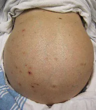
Bulging Flanks7
Report: The abdomen is flat and non-distended. I note no bulging flanks, hernias, or surgical scars.
Why Check: Bulging flanks mildly increase the likelihood of suspected ascites (LR+ 2.0, LR- 0.3). Further, hernias are very common in the general population, with the most common hernias being inguinal hernias, though abdominal hernias can commonly present at surgical sites and the umbilicus. Both a reducible and a strangulated bowel can cause small bowel obstructions. Likelihood of suspected bowel obstruction is greatly increased by visible peristalsis (LR+ 18.8) and distended abdomen (LR+ 9.6, LR- 0.4). Finally, an abdomen not moving with inspiration or spontaneously is suspicious for peritoneal irritation.
Modification for Patients with Obesity: Use fluid wave or shifting dullness tests to appreciate ascites. Bulging flanks due to adipose tissue may be difficult to differentiate from ascites on inspection.
Stigmata of Chronic Liver Disease – Facial
AFMC level Pre-clerkship 2 · Clerkship 4
Say: I will inspect the face for signs of liver disease.
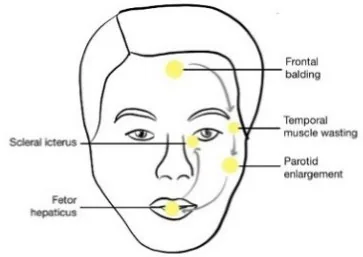
Do: Look for Frontal Balding (especially in females), Temporal Muscle Wasting (temporal regions of head), Parotid Enlargement, Fetor Hepaticus (sour, fecal breath), and Scleral Icterus
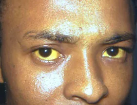
Scleral Icterus8
Report: I note no signs of liver disease in the face including Frontal Balding, Temporal Muscle Wasting, Parotid Enlargement, Fetor Hepaticus, or Scleral Icterus.
Why Check: Liver disease can present with facial findings:
- Jaundice (including scleral icterus) – due to hyperbilirubinemia – jaundice mildly increases the likelihood of suspected cirrhosis in patients with liver disease (LR+ 3.8, LR- 0.82). This can be due to prehepatic causes (e.g. hemolysis), intrahepatic causes (e.g. cirrhosis, hepatitis, liver cancer), or post-hepatic causes (e.g. choledocholithiasis; cholangitis; Mirizzi syndrome where inflamed gallbladder compresses common hepatic duct as a complication of cholecystitis; pancreatitis; cholangiocarcinoma; pancreatic cancer)
- Temporal Muscle Wasting – due to impaired protein synthesis.
- Parotid Enlargement – seen especially in patients with alcohol-related cirrhosis.
- Fetor Hepaticus – due to portal hypertension.
- Frontal balding – look for especially in female patients.
Stigmata of Chronic Liver Disease – Hands
AFMC level Pre-clerkship 2 · Clerkship 4
Say: I will now inspect the hands for signs of liver disease.
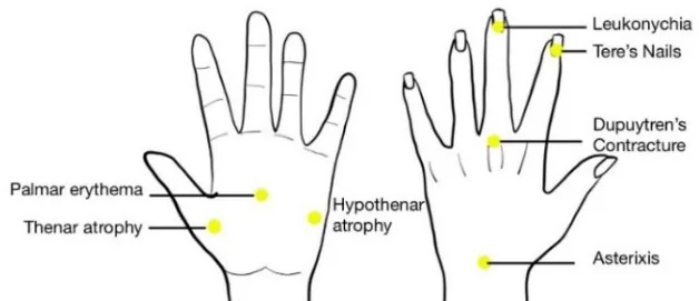
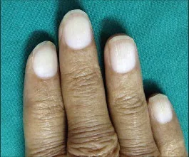
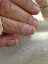
Do: Look for Terry’s Nails (whitening of proximal 2/3rd of nailbed); Palmar Erythema; Thenar Atrophy (Muscle wasting proximal to thumb; DDx includes carpal tunnel syndrome); and Hypothenar Atrophy (Wasting of muscle proximal to little finger).
Say: Can you keep both hands held out with wrist extended? Thank you; now, open both hands fully.
Do: Look for Asterixis – a flapping tremor of the hand with wrist extended (DDx includes Diabetes Mellitus and other causes of myoclonus e.g. neuromuscular disorders). When opening hand, look for Dupuytren’s Contracture – fingers cannot be relaxed from a flexed position.
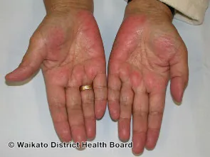
Palmar Erythema11
Report: I note no signs of liver disease in the hands including Terry’s Nails, Palmar Erythema, Thenar Atrophy, Asterixis or Dupuytren’s Contracture.
Why Check: Liver disease can present with physical findings on the hands:
- Terry’s Nails – due to hypo-albuminemia. Terry’s Nails greatly increases the likelihood of cirrhosis in patients with liver disease (LR+ 16-22, LR- 0.57-0.58)
- Palmar Erythema – due to impaired estrogen metabolism – greatly increases the likelihood of suspected hepatocellular disease in patients with jaundice (LR+ 9.8, LR- 0.5). Absence of palmar erythema mildly decreases the likelihood of suspected cirrhosis in patients with known liver disease (LR- 0.59).
- Thenar and Hypothenar atrophy – due to impaired protein synthesis.
- Asterixis – Asterixis is an example of negative myoclonus (transient <0.5s inhibition of tonic muscle contraction) due to impaired metabolism in liver disease (e.g. ammonia metabolism to urea), and part of assessment for hepatic encephalopathy.
- Dupuytren’s Contracture – mainly manifestation of alcohol consumption.
Stigmata of Chronic Liver Disease – Abdomen
AFMC level Pre-clerkship 2 · Clerkship 4
Say: I will now inspect the abdomen (and chest if male) for signs of liver disease.
Do: Look for Spider Angiomas or Caput Medusae in umbilical region. Look for Ascites. In male patients, look for Gynecomastia and Hypogonadism.
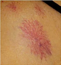
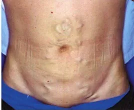
Spider Angiomata12; Caput Medusae13; Bulging Flanks/Ascites7
Report: I note no signs of liver disease in the abdomen including spider angiomas, caput medusae, ascites, or gynecomastia/hypogonadism (if male).
Why Check: Many findings of liver disease present on the abdomen:
- Spider Angiomata – due to impaired estrogen metabolism – slightly increase the likelihood of suspected hepatocellular disease in patients with jaundice (LR+ 4.7, LR- 0.6), and cirrhosis in patients with known liver disease (LR+ 4.3, LR- 0.61).
- Caput Medusae – due to portal hypertension – greatly increase the likelihood of suspected hepatocellular disease in patients with jaundice (LR+ 17.5, LR- 0.6) and cirrhosis in patients with known liver disease (LR+ 11, LR- 0.72)
- Gynecomastia and Testicular Atrophy (in male patients) – due to impaired estrogen metabolism. The likelihood of suspected cirrhosis in patients with known liver disease is moderately increased by the presence of testicular atrophy (LR+5.8, LR- 0.84) or gynecomastia (LR+ 5.8-35, LR- 0.43-0.84)
- Ascites – due to portal hypertension, hypoalbuminemia – moderately increases the likelihood of suspected cirrhosis in patients with known liver disease (LR+ 7.2, LR- 0.69)
Cullen’s Sign and Grey-Turner Sign
AFMC level Pre-clerkship 3 · Clerkship 4
Say: I will now inspect for bruising in the abdomen.
Do: Look for bruising in the subcutaneous tissue around the umbilicus (Cullen’s sign) and in the flanks of the abdomen (Grey-Turner sign)
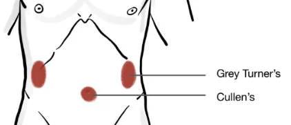
Report: Cullen’s sign and Grey-Turner sign are negative, which is normal.
Why Check: May be seen in patients who experience retroperitoneal bleeds, a complication of several conditions including pancreatitis and AAA rupture.
Auscultation
Bowel Sounds – With Diaphragm
AFMC level Pre-clerkship 3 · Clerkship 4
Say: I will auscultate for bowel sounds using the diaphragm of my stethoscope.
Do: Listen over either the 4 quadrants or 9 regions, depending on time. Stop once you hear them in ONE region, no additional value to doing all of them.
Report: Bowel sounds heard on auscultation within 2 minutes in one of the 9 abdominal regions (epigastric, umbilical, hypogastric, R/L hypochondriac, R/L lumbar, R/L iliac) with diaphragm of stethoscope. Alternatively, can specify by the quadrant – RUQ, RLQ, LLQ, LUQ
Why Check: The likelihood of suspected bowel obstruction is increased moderately by hyperactive bowel sounds (LR+ 5.0, LR- 0.6), and slightly by abnormal bowel sounds (LR+ 3.2, LR- 0.4). Hypoactive or absent bowel sounds may suggest ileus or peritonitis; decreased sounds mildly increase the likelihood of suspected appendicitis in pediatrics (LR+ 2.5, LR- 0.77).
Abdominal Bruits – With Bell
(also explored in Chapter 10 - Renal Exam - MF2)
AFMC level Pre-clerkship 3 · Clerkship 4
Say: I will use the bell of my stethoscope to auscultate key arteries for any signs of turbulent blood flow.
Do: With patient in supine position, auscultate:
- Abdominal Aorta: Slightly left and superior to umbilicus
- R and L Renal: 5cm superior, 5cm lateral to umbilicus
- R and L Iliac: 5cm inferior and 5cm lateral to umbilicus
- R and L Femoral: Midway between ASIS & symphysis pubis
If you detect a renal bruit (“whoosh” sound), clarify if it is systolic or systolic-diastolic (e.g. via timing of carotid upstroke on palpation). Also clarify if it is unilateral vs. bilateral.
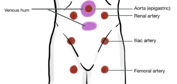
Report: I note no bruits on auscultation in any of the 7 abdominal areas.
Why Check: In a normal vessel, a pulse can be palpated but not auscultated. Bruits occur due to turbulent flow within a vessel, where the pitch and radiation are a function of flow and direction of the turbulent stream. Bruits can be innocent (particularly in young, normotensive, asymptomatic patients), typically described as short, faint and mid-systolic. A systolic-diastolic abdominal bruit greatly increases likelihood of renovascular hypertension (LR+ 39, LR- 0.62) (e.g. renal artery stenosis), a cause of secondary hypertension.
Venous Hum – Liver
AFMC level Pre-clerkship 1 · Clerkship 2
Say: I will use the bell of my stethoscope to auscultate for a venous hum over the epigastric and umbilical areas.
Do: Listen for a humming noise in these areas

Report: I note no venous hum heard over the epigastric and umbilical areas.
Why Check: Portal hypertension can increase collateral circulation between portal and systemic circulation, creating a hum that may be heard on auscultation (especially when listening for esophageal varices and periumbilical veins (Caput Medusae)). A rub may also result from liver metastases/hepatoma/liver mass.
Friction Rub – Peritoneal Irritation
AFMC level Pre-clerkship 1 · Clerkship 2
Say: I will use the diaphragm of my stethoscope to auscultate for a friction rub over the liver and spleen.
Do: Listen for a rough and high-pitched sound when liver and spleen rub peritoneum
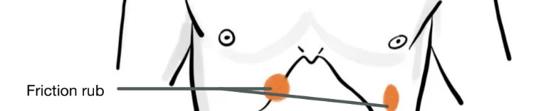
Report: I note no friction rub in the lower right or left costal areas
Why Check: A friction rub can indicate peritoneal irritation, and may be heard with free fluid, hepatic metastases/abscesses, or a splenic infarct/infection/abscess.
Liver Scratch Test (Conditional Test – Consider in Patients with Obesity)
Say: I will use the diaphragm of my stethoscope to auscultate for the lower liver edge. Please breathe normally and let me know if you feel any pain.
Do: Place diaphragm of stethoscope at xiphisternum over the liver, just above the costal margin at the Right Midclavicular Line (MCL). Starting in RLQ, move one finger up the surface of the abdomen, scratching gently. The intensity of the transmitted sound is greatly enhanced once the finger is over the lower border of liver. Look at patient for signs of discomfort and stop the exam if the patient is in distress.
Report: At the right midclavicular line, liver excursion measures 8cm by the liver scratch test, which is normal.
Why Check: Check and comment if you have clinical suspicion of liver disease, hepatomegaly, etc. Evidence14 suggests clinicians should refrain from this test as it “yields a result with no correlation to the actual liver span”, with better evidence for Palpation of the Liver Edge. In patients with obesity15, it is noted that “abdominal fat makes it difficult to palpate deep structures (e.g. liver edge)” with preference for the Liver Scratch test to find the lower liver edge recommended in this population.
Percussion
General
Gen. - [2, 3] Peritonitis – [3, 4]
Say: I will percuss the abdomen in 9 regions. Are you experiencing any pain? If so, I will percuss that area last.
Do: Percuss with a good surface area of hand on patient for max capacity for hearing sounds on percussion.
What is Normal? Expect more tympanic sounds heard in epigastric region, as well as the cecum (re air pockets). Expect variably dull or tympanic sounds at bladder (re bladder distension/pregnancy), liver and spleen may be physiologically dull). Is there any tenderness on percussion?
Report: I note tympanic sounds on percussion of nine abdominal quadrants with no tenderness on percussion.
Why Check: Dullness to percussion may be suspicious of impaction or ascites. Tenderness on percussion mildly increases likelihood of suspected peritonitis (LR+ 2.4, LR- 0.5).
Fluid Wave Test – Ascites – Conditional Test
AFMC level Pre-clerkship 2 · Clerkship 4
Say: I will assess for abdominal fluid using a fluid wave test. Could you place one hand over the umbilical region? (demonstrate with your own hand).
Do: Have patient hold their hand over the midline in umbilical region, put your hands on the patient’s sides, and tap with one side, check to the other side for feeling the impulse sent.
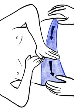
What is Normal: To not feel the impulse sent on the opposite. Pressing on Abdominal wall prevents vibrations from transmitting through adipose tissue, while underlying fluid can transmit the energy.
Report: The fluid wave test for ascites is negative.
Why Check: Ascites may be suspected when the patient presents with abdominal distension, fluid overload, and dullness to percussion in all areas. Positive fluid wave test moderately increases the likelihood of suspected ascites (LR+ 5.3, LR- 0.57). Further, confirmed ascites may represent liver disease, as ascites moderately increases the likelihood of suspected hepatocellular disease in patients with jaundice (LR+ 4.4, LR- 0.6), and of suspected cirrhosis in patients with liver disease (LR+ 7.2, LR- 0.69).
Modification for Patients with Obesity: Use the fluid wave or shifting dullness tests to appreciate ascites, rather than inspecting for bulging flanks. Bulging flanks due to adipose tissue may be difficult to differentiate from ascites on inspection.
Shifting Dullness Test – Ascites – Conditional Test
AFMC level Pre-clerkship 2 · Clerkship 4
Say: I will percuss for abdominal fluid using a shifting dullness test.
Do: With patient starting in supine position, tap from umbilicus laterally until you hear dullness. Ask patient to move into a decubitus position that raises that side of their body, wait a moment for the fluid to move, and see if it’s tympanic
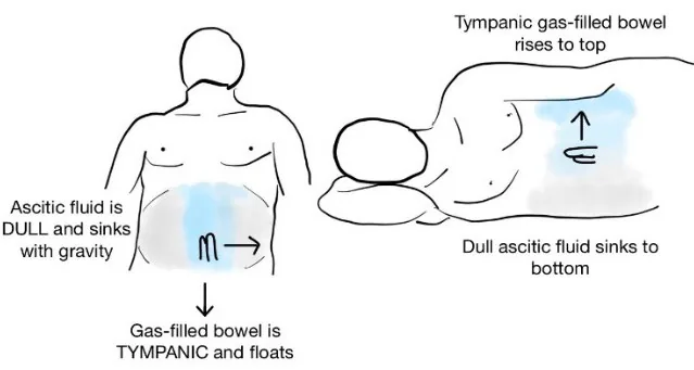
What is Normal? Dullness does not shift
Report: Dullness does not shift with position change. The shifting dullness test for ascites is negative.
Why Check: Ascites may be suspected when the patient presents with abdominal distension, fluid overload, and dullness to percussion in all areas. Shifting dullness mildly increases the likelihood of suspected ascites (LR+ 2.1, LR- 0.40). Further, confirmed ascites may represent liver disease, as ascites moderately increases the likelihood of suspected liver disease in patients with jaundice (LR+ 4.4, LR- 0.6), and of suspected cirrhosis in patients with liver disease (LR+ 7.2, LR- 0.69).
Modification for Patients with Obesity: Use the fluid wave or shifting dullness tests to appreciate ascites, rather than inspecting for bulging flanks. Bulging flanks due to adipose tissue may be difficult to differentiate from ascites on inspection.
Liver Excursion
AFMC level Pre-clerkship 3 · Clerkship 4
Say: I will percuss for the liver span. Please breathe normally.
Do: Beginning at right Anterior superior iliac spine (ASIS) at MCL and percuss up 2 cm with each inspiratory rise of the abdomen. Note progression from tympanic → dull → resonant and landmark the two transition points. The distance between them is liver span assessed via percussion, also assessable at mid-sternal line.
What is Normal? At MCL, liver excursion measures 6-12cm on percussion (at midsternal line, normal 4-8cm)
Report: At the right midclavicular line, liver excursion measures 8 cm on percussion; at midsternal line, the liver excursion measures 6 cm on percussion, which is normal.
Why Check: Can be used to investigate for hepatomegaly, however this finding does not significantly alter the likelihood. Confirmed hepatomegaly mildly increases the likelihood of suspected cirrhosis in patients with liver disease (LR+ 2.4, LR- 0.37).
Castell’s Sign-Spleen
(Also seen in Ch 11 – Lymph Node Exam – MF2)
AFMC level Pre-clerkship 3 · Clerkship 4
Say: I will now be percussing the spleen in the left lower chest. Can you take a deep breath in? And now out?
Do: Percussion of point defined by last intercostal space along the anterior axillary line during full inspiration, looking for shift to dullness. Percuss throughout respiratory cycle. The area should remain tympanic throughout the breath. Any dullness throughout the breath cycle may indicate splenomegaly.
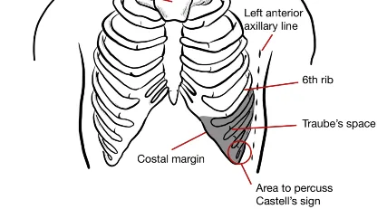
Report: Castell’s sign yields tympanic sounds throughout.
Why Check: Can be used to investigate splenomegaly, or if you’re performing a lymph node exam (Ch 11 – Lymph Node Exam – MF2). Positive Castell’s sign mildly increases likelihood of splenomegaly (LR+ 1.7, LR- 0.7). Castell’s sign is more sensitive and specific than Traube’s space (Sensitivity 82% and Specificity 83% for Castell’s sign, vs. Sensitivity 62% and Specificity 72% for Traube’s space)7. Confirmed splenomegaly mildly increases the likelihood of suspected cirrhosis in patients with known liver disease (LR+ 3.5, LR- 0.74).
Traube’s Space – Spleen
(Also seen in Ch 11 – Lymph Node Exam – MF2)
AFMC level Pre-clerkship 3 · Clerkship 4
Say: I will now be percussing the spleen over a wider area in the left lower chest.
Do: Percussion of the area defined by the sixth rib superiorly, the anterior axillary line laterally, and the left costal margin inferiorly (see diagram above). You are looking for dullness – can be done along a straight line following the Left last intercostal space anteriorly until reaching anterior axillary line.
Report Traube’s space yields tympanic sounds throughout.
Why Check: Can be used to investigate splenomegaly, or if you’re performing a lymph node exam (Ch 11 – Lymph Node Exam – MF2). Dull Traube’s Space mildly increases likelihood of splenomegaly (LR+ 2.1, LR- 0.8). Castell’s sign is more sensitive and specific than Traube’s space (Sensitivity 82% and Specificity 83% for Castell’s sign, vs. Sensitivity 62% and Specificity 72% for Traube’s space)7. Confirmed splenomegaly mildly increases the likelihood of suspected cirrhosis in patients with known liver disease (LR+ 3.5, LR- 0.74).
Palpation
Light Palpation
AFMC level Pre-clerkship 3 · Clerkship 4
Say: I will now palpate the abdomen in 9 regions using light palpation Are you experiencing any pain right now? If so, I will palpate that area last.
Do: Using the palm standing at patient’s side, palpate the abdomen (should be good surface contact), and progress along in a systematic way. Should be good amount of pressure, it is palpated using the fingers. Assess for masses, tenderness, distension, and hardness/rigidity (involuntary guarding). While doing this, look at patients’ face for signs of pain.
Report: No evidence of guarding, facial discomfort, or masses on light palpation in 9 abdominal regions
Why Check: Can identify tenderness, masses, hernias, and peritonitis. In patients with acute abdominal pain, the likelihood of suspected peritonitis increases slightly with rigidity (LR+ 3.9) and guarding (LR+ 2.6, LR- 0.6), and decreases significantly with positive abdominal wall tenderness test (LR+ 0.1). Further, the likelihood of suspected acute appendicitis increases moderately with RLQ pain (LR+ 7.3-8.5, LR- 0-0.28), and slightly with rigidity (LR+ 3.8, LR- 0.82) and guarding (LR+ 1.7-1.8, LR- 0-0.54). The likelihood of suspected acute cholecystitis may be slightly increased by RUQ tenderness (LR+ 1.6); however this finding is not statistically significant. Additionally, Courvoisier’s Sign states that a patient with painless jaundice and a RUQ mass/palpable gallbladder has a pancreatobiliary neoplasm until proven otherwise. A palpable gallbladder/RUQ mass (Courvoisier's Sign) greatly increases the likelihood of suspected obstructive bile ducts in patients with jaundice (LR+ 26, LR- 0.7), mildly increases the likelihood of suspected malignant obstruction in patients with obstructive jaundice (LR+ 2.6, LR- 0.7), and significantly decreases the likelihood of suspected hepatocellular disease in patients with jaundice (LR+ 0.04, LR- 1.4).
Deep Palpation
AFMC level Pre-clerkship 3 · Clerkship 4
Say: I will now palpate the abdomen in 9 regions using deep palpation Are you experiencing any pain right now? If so, I will palpate that area last.
Do: Similar to Light General Palpation, proceeding in a systematic way with deeper pressure (visibly depressed into abdomen). Palpate for masses, tenderness, distension, and hardness/rigidity (involuntary guarding).
Note: May be able to palpate cecum in right iliac fossa if patient very thin.
Report: No evidence of tenderness, masses or organomegaly on deep palpation in 9 abdominal regions.
Why Check: Can identify tenderness, masses, hernias, and peritonitis. In patients with acute abdominal pain, the likelihood of suspected peritonitis increases slightly with rigidity (LR+ 3.9) and guarding (LR+ 2.6, LR- 0.6), and decreases significantly with positive abdominal wall tenderness test (LR+ 0.1). Further, the likelihood of suspected acute appendicitis increases moderately with RLQ pain (LR+ 7.3-8.5, LR- 0-0.28), and slightly with rigidity (LR+ 3.8, LR- 0.82) and guarding (LR+ 1.7-1.8, LR- 0-0.54). The likelihood of suspected acute cholecystitis is slightly increased by RUQ tenderness (LR+ 1.6); however this finding is not statistically significant. Additionally, Courvoisier’s Sign states that a patient with painless jaundice and a RUQ mass/palpable gallbladder) has a pancreatobiliary neoplasm until proven otherwise. A palpable gallbladder/RUQ mass (Courvoisier's Sign) greatly increases the likelihood of suspected obstructive bile ducts in patients with jaundice (LR+ 26, LR- 0.7), mildly increases the likelihood of suspected malignant obstruction in patients with obstructive jaundice (LR+ 2.6, LR- 0.7), and decreases the likelihood of suspected liver disease in patients with jaundice (LR+ 0.04, LR- 1.4).
Rebound Tenderness – Conditional Test
AFMC level Pre-clerkship 3 · Clerkship 4
Say: I will now assess for rebound tenderness in the area of the abdomen you felt tenderness on palpation earlier. Tell me if it hurts more when I press down with my hand, or when I remove it.
Do: Only assess for Rebound Tenderness in areas of tenderness identified. Using the tips of the fingers, press perpendicularly into the abdomen slowly. Hold this position until the patient’s tenderness appears to have calmed. Then quickly release your hand from the abdomen and assess for signs of pain in the patient’s face.
Report: Patient shows no rebound tenderness and does not appear peritonitic.
Why Check: The stretching and movement produced by rebounding can elicit pain when peritonitis is present. Rebound tenderness mildly increases likelihood of suspected peritonitis (LR+ 2.1, LR- 0.5). Positive rebound tenderness mildly increases likelihood of suspected appendicitis (LR+ 1.1-6.3, LR- 0-0.86 in adults; LR+ 3.0, LR- 0.28 in pediatrics). Further, diffuse peritonitis largely increases the likelihood of suspected appendicitis (LR+ 25, LR- 0.76 in pediatrics).
Shake Test – Conditional Test
Say: I will now shake the abdomen (or the exam table). Please let me know if this causes pain or discomfort.
Do: Grasp the patient’s hips, and gently shake the patient in order to shake the abdomen. Alternatively, using your hands or your knees, gently shake the patient’s exam table.
Report: Patient shows to pain on shake test and does not appear peritonitic.
Why Check: Pain on shaking the abdomen may be suggestive of peritonitis. Similarly, you can ask the patient if they had pain on the road while riding to the hospital, or if they have pain with coughing. Positive cough test mildly increases likelihood of suspected peritonitis (LR+ 1.8, LR- 0.4). Further, diffuse peritonitis greatly increases the likelihood of suspected appendicitis in pediatrics (LR+ 25, LR- 0.76).
Palpation of Liver Edge
Gen. - [3, 4] Consistency - [1, 2] Tenderness - [2, 2] Pulsatility – [1, 1]
Say: I will palpate for the lower liver edge. Please breathe normally and let me know if you feel any pain.
Do: Make an L shape with your right hand in patient’s Right Lower Quadrant along MCL and use hand to mimic the liver edge you’re palpating for. Move up 2 cm with each rise of the patients’ belly (to keep it consistent due to liver moving down with inspiration). Your hand’s orientation should be parallel with the rectus abdominus muscle. Check for pulsatility. Look at patient for signs of discomfort and stop the exam if the patient is in distress.
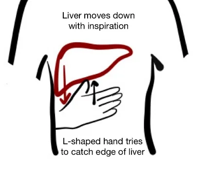
Report: (If you can feel liver) The liver is palpable less than 2 cm below the right costal margin along the MCL with rest inspiration. Liver edge is smooth and non-nodular, and non-tender to palpation.
Why Check: Palpation of the liver edge essentially confirms hepatomegaly (LR+ 233.7, LR- 0.5). The likelihood of suspected cirrhosis in patients with known liver disease is slightly increased by hepatomegaly (LR+ 2.4, LR- 0.37) and by firm liver edge (LR+ 3.3, LR- 0.37).
- Consistency: Nodularity is indicative of cirrhosis.
- Tenderness: Indicative of inflammation.
- Pulsatility: A specific finding of severe Tricuspid Regurgitation (TR).
Splenic Palpation
(Also seen in Ch 11 – Lymph Node Exam – MF2)
AFMC level Pre-clerkship 3 · Clerkship 4
Say: I will palpate for an enlarged spleen in the abdomen. Please take a deep breath each time there is pressure on the abdomen.
Do: Use a deep palpation technique, where the spleen should not be palpable. The spleen moves down on inspiration due to the displacement of the diaphragm, so time your deep palpation during inspiration. Use your findings on percussion (Castell’s Sign, Traube’s Space) to guide the starting point of palpation:
- If Castell’s Sign/Traube’s Space yield tympanic sounds: Start palpation close to the left costal margin. You can pull the left side of the rib cage towards your right hand as you palpate for the spleen.
- If Castell’s Sign/Traube’s Space yield dull sounds (increasing likelihood of splenomegaly): Start palpation in RLQ along right MCL, moving slowly towards LUQ through umbilicus.
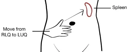
Report: The spleen is non-palpable, including on inspiration.
Why Check: In cases of splenomegaly, the spleen will grow diagonally, so it is important to start at the RLQ and work towards the LUQ. A palpable spleen moderately increases the likelihood of suspected splenomegaly (LR+ 8.5, LR- 0.5). Additionally, splenomegaly mildly increases the likelihood of suspected hepatocellular disease in patients with jaundice (LR+ 2.9, LR- 0.7), and of suspected cirrhosis in patients with known liver disease (LR+ 3.5, LR- 0.74).
Digital Rectal Exam (DRE)
Inspect – [3, 4] Palpate – [2, 4]
In an OSCE: Always OFFER to do a DRE if abdominal exam is required (rule of thumb is every abdominal exam requires a DRE). You will likely not have to perform a DRE in the OSCE, as you would in clinical practice.
Say: Based on your findings on history and physical exam, a rectal exam is important to determine next best steps in your care. This is something we do with all our patients who come in with ___ (e.g. abdominal pain and blood in the stool) The exam should take under 5 minutes. How does that sound to you?
Setup: Offer a chaperone (see Ch 4 – Vital Signs and Introduction to the Physical Exam – MF1). With patient providing informed consent, in a private exam room space, give the patient a gown and drape, and ask them to change with undergarments off after leaving the room. Let them know you will return in 5 minutes. Knock on door to ask for permission to re-enter room. Put gloves on and lubricate gloved finger (if stock, DROP from nozzle – avoid contamination of stock) with good lighting. The patient lies on their left side, both knees flexed towards the chest. Spread the buttocks, and inspect for external hemorrhoids, fissures, rectal prolapse, perianal abscesses, or anal fistula.
Do: Next, soft rounded pad of your finger should go through; stop for a moment at the anal verge then continue; make note of anal sphincter tone (re: Neurology, Upper motor neuron lesion vs. Lower motor neuron lesion differentiation – explored in Ch 31 – Neurology Exam – MF4). Rotate hand to feel all sides of walls, checking for any irregularity in thickness of the wall, hemorrhoids, abscesses, or fecal impaction. May also check for swelling or tenderness of rectovesical pouch in males or rectouterine pouch (Pouch of Douglas) in females. In male exam, may also check the prostate for size, swelling, tenderness, consistency symmetry, and nodularity (do not need to palpate prostate in DRE specifically, but may be combined as part of exam if indicated). There should not be a sensation for patient, except the urge to pass urine when palpating around the prostate in male exam. After completing exam, inspect the gloved finger for any blood or melena stool.
Report: There is no evidence of external or internal hemorrhoids, fissures, fistula, rectal prolapse, perianal abscesses, or inflammation on DRE, which is reassuring. (If prostate was palpated, report if enlarged, nodularity, firm or asymmetrical).
Why Check: A DRE is distinct from Bimanual Exam or Male Genital Exam (Chapter 19 – Sensitive Pelvic Exams). It is a routine part of abdominal exam (hence “every abdominal exam requires a DRE”), though it is particularly important if patient complains of BRBPR (Hematochezia) or Melena. Melena stool on exam greatly increases the likelihood of suspected upper GI bleed (LR+ 25, LR-0.52). A DRE can also help with the assessment of internal and external hemorrhoids, which can be a cause of BRBPR. For patients with constipation refractory to laxatives, a DRE can be helpful to note rectal tone, masses, consistency of stool and stool impaction. A ↓ rectal tone can be helpful in assessment for spinal cord compression (explored in Ch 31 – Screening Neuro Exam – MF4).
McBurney’s Point (Appendicitis Special Test)
AFMC level Pre-clerkship 3 · Clerkship 4
Say: I will palpate for signs of appendicitis.
Do: McBurney’s Point is 1/3rd the distance from the ASIS to the umbilicus on side of ASIS. Palpate at this point using deep palpation. While doing this, you look at patients’ face for signs of pain.
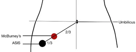
Report: No evidence of discomfort on palpation of McBurney’s Point
Why Check: If present, would raise suspicion of inferior appendicitis. RLQ pain moderately increases the likelihood of acute appendicitis (LR+ 7.3-8.5, LR- 0-0.28 in adults; LR+ 1.3, LR- 0.45 in pediatrics).
Rovsing’s Sign (Appendicitis Special Test)
Peds - [2, 4]
Say: I will palpate for signs of appendicitis.
Do: Palpate at the LLQ, and look at the patient’s face for signs of pain in RLQ
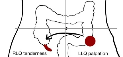
Report: No evidence of tenderness in RLQ on LLQ deep palpation.
Why Check: If present, would raise suspicion of inferior appendicitis (LR+ 1.9, LR- 0.83 in pediatrics).
Psoas’ Sign (Appendicitis Special Test)
Peds - [2, 4]
Say: I will palpate for signs of appendicitis. Please lie down on the exam table and relax the right leg. I will maneuver the right leg, so the right knee and hip are flexed. When you feel my hand pressing down on the right thigh, could you push against it? Did you feel any pain?
Do: Patient in supine position, right leg relaxed and then manipulate so knee in approximately 90 degrees flexion. Press against thigh with resistance from patient. Look at patient’s face during the maneuver for signs of pain. Leave right leg in same position (to check for Obturator Sign).
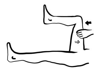
Report: No evidence of RLQ discomfort on flexion of hip against resistance.
Why Check: Named after the Psoas muscle. If present, would slightly raise suspicion of retrocecal appendicitis (LR+ 2.4, LR- 0.9).
Obturator Sign (Appendicitis Special Test)
Say: I will continue to assess for appendicitis by rotating the right leg. Could you continue to keep the right leg relaxed? Let me know if you feel any pain as I rotate.
Do: Grasp patient’s right leg below knee and ankle, hip and knee both flexed at 90°. Internally rotate the hip. Look at patient’s face during the maneuver for signs of pain.
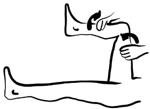
Report: No evidence of RLQ discomfort on right hip flexion and internal rotation.
Why Check: If present, would raise suspicion of pelvic appendicitis - Named after Obturator muscle
Murphy’s Sign (Cholecystitis Special Test)
Murphy- [3, 4] Courvoisier - [1, 3]
Say: I will now palpate for the gallbladder. Please take a deep breath in.
Do: As patient begins deep inspiration, hook right hand under right costal margin at MCL. Look at patient to assess whether inspiration has been arrested.
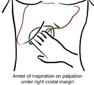
Report: On palpating underneath the right costal margin during deep inspiration, the gallbladder is non-palpable with no signs of acute pain or arrested inspiration. Murphy’s sign is negative.
Why Check: A positive Murphy’s sign is 87% specific for acute cholecystitis. The likelihood of acute cholecystitis may be slightly increased by positive Murphy’s Sign (LR+ 2.8, LR- 0.5) and by general RUQ pain (LR+ 1.5, LR- 0.7) and tenderness (LR+ 1.6). However, none of these likelihood ratios were statistically significant, limiting their utility.
Note: Sonographic Murphy’s Sign may be completed during ultrasound when assessing for cholecystitis.
Note: Courvoisier’s Law – A palpable non-tender gallbladder with mild jaundice is cancer of the gallbladder or pancreas until proven otherwise. A palpable gallbladder is also specific for obstructive bile ducts in patients with jaundice (LR+ 26.0, LR- 0.70)
Helpful Stethopedia Videos to Review:
- Video 1 – Abdominal Exam OSCE Style (Link: https://videopress.com/v/HPWUhrfC https://videopress.com/v/ZMrJEAXn)
- Video 2 – Abdominal Exam Special Tests (Link: https://videopress.com/v/51lW6iwK)
Part 4: Relevant Likelihood Ratios – from Rational Clinical Examination JAMA series
Clinical Question 1: Does this patient have Acute Cholecystitis?16
| Procedure | Sensitivity | Specificity | LR+ (95% CI) | LR- (95% CI) |
|---|---|---|---|---|
| Murphy Sign | 0.65 (0.58-0.71) | 0.87 (0.85-0.89) | 2.8 (0.8-8.6) (no change: 95% CI includes 1) | 0.5 (0.2-1.0) (no change: 95% CI includes 1) |
| RUQ Tenderness | 0.77 (0.73-0.81) | 0.54 (0.52-0.56) | 1.6 (1.0-2.5) (no change: 95% CI includes 1) | 1.0-1.2 (no change) |
| Emesis | 0.71 (0.65-0.76) | 0.53 (0.52-0.55) | 1.5 (1.1-2.1) (about +15%) | 0.6 (0.3-0.9) (about −15%) |
| Fever (>35C) | 0.35 (0.31-0.38) | 0.80 (0.78-0.82) | 1.5 (1.0-2.3) (no change: 95% CI includes 1) | 0.9 (0.8-1.0) (no change: 95% CI includes 1) |
| RUQ Pain | 0.81 (0.78-0.85) | 0.67 (0.65-0.69) | 1.5 (0.9-2.5) (no change: 95% CI includes 1) | 0.7 (0.3-1.6) (no change: 95% CI includes 1) |
| Guarding | 0.45 (0.37-0.54) | 0.70 (0.69-0.71) | 1.1-2.8 (about +15%) | 0.5-1.0 (about −15%) |
| Anorexia | 0.65 (0.57-0.73) | 0.50 (0.49-0.51) | 1.1-1.7 (about +15%) | 0.5-0.9 (about −15%) |
| Nausea | 0.77 (0.69-0.83) | 0.36 (0.34-0.38) | 1.0-1.2 (no change) | 0.6-1.0 (about −15%) |
| Rebound Tenderness | 0.30 (0.23-0.37) | 0.68 (0.67-0.69) | 1.0 (0.6-1.7) (no change: 95% CI includes 1) | 1.0 (0.8-1.4) (no change: 95% CI includes 1) |
| RUQ Mass | 0.21 (0.18-0.23) | 0.80 (0.75-0.85) | 0.8 (0.5-1.2) (no change: 95% CI includes 1) | 1.0 (0.9-1.1) (no change: 95% CI includes 1) |
| Rigidity | 0.11 (0.06-0.18) | 0.87 (0.86-0.87) | 0.50-2.32 (about −15%) | 1.0-1.2 (no change) |
| Rectal Tenderness | 0.08 (0.04-0.14) | 0.82 (0.81-0.83) | 0.3-0.7 (about −30%) | 1.0-1.3 (no change) |
Clinical Question 2: Does this patient have Acute Appendicitis?17
| Procedure | Sensitivity | Specificity | LR+ (95% CI) | LR- (95% CI) |
|---|---|---|---|---|
| RLQ Pain | 0.84 | 0.90 | 7.3-8.5 (about +30%) | 0-0.28 (about −45%) |
| Rigidity | 0.20 | 0.89 | 3.8 (3.0-4.8) (about +15%) | 0.82 (0.79-0.85) (about −15%) |
| Migration of Pain | 0.64 | 0.82 | 3.2 (2.4-4.2) (about +15%) | 0.50 (0.42-0.59) (about −15%) |
| Pain before vomiting | 1.0 | 0.64 | 2.8 (1.9-3.9) (about +15%) | N/A |
| Psoas Sign | 0.16 | 0.95 | 2.4 (1.2-4.7) (about +15%) | 0.90 (0.83-0.98) (about −15%) |
| Fever | 0.67 | 0.79 | 1.9 (1.6-2.3) (about +15%) | 0.58 (0.51-0.67) (about −15%) |
| Rebound Tenderness | 0.63 | 0.69 | 1.1-6.3 (about +15%) | 0-0.86 (about −45%) |
| Guarding | 0.73 | 0.52 | 1.7-1.8 (about +15%) | 0-0.54 (about −45%) |
| No similar pain previous | 0.86 | 0.40 | 1.50 (1.46-1.7) (about +15%) | 0.32 (0.25-0.42) (about −30%) |
| Rectal tenderness | 0.41 | 0.77 | 0.83-5.3 (about −15%) | 0.36-1.1 (about −30%) |
| Anorexia | 0.68 | 0.36 | 1.3 (1.2-1.4) (about +15%) | 0.64 (0.54-0.75) (about −15%) |
| Nausea | 0.58 | 0.37 | 0.69-1.2 (about −15%) | 0.70-0.84 (about −15%) |
| Vomiting | 0.51 | 0.45 | 0.92 (0.82-1.0) (no change: 95% CI includes 1) | 1.1 (0.95-1.3) (no change: 95% CI includes 1) |
Clinical Question 3: Does this child have Appendicitis?18
| Procedure | LR+ (95% CI) | LR- (95% CI) | |
|---|---|---|---|
| History | Fever | 3.4 (2.4-4.8) (about +15%) | 0.32 (0.16-0.64) (about −30%) |
| Diarrhea | 2.6 (1.3-4.9) (about +15%) | 0.77 (0.58-1.0) (no change: 95% CI includes 1) | |
| Vomiting | 2.2 (1.7-2.9) (about +15%) | 0.33 (0.15-0.71) (about −30%) | |
| Abdominal Pain migrating to RLQ | 1.9-3.1 (about +15%) | 0.41-0.72 (about −30%) | |
| Unable to walk normally | 1.7 (1.4-2.0) (about +15%) | 0.38 (0.27-0.54) (about −30%) | |
| Nausea | 1.4 (0.70-2.7) (no change: 95% CI includes 1) | 0.90 (0.69-1.2) (no change: 95% CI includes 1) | |
| Right Lower Quadrant Pain | 1.2 (1.0-1.5) (no change: 95% CI includes 1) | 0.56 (0.43-0.73) (about −15%) | |
| Constipation | 1.2 (0.53-2.7) (no change: 95% CI includes 1) | 1.0 (0.81-1.1) (no change: 95% CI includes 1) | |
| Abrupt (vs. gradual) pain onset | 1.0 (0.82-1.3) (no change: 95% CI includes 1) | 0.98 (0.82-1.2) (no change: 95% CI includes 1) | |
| Lethargy | 0.84 (0.11-6.2) (no change: 95% CI includes 1) | 1.0 (0.92-1.1) (no change: 95% CI includes 1) | |
| Symptom Duration <24h | 0.83 (0.55-1.2) (no change: 95% CI includes 1) | 1.3 (0.82-1.9) (no change: 95% CI includes 1) | |
| Anorexia | 0.77 (0.34-1.7) (no change: 95% CI includes 1) | 1.1 (0.87-1.35) (no change: 95% CI includes 1) | |
| Dysuria | 0.59 (0.03-10.1) (no change: 95% CI includes 1) | 1.0 (0.95-1.1) (no change: 95% CI includes 1) | |
| Intermittent (vs. persistent) pain | 0.48-0.75 (about −30%) | 1.3-1.4 (about +15%) | |
| Physical | Diffuse Peritonitis | 25.0 (1.5-414.1) (about +45%) | 0.76 (0.63-0.90) (about −15%) |
| Abdominal Mass | 6.9 (0.43-111.0) (no change: 95% CI includes 1) | 0.90 (0.83-0.97) (about −15%) | |
| Rebound Tenderness | 3.0 (2.3-3.9) (about +15%) | 0.28 (0.14-0.55) (about −30%) | |
| Bowel Sounds Decreased | 2.5 (1.6-3.7) (about +15%) | 0.77 (0.68-0.88) (about −15%) | |
| Abdominal Distention | 2.3-6.5 (about +15%) | 0.80-0.90 (about −15%) | |
| Rectal Tenderness | 2.3 (1.3-4.1) (about +15%) | 0.70 (0.56-0.87) (about −15%) | |
| Obturator Sign | 2.2 (1.4-3.4) (about +15%) | 0.82 (0.73-0.93) (about −15%) | |
| Psoas Sign | 2.0-2.5 (about +15%) | 0.75-0.86 (about −15%) | |
| Pain with percussion, hopping, cough | 2.0 (1.7-2.4) (about +15%) | 0.36 (0.26-0.50) (about −30%) | |
| Rovsing Sign | 1.9 (1.3-2.8) (about +15%) | 0.83 (0.74-0.93) (about −15%) | |
| Guarding | 1.6-2.6 (about +15%) | 0.21-0.61 (about −30%) | |
| RLQ Tenderness | 1.3 (1.1-1.4) (about +15%) | 0.45 (0.35-0.59) (about −30%) | |
| Localized Abdominal Tenderness | 1.1 (0.47-2.4) (no change: 95% CI includes 1) | 1.0 (0.79-1.2) (no change: 95% CI includes 1) | |
| CVA Tenderness | 0.87 (0.45-1.7) (no change: 95% CI includes 1) | 1.0 (0.95-1.1) (no change: 95% CI includes 1) |
Clinical Question 4: Does this patient with Acute Abdominal Pain have Peritonitis?19
| Procedure | LR+ (95% CI) | LR- (95% CI) |
|---|---|---|
| Rigidity | 3.9 (2.2-6.7) (about +15%) | 0.8 (0.7-1.0) (no change: 95% CI includes 1) |
| Guarding | 2.6 (1.8-3.9) (about +15%) | 0.6 (0.5-0.7) (about −15%) |
| Percussion Tenderness | 2.4 (1.5-3.9) (about +15%) | 0.5 (0.3-0.7) (about −15%) |
| Abnormal Bowel Sounds | 2.2 (0.5-9.7) (no change: 95% CI includes 1) | 0.8 (0.7-0.9) (about −15%) |
| Rebound Tenderness | 2.1 (1.7-2.5) (about +15%) | 0.5 (0.4-0.6) (about −15%) |
| Positive cough test | 1.8 (1.4-2.2) (about +15%) | 0.4 (0.3-0.5) (about −30%) |
| Rectal Tenderness | 1.2 (1.0-1.6) (no change: 95% CI includes 1) | 0.9 (0.9-1.0) (no change: 95% CI includes 1) |
| Positive abdominal wall tenderness test | 0.1 (0-0.7) (about −45%) | 1.9 (0.9-4.4) (no change: 95% CI includes 1) |
Clinical Question 5: Does this patient with Acute Abdominal Pain have a Bowel Obstruction?19
| Procedure | LR+ (95% CI) | LR- (95% CI) |
|---|---|---|
| Visible peristalsis | 18.8 (4.3-81.9) (about +45%) | 0.9 (0.9-1.0) (no change: 95% CI includes 1) |
| Distended abdomen | 9.6 (5.0-18.6) (about +30%) | 0.4 (0.3-0.5) (about −30%) |
| Hyperactive bowel sounds | 5.0 (2.4-10.6) (about +15%) | 0.6 (0.5-0.8) (about −15%) |
| Abnormal bowel sounds | 3.2 (1.7-6.1) (about +15%) | 0.4 (0.3-0.5) (about −30%) |
| Rigidity | 1.2 (0.4-3.6) (no change: 95% CI includes 1) | 1.0 (0.9-1.2) (no change: 95% CI includes 1) |
| Guarding | 1.0 (0.6-1.7) (no change: 95% CI includes 1) | 1.0 (0.7-1.4) (no change: 95% CI includes 1) |
| Rebound Tenderness | 0.9 (0.7-1.1) (no change: 95% CI includes 1) | 1.1 (1.0-1.2) (no change: 95% CI includes 1) |
| Rectal tenderness | 0.9 (0.6-1.5) (no change: 95% CI includes 1) | 1.0 (1.0-1.1) (no change: 95% CI includes 1) |
Clinical Question 6: Does this patient have an Abdominal Aortic Aneurysm (AAA)?20
| Ability to detect an Asymptomatic Aneurysm on Palpation According to Size | LR+ (95% CI) | LR- (95% CI) |
|---|---|---|
| Aneurysm >4.0cm (n=12 studies) | 16 (8.6-29) (about +45%) | 0.51 (0.38-0.67) (about −15%) |
| Aneurysm >3.0cm (n=15 studies) | 12 (7.4-20) (about +45%) | 0.72 (0.65-0.81) (about −15%) |
Clinical Question 7: Does this patient with Chronic Liver Disease have Cirrhosis?21
| Procedure | Sensitivity | Specificity | LR+ (95% CI) | LR- (95% CI) | |
|---|---|---|---|---|---|
| History | Past History of Diabetes Mellitus | 0.34 | 0.88 | 2.8 (1.5-4.0) (about +15%) | 0.78 (0.58-0.91) (about −15%) |
| Past History of Minor Nose or Gum Bleeding | 0.25 | 0.84 | 1.6 (0.99-2.6) (no change: 95% CI includes 1) | 0.89 (0.79-1.0) (no change: 95% CI includes 1) | |
| Past History of Alcohol Use | 0.47 | 0.66 | 1.5 (1.0-2.0) (no change: 95% CI includes 1) | 0.76 (0.52-1.0) (no change: 95% CI includes 1) | |
| Past History of Upper GI Tract Bleed | 0.22-0.65 | 0.07-0.84 | 0.70-1.4 (about −15%) | 0.92-4.9 (about −15%) | |
| Fatigue | 0.63 | 0.51 | 1.3 (1.1-1.6) (about +15%) | 0.89 (0.53-1.2) (no change: 95% CI includes 1) | |
| Weakness | 0.40-0.80 | 0.31-0.64 | 1.1-1.2 (about +15%) | 0.64-0.94 (about −15%) | |
| Pruritus | 0.14-0.23 | 0.65-0.93 | 0.69-2.0 (about −15%) | 0.92-1.2 (about −15%) | |
| Anorexia | 0.37-0.43 | 0.23-0.68 | 0.56-1.2 (about −15%) | 0.93-2.5 (about −15%) | |
| Physical | Terry Nails | 0.43-0.44 | 0.97-0.98 | 16-22 (about +45%) | 0.57-0.58 (about −15%) |
| Gynecomastia | 0.18-0.58 | 0.98-0.98 | 5.8-35 (about +30%) | 0.43-0.84 (about −30%) | |
| Distended abdominal veins | 0.31 | 0.98 | 11 (2.7-44) (about +45%) | 0.72 (0.57-0.91) (about −15%) | |
| Encephalopathy | 0.16 | 0.98 | 10 (1.5-77) (about +45%) | 0.86 (0.76-0.95) (about −15%) | |
| Decreased body hair | 0.36 | 0.97 | 9.0 (6.4-13) (about +30%) | 0.65 (0.51-0.84) (about −15%) | |
| Ascites | 0.35 | 0.95 | 7.2 (2.9-12) (about +30%) | 0.69 (0.59-0.78) (about −15%) | |
| Facial telangiectasia | 0.73-0.82 | 0.88-0.92 | 5.9-10 (about +30%) | 0.20-0.31 (about −30%) | |
| Testicular atrophy | 0.18 | 0.97 | 5.8 (2.4-14) (about +30%) | 0.84 (0.74-0.96) (about −15%) | |
| Palmar erythema | 0.46 | 0.91 | 5.0 (0.80-9.1) (no change: 95% CI includes 1) | 0.59 (0.39-0.79) (about −15%) | |
| Spider nevi | 0.46 | 0.89 | 4.3 (2.4-6.2) (about +15%) | 0.61 (0.54-0.68) (about −15%) | |
| Jaundice | 0.28 | 0.93 | 3.8 (2.0-7.2) (about +15%) | 0.82 (0.77-0.88) (about −15%) | |
| Splenomegaly | 0.34 | 0.90 | 3.5 (1.8-5.2) (about +15%) | 0.74 (0.61-0.86) (about −15%) | |
| Firm liver | 0.73 | 0.81 | 3.3 (2.3-4.7) (about +15%) | 0.37 (0.31-0.43) (about −30%) | |
| Peripheral edema | 0.37 | 0.90 | 3.0 (1.9-4.8) (about +15%) | 0.71 (0.56-0.91) (about −15%) | |
| Hepatomegaly | 0.74 | 0.69 | 2.4 (1.2-3.6) (about +15%) | 0.37 (0.24-0.51) (about −30%) | |
| Obesity | 0.64 | 0.52 | 1.3 (1.1-1.6) (about +15%) | 0.76 (0.49-1.2) (no change: 95% CI includes 1) |
Clinical Question 8: Does this patient with Jaundice have Hepatocellular Disease (non-obstructive jaundice)?19
| Procedure | LR+ (95% CI) | LR- (95% CI) |
|---|---|---|
| Distended Abdominal Veins | 17.5 (1.1-277) (about +45%) | 0.6 (0.5-0.8) (about −15%) |
| Palmar Erythema | 9.8 (1.4-67.6) (about +30%) | 0.5 (0.4-0.7) (about −15%) |
| Spider Angiomata | 4.7 (1.1-22.4) (about +15%) | 0.6 (0.5-0.9) (about −15%) |
| Ascites | 4.4 (1.1-17.1) (about +15%) | 0.6 (0.5-0.8) (about −15%) |
| Palpable Spleen | 2.9 (1.2-6.8) (about +15%) | 0.7 (0.6-0.9) (about −15%) |
| Liver Tenderness | 1.4 (0.8-2.6) (no change: 95% CI includes 1) | 0.8 (0.7-1.1) (no change: 95% CI includes 1) |
| Palpable Liver | 0.9 (0.8-1.1) (no change: 95% CI includes 1) | 1.4 (0.6-3.4) (no change: 95% CI includes 1) |
| Weight loss | 0.8 (0.2-3.2) (no change: 95% CI includes 1) | 1.3 (0.5-3.3) (no change: 95% CI includes 1) |
| Palpable Gallbladder | 0.04 (0-0.7) (about −45%) | 1.4 (1.1-1.9) (about +15%) |
Clinical Question 9: Does this patient have Ascites?22
| Procedure | LR+ (95% CI) | LR- (95% CI) | |
|---|---|---|---|
| History | Increased Abdominal Girth | 4.1 (2.3-4.7) (about +15%) | 0.17 (0.05-0.62) (about −30%) |
| Recent Weight Gain | 3.2 (1.7-6.2) (about +15%) | 0.42 (0.20-0.87) (about −30%) | |
| Ankle Swelling | 2.8 (1.8-4.3) (about +15%) | 0.10 (0.01-0.67) (about −45%) | |
| Physical | Fluid Wave | 5.3 (2.9-9.5) (about +30%) | 0.57 (0.38-0.85) (about −15%) |
| Peripheral Edema | 3.8 (2.2-6.8) (about +15%) | 0.17 (0.05-0.50) (about −30%) | |
| Shifting Dullness | 2.1 (1.6-2.9) (about +15%) | 0.40 (0.21-0.78) (about −30%) | |
| Bulging Flanks | 1.8 (1.4-2.5) (about +15%) | 0.48 (0.28-0.83) (about −30%) | |
| Flank Dullness | 1.7 (1.0-2.7) (no change: 95% CI includes 1) | 0.44 (0.20-1.0) (no change: 95% CI includes 1) | |
| Auscultatory Percussion | 1.3 (0.85-2.00) (no change: 95% CI includes 1) | 0.71 (0.39-1.3) (no change: 95% CI includes 1) |
Clinical Question 10: Does this patient have Hepatomegaly or Splenomegaly?19
| Procedure | LR+ (95% CI) | LR- (95% CI) |
|---|---|---|
| Detecting liver edge below costal margin on palpation | 233.7 (14.6-3737) (about +45%) | 0.5 (0.5-0.6) (about −15%) |
| Detecting enlarged liver on palpation | 1.8 (1.5-2.2) (about +15%) | 0.6 (0.5-0.8) (about −15%) |
| Liver Percussion Span >10cm in MCL | 1.2 (1.0-1.5) (no change: 95% CI includes 1) | 0.5 (0.2-1.7) (no change: 95% CI includes 1) |
| Palpable Spleen | 8.5 (6.2-11.8) (about +30%) | 0.5 (0.4-0.7) (about −15%) |
| Traube’s Space Dullness | 2.1 (1.7-2.6) (about +15%) | 0.8 (0.6-0.9) (about −15%) |
| Nixon’s Sign | 2.0 (1.2-3.5) (about +15%) | 0.7 (0.6-0.9) (about −15%) |
| Castell’s Sign | 1.7 (1.2-2.2) (about +15%) | 0.7 (0.5-0.9) (about −15%) |
Clinical Question 11: What is the potential clinical significance of a Palpable Gallbladder (Courvoisier’s Sign)?19
| Interpretation | Sensitivity | Specificity | LR+ (95% CI) | LR- (95% CI) |
|---|---|---|---|---|
| Obstructive bile ducts in patients with jaundice | 0.31 | 0.99 | 26.0 (about +45%) | 0.70 (about −15%) |
| Malignant obstruction in patients with obstructive jaundice | 0.26-0.55 | 0.83-0.90 | 2.6 (about +15%) | 0.70 (about −15%) |
Clinical Question 12: Does this patient have a Severe Upper Gastrointestinal Bleed?23
Table 1: Clinical Factors of Bleeding Location (Upper vs. Lower GI Bleed)
| Type | Finding | Sensitivity (95% CI) | Specificity (95% CI) | LR+ (95% CI) | LR- (95% CI) |
|---|---|---|---|---|---|
| Risk Factor | Prior History of Upper GI Bleed | 22 (18-25) | 96 (94-98) | 6.2 (2.8-14.0) (about +30%) | 0.81 (0.74-0.89) (about −15%) |
| Age < 50 years old | 27 (22-31) | 92 (89-95) | 3.5 (2.0-6.1) (about +15%) | 0.80 (0.71-0.89) (about −15%) | |
| Cirrhosis | 5 (3-6) | 99 (97-99.4) | 3.1 (0.78-12.0) (no change: 95% CI includes 1) | 0.97 (0.93-1.00) (no change: 95% CI includes 1) | |
| Warfarin Use | 12 (8-15) | 95 (93-97) | 2.3 (1.1-5.0) (about +15%) | 0.93 (0.87-1.00) (no change: 95% CI includes 1) | |
| Iron Use | 6 (3-8) | 98 (96-99) | 2.2 (0.7-6.6) (no change: 95% CI includes 1) | 0.97 (0.93-1.00) (no change: 95% CI includes 1) | |
| History of Lower GI Bleed | 6 (3-11) | 64 (62-67) | 0.17 (0.09-0.35) (about −30%) | 1.5 (1.3-1.6) (about +15%) | |
| Symptoms | Black stool history (melena) | 77-95 | 81-87 | 5.1-5.9 (about +30%) | 0.06-0.27 (about −45%) |
| Epigastric pain | 17 (12-21) | 93 (90-95) | 2.3 (1.2-4.4) (about +15%) | 0.90 (0.82-0.98) (about −15%) | |
| Signs | Melena stool on examination | 49 (45-50) | 98 (91-99.6) | 25 (4-174) (about +45%) | 0.52 (0.42-0.64) (about −15%) |
| Nasogastric lavage: Blood or coffee grounds | 44 (39-48) | 95 (90-98) | 9.6 (4.0-23.0) (about +30%) | 0.58 (0.49-0.70) (about −15%) | |
| Clots in stool | 15 (14-15) | 99.2 (96.0-99.9) | 0.05 (0.01-0.38) (about −45%) | 1.2 (1.1-1.2) (about +15%) | |
| Serum Lab Values | Urea Nitrogen: Creatinine Ratio >30 (Canadian units = Urea (mmol/l): Creatinine (µmol/L) >0.12) | 51 (26-75) | 93 (87-99) | 7.5 (2.8-12.0) (about +30%) | 0.53 (0.28-0.78) (about −15%) |
| Hematocrit ≤20% | N/A | N/A | 2.6 (1.4-4.6) (about +15%) | ||
| Hematocrit 21-29% | N/A | N/A | 1.9 (1.4-2.5) (about +15%) | ||
| Hematocrit 30-39% | N/A | N/A | 0.46 (0.32-0.65) (about −30%) | ||
| Hematocrit ≥40% | N/A | N/A | 0.26 (0.10-0.67) (about −30%) |
Table 2: Clinical Factors of Bleeding Severity to Determine Need for Urgent Evaluation of Upper GI Bleed
| Type | Finding | Sensitivity (95% CI) | Specificity (95% CI) | LR+ (95% CI) | LR- (95% CI) |
|---|---|---|---|---|---|
| Risk Factor | History of malignancy or cirrhosis | 22 (14-28) | 94 (92-96) | 3.7 (1.6-8.8) (about +15%) | 0.83 (0.72-0.97) (about −15%) |
| Cirrhosis | 15 (12-18) | 95 (94-97) | 3.2 (2.1-4.9) (about +15%) | 0.89 (0.85-0.94) (about −15%) | |
| Syncope | 8 (6-10) | 98 (97-98) | 3.0 (1.7-5.4) (about +15%) | 0.95 (0.91-0.98) (about −15%) | |
| Analgesic use | 13 (8-19) | 95 (94-96) | 2.6 (1.3-5.2) (about +15%) | 0.92 (0.84-0.99) (about −15%) | |
| Coffee ground vomiting | 7 (4-10) | 83 (82-84) | 0.41 (0.26-0.64) (about −30%) | 1.1 (1.1-1.2) (about +15%) | |
| Hematochezia | 2 (1-4) | 92 (91-93) | 0.22 (0.09-0.53) (about −30%) | 1.1 (1.0-1.1) (no change: 95% CI includes 1) | |
| Signs | Pulse rate >100/minute | 71 (60-79) | 86 (82-89) | 4.9 (3.2-7.6) (about +15%) | 0.34 (0.22-0.53) (about −30%) |
| Nasogastric lavage, red blood | 77 (57-90) | 76 (32-95) | 3.1 (1.2-14.0) (about +15%) | 0.32 (0.17-0.57) (about −30%) | |
| Shock (Systolic BP <100mmHg, HR >100bpm, Orthostatic ↓ systolic BP >10% or ↑systolic BP >10% between sitting vs. supine position) | 78 (56-90) | 71 (46-88) | 2.8 (1.1-7.2) (about +15%) | 0.32 (0.10-0.96) (about −30%) | |
| Nasogastric lavage, red blood or coffee grounds | 81 (67-89) | 55 (19-87) | 2.0 (1.0-4.0) (no change: 95% CI includes 1) | 0.40 (0.20-0.81) (about −30%) | |
| Hypotension | 55-59 | 53-89 | 1.2-4.8 (about +15%) | 0.51-0.78 (about −15%) | |
| Serum Lab Values | Hemoglobin <80 g/L | 65-68 | 86-89 | 4.5-6.2 (about +15%) | 0.36-0.41 (about −30%) |
| Serum urea nitrogen >32.14 mmol/L (90mg/dL) | 63 (52-72) | 83 (79-86) | 3.6 (2.4-5.5) (about +15%) | 0.45 (0.31-0.65) (about −30%) | |
| White Blood Cell Count > 12 x 10⁹ /L | 61 (50-71) | 82 (78-86) | 3.4 (2.2-5.1) (about +15%) | 0.48 (0.34-0.68) (about −30%) |
References
-
Wilkinson JM, et al. Dysphagia: evaluation and collaborative management. Am Fam Physician. 2021;103(2):97-106. ↩
-
Recommendations. Choosing Wisely Canada. Accessed August 1, 2022. https://choosingwiselycanada.org/recommendations/#list ↩ ↩2
-
Centers for Disease Control and Prevention. Accessed March 11, 2019. https://www.cdc.gov/std/treatment/sexualhistory.pdf ↩
-
National LGBT Health Education Center. Taking Routine Histories of Sexual Health: A Systems Wide Approach for Health Centers. 2015. ↩ ↩2
-
Canadian Public Health Association. Discussing Sexual Health, Substance Use and STBBIs. 2017. ↩
-
Evans L, et al. Surviving Sepsis Campaign: international guidelines for management of sepsis and septic shock 2021. Crit Care Med. 2021;49(11):e1063-e1143. ↩
-
File:Hepaticfailure.jpg. Wikimedia Commons. Accessed April 1, 2023. https://commons.wikimedia.org/wiki/File:Hepaticfailure.jpg ↩ ↩2 ↩3 ↩4
-
Centers for Disease Control and Prevention. Accessed February 21, 2023. https://phil.cdc.gov/Details.aspx?pid=2860 ↩
-
Lakshmi BS, Ram R, Kumar VS. Terry’s nails. Images in Nephrology. 2015;25(3):184. ↩
-
File:Terry's nails.jpg. Wikimedia Commons. Accessed May 2, 2023. https://commons.wikimedia.org/wiki/File:Terry%27s_nails.jpg ↩
-
Cutaneous adverse effects of alcohol. DermNet NZ. Accessed April 1, 2023. https://dermnetnz.org/topics/cutaneous-adverse-effects-of-alcohol ↩
-
File:Spider nevus.jpg. Wikimedia Commons. Accessed May 2, 2023. https://commons.wikimedia.org/wiki/File:Spider_nevus.jpg ↩
-
File:Caput medusae.jpg. Wikimedia Commons. Accessed April 1, 2023. https://commons.wikimedia.org/wiki/File:Caput_medusae.jpg ↩
-
Does this patient have hepatomegaly. In: Rational Clinical Examination Series. Chapter 22. 2009:289-300. ↩
-
Silk AW, McTigue KM. Re-examining the physical examination for obese patients. JAMA. 2011;305(2):193-194. ↩
-
Does this patient have acute cholecystitis. In: Rational Clinical Examination Series. Chapter 12. 2009:140. ↩
-
Does this adult patient have appendicitis. In: Rational Clinical Examination Series. Chapter 5. 2009:57. ↩
-
Bundy DG, Beverley JS, Liles EA, Perrin EM, Katznelson J, Rice HE. Does this child have appendicitis? JAMA. 2007;298(4):438-451. ↩
-
McGee S. Evidence Based Physical Diagnosis. Saunders; 2012:425-456. ↩ ↩2 ↩3 ↩4 ↩5
-
Does this patient have an abdominal aortic aneurysm. In: Rational Clinical Examination Series. Chapter 2. 2009:27. ↩
-
Udell JA, et al. Does this patient with liver disease have cirrhosis? JAMA. 2012;307(8):832-842. ↩
-
Does this patient have ascites. In: Rational Clinical Examination Series. Chapter 6. 2009:68-69. ↩
-
Srygley FD, et al. Does this patient have a severe upper gastrointestinal bleed? JAMA. 2012;307(10):1072-1079. ↩


