Medical Foundations 3
Chapter 20Obstetrics Visit Notes
Part 1: Focused History
Focus of Scheduled Visits – First Trimester (Weeks 1-13) – Visits every 4 weeks
Visit 1: Confirmation of Pregnancy
- Pro Comp Connection – To Open Discussion, Ask: What does this pregnancy mean for you? About half of pregnancies are unplanned, and it is vital to not assume that patients want to carry pregnancy to term – beginning with an open question instead of “Congratulations” gives space to explore pregnancy options with the patient in a patient-centred way.
- Common Symptoms of Early Pregnancy: Amenorrhea; Nausea/Vomiting; Breast enlargement and tenderness; Urinary frequency without dysuria; and Fatigue
- Focus: Confirmation of pregnancy (including considering 1st trimester ultrasound for dating gestational age), and counseling re. lifestyle recommendations, Folic Acid supplementation, etc.
- Note: May do urine pregnancy test to confirm before rooming patient for clinic visit.
- Pregnancy Dating – Gold Standard – 1st Trimester Ultrasound (T1US): Most accurate dating method for gestational age, least genetic impact on the size of the fetus (which introduces inaccuracy for dating gestational age in T2US and T3US)
- Dating by Last Menstrual Period (LMP) – Naegele’s Rule: From 1st date of LMP, add 1 year, subtract 3 months, and add 7 days. Based on 28-day cycle, less accurate vs. T1US
Visit 2: First Prenatal (Often Longer Visit at 10-12 weeks)
- Ask: Pelvic Pain; Vaginal Bleeding; Discharge; Nausea/Vomiting
- Detailed history (e.g. PMHx, past obstetrical Hx, FMHx, Meds, Allergies, Social history)
- Detailed physical exam (head to toe; pelvic exam including Pap smear if indicated; fetal heart rate detection –detectable at 6.5-7 weeks gestation)
- Counseling re. First Trimester Screen (offered at 10-13 weeks; includes Ultrasound for Nuchal Translucency and bloodwork e.g. PAPP-A, Free β-HcG)
- Non-Invasive Prenatal Testing (NIPT): Screening test offered from 10+ weeks; cell-free DNA screening; in Ontario, OHIP-funded in multi-gestation pregnancy, history of previous fetus with Trisomy 13, 18 or 21; maternal age 40+ years old; or positive nuchal translucency test e.g. on First Trimester screen
- If Positive Screen, can refer for Chorionic Villous Sampling (CVS – 10-13 weeks) or Amniocentesis (15+ weeks) for diagnostic testing – important to counsel procedural risk of pregnancy loss of 0.5-1%.
- Start discussing breastfeeding in patient-centred shared decision-making model (ongoing dialogue):
- What have you heard about breastfeeding?
- What have you heard from friends or family members about feeding their babies?
- Tell me any concerns you may have about feeding your baby?
- Multiparous pts: Do you have any previous experience with breastfeeding? What was that like for you?
- Baseline investigations:
- Bloodwork: CBC, Ferritin, Blood group, Rh type and antibody screen; Serology e.g. Syphilis, Hepatitis B Surface Antigen, Rubella, HIV Serology, Varicella (if unknown)
- Additional Select Bloodwork to consider: TSH (controversial); Hemoglobin A1c or Fasting Blood Glucose (if ↑ risk of diabetes history of gestational DM, pre-diabetes, BMI >30, PCOS, >35 years old); Hgb Electrophoresis for hemoglobinopathies (if ↑ risk e.g. Thalassemia in Mediterranean population, Sickle Cell Anemia in black population); Cystic Fibrosis mutation (if +ve FMHx); Tay-Sachs Disease (if +ve FMHx e.g. in French-Canadian patients); Ashkenazi Jewish Screening Panel (genetic screening if indicated)
- Urinalysis to screen for asymptomatic bacteriuria and proteinuria screen
- Gonorrhea, Chlamydia screening (vaginal swab or urine nucleic acid test)
Focus of Scheduled Visits – Second Trimester (Weeks 14-26) – Visits every 4 weeks
Visit 3: Prep for Anatomy U/S, Counseling re: Preterm Labour (e.g. at 16 weeks)
- Ask: Pelvic Pain; Vaginal Bleeding; Discharge; Nausea/Vomiting
- Referral to T2US i.e. Anatomy Ultrasound (typically at 18-20 weeks): Including for placental location (assess for placenta previa), fetal anatomy, neural tube defects.
- Counseling re. Second trimester prenatal screening - Quad Screening: May be offered in 2nd trimester for women who missed 1st trimester screening. Includes maternal serum AFP, uE3, free β-HcG, and Inhibin A. May also be offered as part of Integrated Prenatal Screening (either sequential, or conditional based on results of 1st trimester screen)
- If Positive Screen, can refer for Amniocentesis (15+ weeks) for diagnostic testing – important to counsel procedural risk of pregnancy loss of 0.5-1%.
- Counseling re Preterm Labour (defined as regular contractions and evidence of cervical change from 20 weeks to 37 weeks) – including signs and symptoms to watch for, and to present to care if concern for preterm labour.
Visit 4: Review Anatomy U/S, FM (e.g. at 20 weeks)
- Ask: Pelvic Pain; Vaginal Bleeding; Discharge; Nausea/Vomiting; Fetal movements (FM)
- Review Anatomy Ultrasound, Prenatal Screening results, and refer if needed.
Visit 5: Bloodwork Prep (e.g. at 24 weeks)
- Ask: Pelvic Pain; Vaginal Bleeding; Discharge; Nausea/Vomiting; Fetal Movements
- Investigations: CBC, Ferritin, Syphilis Ab.
- Gestational Diabetes Screen: At 24-28 weeks with 50g Oral Glucose Tolerance Test
- Diagnosis: If screening positive, follow-up 75g Oral Glucose Tolerance Test
- Repeat Blood Type/Antibody Screen if Primiparous or Rh-ve patient: if patient is Rh-ve, and fetal Rh status unknown or paternal Rh+ve – give RhoGAM at 28 weeks and <72 hours post-partum
- Gestational Diabetes Screen: At 24-28 weeks with 50g Oral Glucose Tolerance Test
Focus of Scheduled Visits – Third Trimester (Weeks 28-40) – Visits q2weeks from 30-37 weeks, weekly after
Visit 6: RhoGAM, GDM Screen Results (e.g. at 28 weeks)
- Ask: Pelvic Pain; Vaginal Bleeding; Discharge; Nausea/Vomiting; Fetal Movements
- Review Gestational Diabetes screen – referral to Endocrinology indicated if 75g OGTT is positive. Also review other bloodwork ordered at 24-week visit.
- Review Blood Type/Antibody Screen if Primiparous or Rh-ve patient: if patient is Rh-ve, and fetal Rh status unknown or paternal Rh+ve – give RhoGAM at 28wks and <72 hours post-partum
Other: Highlights in 3rd Trimester Visits
- Ask: Pelvic Pain; Vaginal Bleeding; Discharge; Nausea/Vomiting; Fetal Movements
- Note: Choosing Wisely recommends against routine urine dip screens (e.g. for proteinuria) for low-risk pregnancies.
- 30 Week Visit: Discuss Fetal Movement Counting (Normal: 6+ movements in 2 hours); and when to present to ER (e.g. pre-term labor; vaginal bleeding; rupture of membranes, or worsening abdominal pain/contractions)
- 32 Week Visit: Nothing specific at this visit.
- 34 Weeks: Discuss Group B Strep (GBS) Swab; check cephalic position on exam
- T3US: Not routinely indicated unless specific clinical question (e.g. cephalic position).
- 36 Weeks: Perform GBS Swab (with sensitivities if patient is penicillin-allergic – GBS Swab is valid for 5 weeks from delivery re. why screening swab not performed earlier)
- 37 Weeks: Review GBS results, and when to go to hospital/signs of labour (now at term)
- 38+ Weekly Visits: Offer Cervical Stretch and Sweep (3 passes in external cervical os on cervical check to ↑ local prostaglandin production); prepare for Induction of Labour (IOL) at 41+ weeks (earlier if high risk pregnancy where risks of continuing pregnancy outweigh risk of IOL e.g. GBS+ve term prelabour rupture of membranes, severe pre-eclampsia)
Focus of Intrapartum Visits
4 essential questions to always ask at L&D triage
- Contractions/labour: When did they start? How often (X minutes)? How long do they last? Getting bigger or closer together?
- Fluid/rupture of membranes: Any water breaking? When? Big gush or small trickle? What colour (clear vs. green vs. red)?
- Bleeding: When did it start? How many pads? What colour (red vs. brown vs. pink)? Any pain? Any trauma, intercourse or instrumentation? Last ultrasound + placenta location?
- Fetal movements: Still feeling kicks? Any change to FM? Kick count okay (≥6 in 2h)?
- Common Reasons for Presentation to Labour and Delivery Triage: Rupture of Membranes (ROM), Labour (regular contractions with evidence of cervical change), Vaginal Bleeding, ↓ Fetal Movement, Fever; or planned presentation e.g. for induction of labour (IOL) or planned C-section
Stages of Labour
1st Stage: Latent Phase
- Mean 6.8 hours in a G1P0 patient, 4.8 hours in multiparous patient.
- Regular contractions with evidence of Cervical change. Cervix is <4cm dilated and <1cm in length on cervical check → not admitted to labour and delivery due to variable duration of latent phase of labour
- Cannot diagnose dystocia (abnormal labour progression) in the Latent Phase of 1st stage of labour
1st Stage: Active Phase
- Mean dilation 3cm/hour in G1P0 patient, 5.7cm/hour in multiparous patient.
- Regular contractions, with evidence of Cervical change, where Cervix is 4-10cm dilated
- Dystocia in Active Phase, 1st Stage of Labour: <2cm in 4 hours or no change in 2 hours
- Dystocia Causes – the 4 Ps:
- Power: Contractions (tone, frequency, coordination) or Expulsive Effort (efficacy, or contraindicated)
- Passenger: Position, attitude, size, abnormalities (most commonly malposition)
- Passage: Pelvic bony structures, soft tissue (e.g. full bladder/rectum, tumors)
- Psyche: Pain or anxiety (inhibiting uterine smooth muscle contractions)
2nd Stage: Passive Phase
- Mean 2nd stage 1.1 hours in G1P0 patient, 0.4 hours in multiparous patient, epidural analgesia adds 1 hour.
- Full dilatation, without active pushing (e.g. no passive descent of fetus or urge to push).
- Dystocia in Passive Phase, 2nd Stage of Labour: No urge to push in 1 hour (2 hours with epidural)
2nd Stage: Active Phase
- Upper limit of normal is 1 hour of active pushing
- Full dilatation, with active pushing → delivery
- Dystocia in Active Phase, 2nd Stage of Labour: Over 1 hour of active pushing
3rd Stage
- 90% of patients complete 3rd Stage of Labour in <15 minutes
- From delivery of baby to delivery of placenta → focus on post-partum hemorrhage
4th Stage
- 1 hour post-delivery of placenta – includes perineal tear repairs, and focus on post-partum hemorrhage
- 1st Degree Perineal Tear: Involves skin and subcutaneous tissue of perineum and vaginal epithelium only
- 2nd Degree Perineal Tear: Extends to involve fascia and musculature of perineal body, anal sphincter muscles intact
- 3rd Degree Perineal Tear: Extends to involve fibers of external anal sphincter and/or internal anal sphincter
- 4th Degree Perineal Tear: Extends to involve anal sphincter complex (internal + external) and anal mucosa
Focus of Postpartum Visits12 (B Questions)
- Recommended Post-Partum Visits (WHO): At 48-72 hours post-partum; at 1-2 weeks; and at 6 weeks
Baby
- How is the baby doing? Any particular concerns you have about your baby?
- Recommend a systematic approach e.g. the Rourke Baby Record to record relevant history, developmental milestones, focused physical exam, growth chart, and education topics for parents – also explored in Ch 17 – Select Topics in Pediatric Growth and Development – MF3
Breast
- WHO Recommendation: Early initiation of breastfeeding within 1 hour of birth; exclusive breastfeeding for the first 6 months of life; and introduction of nutritionally adequate and safe complementary (solid) foods at 6 months together with continued breast feeding up to 2 years of age or beyond.
- Continuing discussion of infant feeding in patient-centred shared decision-making model3:
- For breastfeeding:
- You have indicated you wish to breastfeed. What have you learned about breastfeeding?
- What do you expect breastfeeding to be like? What concerns do you have about breastfeeding?
- If concerns identified: Would you like help with breastfeeding? I might be able to suggest some strategies to ensure that your baby is feeding as well as possible.
- For formula feeding:
- What is your experience preparing formula? What is your experience feeding a baby formula?
- What concerns do you have about formula feeding?
- What equipment do you already have ready for safely preparing and feeding formula?
- Helpful resource to discuss infant feeding: Informed Decision Making: Having Meaningful Conversations Regarding Infant Feeding
- For breastfeeding:
- For breastfeeding: How is supply, milk transfer, latch, swallow, nipple condition and hold of infant?
- Referral to lactation specialist may be helpful.
- If breast is painful, swollen and erythematous – concern for lactational mastitis (concern for bacterial infection if persistent and with fever), plugged duct (common issue, may be helped by optimizing feeding technique, warm compresses and manual massage), breast abscess (tender and fluctuant area), or inflammatory breast cancer (if not resolving with treatment)
- See Breastfeeding history in Ch 17 – Select Topics in Pediatric Growth and Development – MF3
Bowels
- Constipation? – common, can increase perineal pain; iron supplements can be a contributing factor
- Recommend high fiber diet, ↑ water intake, laxatives as needed (esp. osmotic laxatives e.g. PEG, lactulose)
- Diarrhea? Incontinence? Passing gas? Any improvement?
Bladder
- Urinary Incontinence? – common complication in 1st year post-partum
- Bladder training, fluid management, body weight loss, pelvic floor muscle exercises can be helpful here.
- Any dysuria, or other urinary symptoms? Any improvement?
Belly
- Belly pain? C-section scar (if applicable)? Is the uterus firm or boggy?
- Post-partum endometritis? – Maternal fever, maternal tachycardia, ↑ uterine tenderness, vaginal discharge
Bottom
- Perineal pain? Healing of lacerations?
- Hemorrhoids? – secondary to constipation, or due to active pushing in 2nd stage active phase of labour
Bleeding
- Lots of bleeding? – Ask about frequency of pad changes
- Re: secondary post-partum hemorrhage; occurs >24 hours post-partum, often due to infection, retained placental tissue
- Normal lochia (light/brown from weeks 2 until weeks 6-8)?
- Any four odour? Any fevers?
- Post-partum endometritis? - maternal fever, maternal tachycardia, ↑ uterine tenderness, vaginal discharge
Blood work
- Normal TSH, CBC, Fe and glucose (re: GDM)?
- Monitor for signs of hyperthyroidism or hypothyroidism (See Ch 15 – Thyroid Exam – MF3)
- Patients with Type 1 Diabetes: Check TSH and Free T4 screen at 3-6 months post-partum (↑ thyroiditis risk)
- Patients with Gestational Diabetes Mellitus: Will need ongoing follow-up and screening for diabetes.
Blood pressure
- How has your blood pressure been? Any headaches, changes to vision, or RUQ pain? Seizures? (re: pre-eclampsia, eclampsia)
- Recommend check BP within 7 days of delivery; highest risk for women with Hypertensive Disorders in pregnancy is <48 hours post-partum.
- If gestational hypertension persists beyond 12 weeks, it is considered chronic hypertension
Blood clots
- Any unilateral leg swelling? Sudden shortness of breath or pleuritic chest pain? (re: DVT/PE)
- 5x risk of Venous Thromboembolism (VTE) in post-partum vs. in pregnancy
Blues
- PHQ-2 Quick Question Screen (recommended by Canadian Network of Mood and Anxiety Treatments):
- In the last month, have you been bothered by little interest or pleasure in doing things?
- In the last month, have you been feeling down, depressed or hopeless?
- Screening tool for Peripartum Depression (PPD) – Edinburgh Postnatal Depression Scale
- Note: Canadian Task Force on Preventive Healthcare recommends against instrument-based depression screening. Instead, providers “will inquire about and be attentive to mental health and well-being”.
- Note on Postpartum Blues: Postpartum blues is a transient condition characterized by mild depressive symptoms (e.g. sadness, crying, irritability, anxiety, insomnia, exhaustion, decreased concentration). It tends to develop 2-3 days after delivery, peak in the following days, and resolve within 2 weeks. While non-pathological, it is important to assess for postpartum blues as a risk factor for peripartum depression (PPD).
Birth control
- Are you considering contraception? We can talk about options safe for breastfeeding if needed.
- Limitations of lactational amenorrhea method: Needs to be breast-feeding exclusive on demand, be amenorrheic, with infant < 6 months old to be effective. This method becomes less reliable once infant starts eating solid food. With perfect use, lactational amenorrhea is 98% effective against pregnancy for first 6 months post-partum.
- Can offer progestin-only methods immediately regardless of lactation
- Assess post-partum libido (May be ↓ up to 1-year post-partum due to lower estrogen levels) and sexuality
- Assess for Intimate Partner Violence: May use general screening tools e.g. HITS questionnaire4
- Hurt – How often does your partner physically hurt you?
- Insult – How often does your partner insult or talk down to you?
- Threaten – How often does your partner threaten you with physical harm?
- Scream – How often does your partner scream at you?
Body5
- Monitor weight, and if helpful, offer counseling re. dietary modifications or exercise modifications (See Ch 38 – Motivational Interviewing and Lifestyle Interventions – IF).
- Pregnant patients who gain more weight than recommended range for their pre-pregnancy BMI are more likely to keep excess weight up to 3 years after birth. Many other factors can also affect efforts to lose weight after pregnancy (e.g. pre-pregnancy BMI, household income, ethnicity, and infant feeding practices).
- Patients who retain excess weight or gain weight in postpartum period at ↑ risk of complications during next pregnancy, and ↑ risk of long-term health complications. These patients may need extra time and support to lose weight (e.g. counselling with Registered Dietitian, peer support, or improved access to opportunities for physical activity including access to programs providing childcare at no or low cost)
Prenatal and Intrapartum Presentations:
HPI
Patient-Centred Weight Discussion
- Sample Questions to Begin Conversation with Pregnant Patients about Weight:
- Could we discuss your thoughts and feelings regarding weight gain during pregnancy?
- Are you concerned about weight gain during pregnancy?
- Would you be interested in information about weight gain during pregnancy?
*Source: 5As of Healthy Pregnancy Weight Gain. Canadian Obesity Network. www.obesitynetwork.ca
Pre-Pregnancy BMI and Recommended Total Pregnancy Weight Gain:
| Pre-Pregnancy BMI | < 18.5 | 18.5 – 24.9 | 25 – 29.9 | ≥ 30 |
|---|---|---|---|---|
| Recommended Total Weight Gain in Singleton Pregnancy (Note – weekly amounts listed in 2nd and 3rd trimesters are mean estimates) | 28 – 40 lbs (1 lb/week in 2nd & 3rd trimesters) | 25 – 30 lbs (1 lb/week in 2nd & 3rd trimesters) | 15 – 25 lbs (0.6 lb/week in 2nd & 3rd trimesters) | 11 – 20 lbs (0.5 lb/week in 2nd & 3rd trimesters) |
Please see Chapter 4 (Vital Signs) on a detailed weight history.
Pelvic Pain – Prenatal and Intrapartum
Common Obstetric/Gynecologic Causes of Pelvic Pain in Pregnancy:
- Round Ligament Pain: Commonly right-sided lower abdominal/pelvic pain, occurring on waking, rolling over in bed, or with vigorous activity, where change in position may relieve pain. Not an episodic contraction pattern. This pain is usually mild and self-limited, no treatment required – most commonly felt < 20 weeks gestation.
- Braxton-Hicks Contractions (commonly felt in 2nd and 3rd trimester)
- No cervical change (vs. cervical dilatation in true contractions)
- Irregular (vs. regular and progressively closer together in true contractions)
- Mild/non-painful (vs. strong in true contractions)
- Variable length of contractions (vs. 30-90 seconds and progressively longer in true contractions,
- Affected by movement (vs. unaffected by movement in true contractions)
Antepartum Hemorrhage
Nausea/Vomiting of Pregnancy
- Definitions:
- Nausea: Unpleasant sensation with urge to vomit
- Vomiting: Forced, retrograde expulsion of gastric contents (vs. retching – no expulsion)
- Nausea/Vomiting in Pregnancy: Often mild and self-limited, starting ≤4 weeks after LMP, peak at 9 weeks, with 60% resolved after T1, and 90% resolving by 20 weeks.
- Hyperemesis Gravidarum: Persistent vomiting, with 5% weight loss, orthostatic hypotension, and abnormal electrolytes.
- Red Flags to Prompt close assessment: Fever, abdominal tenderness, peritoneal signs on exam.
- For more fulsome assessment of Nausea/Vomiting, see Chapter 13 – Abdo Exam – MF3
Decreased Fetal Movement
- Assessment: Timing of counting; method of counting; when did you notice ↓ fetal movements?
- Associated Symptoms: Vaginal bleeding? Rupture of Membranes? Contractions/Pain?
- Technique: If pregnant patient is counting fetal movements (particularly in 3rd trimester), done with patient in a reclined (not supine) position, with patient concentrating on the movements.
- Normal: 6+ movements in 2 hours are normal, particularly in 3rd trimester; transient decrease in fetal movements may be normal (e.g. as part of fetal sleep-wake cycle)
- ↓ Fetal Movements: Urgent assessment of fetal wellbeing e.g. Non-Stress Test (NST) followed by a Biophysical Profile (BPP) if abnormal NST or risk of Intrauterine Growth Restriction (IUGR)
Rupture of Membranes (ROM)
- Assessment:
- Time of onset? Amount of fluid? (e.g. “gush”/leak of fluid?) Any bleeding?
- Contractions: Time of onset? Regular or irregular? Time between contractions? Length of each contraction? Quality and severity of pain (e.g. mild vs. severe, need to stop activity)?
- Fetal Movements: Are you feeling baby move regularly? Any changes?
- GBS (Group B Strep) Status: If GBS positive, unknown or prolonged ROM (>18h), may require prophylaxis with IV Penicillin G during labour
- Definitions:
- Preterm Prelabour Rupture of Membranes (PPROM): ≥1 hour between rupture of membranes (ROM) and onset of contractions in pre-term fetus (<37 weeks gestation)
- Prelabour Rupture of Membranes (PROM): ≥1 hour between ROM and onset of contractions in a term fetus (≥37 weeks gestation)
- Spontaneous Rupture of Membranes (SROM): <1 hour between ROM and onset of contractions in a term fetus (≥37 weeks gestation)
Post-Partum Hemorrhage
- Definition: 500cc blood loss for spontaneous vaginal delivery, 1000cc for C-section, or any amount of blood loss producing hemodynamic instability. Important cause of maternal mortality.
- Etiologies – the 4 T’s:
- Tone – Uterine atony (most common – poor myometrial contraction to occlude blood vessels), distended bladder, infection
- Trauma – Uterine, Cervical, Vaginal (e.g. laceration) - ↑ risk if precipitous delivery, operative delivery, uterine rupture
- Tissue – Retained products of conception (e.g. vasa previa), clots
- Thrombin – Pre-existing (e.g. Hemophilia, Von-Willebrand Factor, Anticoagulation) or Acquired (e.g. Idiopathic Thrombocytopenia Purpura, HELLP, Disseminated Intravascular Coagulopathy)
Pelvic Pain – Post-Partum
Common Obstetrical/Gynecologic Causes in Post-Partum Patients:
- Uterine Involution (Afterpains due to hypertonic uterine contractions): Midline, intermittent lower abdo/back pain, can be worse with breastfeeding; commonly resolves after 1st week post-partum.
- Incisional Pain (e.g. hematoma, seroma, infection, wound dehiscence): Localized pain
- Urinary Retention (more common with C-section, epidural anesthesia, and forceps-assisted deliveries): Suprapubic and abdominal pain with varying bladder emptying; fundal height may be well above umbilicus due to bladder distension.
- Endometritis (Post-partum – risk of sepsis): Acute fever, and midline lower abdominal pain with uterine tenderness in a post-partum patient. May present with malodorous vaginal discharge (lochia) and chills.
Diabetes Mellitus (including gestational diabetes)
- Definitions:
- Pregestational Diabetes: Type 1 or Type 2 Diabetes Mellitus diagnosed pre-conception
- Screening: With A1c at initial prenatal visit if ↑ risk (e.g. history of gestational diabetes, pre-diabetes, BMI >30, Polycystic Ovary Syndrome, >35 years old)
- Gestational Diabetes: Diabetes first recognized at any time during pregnancy.
- Screening: At 24-28 weeks with 50g Oral Glucose Tolerance Test
- Diagnosis: If screening positive, follow-up with 75g Oral Glucose Tolerance Test for diagnosis
- Pregestational Diabetes: Type 1 or Type 2 Diabetes Mellitus diagnosed pre-conception
- Assessment (see Chapter 14 – Diabetes Exam – MF3 for full Diabetes Exam)
- Tracking Sugars: Do you keep track of your sugars? When & how often do you check them? Do your sugars tend to be high or low (ask if patient can share specific values from logbook of measurements)? When are they high or low?
- Targets in Gestational Diabetes: Fasting Blood Glucose <5.3 mmol/L; One-Hour post-prandial blood glucose <7.8 mmol/L; Two-Hour post-prandial blood glucose <6.7 mmol/L
- High Sugar Level Symptoms: Excessive thirst (Polydipsia), polyuria, polyphagia, hospitalizations
- Low Sugar Level Symptoms: Shakiness, dizziness, sweating, hunger, palpitations, seizures? Are you able to manage lows by yourself?
- When was your last follow-up with endocrinology? Did they recommend diet-control, or start you on insulin therapy for your diabetes?
- Tracking Sugars: Do you keep track of your sugars? When & how often do you check them? Do your sugars tend to be high or low (ask if patient can share specific values from logbook of measurements)? When are they high or low?
- Post-partum care for patients with gestational and pregestational diabetes: Will need ongoing follow-up and screening for diabetes.
- Patients with Type 1 Diabetes – Unique Consideration: Check TSH and Free T4 screen at 3-6 months post-partum (↑ thyroiditis risk)
Hypertensive Disorders in Pregnancy
- Definitions:
- Gestational HTN: Onset ≥ 20 weeks gestation, with no proteinuria or end-organ dysfunction
- Blood pressure in pregnancy is lowest at 20 weeks due to establishment of uteroplacental circulation. It later rises with cardiovascular compensation
- Chronic HTN (i.e. non-gestational): Onset pre-pregnancy, onset <20 weeks gestation, or persisting >12 weeks postpartum (e.g. gestational hypertension that persists)
- Pre-Eclampsia: Onset ≥ 20 weeks gestation with proteinuria or end-organ dysfunction. May develop postpartum. May also be superimposed on chronic HTN.
- Eclampsia: Grand mal seizure in patient with pre-eclampsia in absence of other causes
- HELLP (Hemolysis, Elevated Liver Enzymes, Low Platelets): Likely subtype of Pre-eclampsia; ↑ risk of Postpartum Hemorrhage (due to coagulopathy of HELLP)
- Gestational HTN: Onset ≥ 20 weeks gestation, with no proteinuria or end-organ dysfunction
- Assessment: Measuring BP at each visit is part of screening for hypertensive disorders in pregnancy
- Alarm Features for Pre-Eclampsia:
- Upper abdominal, retrosternal or epigastric pain → do not always attribute this to gastroesophageal reflux disease (GERD; common in pregnancy especially at night).
- Persistent/Severe Headache
- Visual abnormalities (blurry vision, temporary blindness, photophobia, scotoma)
- Altered Mental Status (confusion, agitation)
- New onset dyspnea, orthopnea
- HELLP Symptoms: Symptoms often rapidly onset and progressively worsen.
- Right Upper Quadrant/Epigastric Pain (most common)
- Proteinuria (e.g. frothy urine)
- Nausea/vomiting
- Headache
- Visual changes (blurry vision, temporary blindness, photophobia, scotoma)
- Others (uncommon): Jaundice; Coagulopathy (e.g. hematuria, petechia, ecchymosis
- Alarm Features for Pre-Eclampsia:
- Remember: Patients can rarely present with pre-eclampsia in the post-partum period. Usually pre-eclampsia resolves with placental delivery. If gestational hypertension persists beyond 12 weeks post-partum, it is then considered chronic hypertension.
Past Medical History
Select Risk Factors in Pregnancy
- Rh Status: If mom is Rh-ve, this creates risk of Rh Isoimmunization if fetus is Rh+ve with maternal-fetal blood mixing (e.g. delivery, post-partum hemorrhage, amniocentesis/chorionic villus sampling, early pregnancy losses) which can cause fetal immune-mediated hemolytic anemia in subsequent pregnancies (if subsequent pregnancies also have Rh+ve fetus) → important to give RhoGAM
- GBS Status: Important risk factor for early sepsis in term infants – screen for GBS with Rectovaginal Swab in routine prenatal care (typically at 35+ weeks – results valid for 5 weeks) and treat in labour if positive.
- For pregnancy at term (≥37 weeks gestation) with positive GBS or unknown GBS status AND Prolonged ROM (>18 hours) treated with IV Penicillin G.
- For preterm prelabour rupture of membranes (PPROM – <37 weeks gestation) with positive GBS or unknown GBS status, treated with IV Penicillin G.
- ↑ Risk Hypertensive Disorders in Pregnancy: Hx Pre-eclampsia in prior pregnancy; Maternal Age 35+ years; Nulliparity; Multi-gestation; Chronic hypertension; Diabetes; Antiphospholipid syndrome; Systemic Lupus Erythematosus; Chronic Kidney Disease; Class II-III Obesity
- ↑ Risk Gestational Diabetes: Maternal age >40 years old; Polycystic Ovarian Syndrome; History of glucose intolerance, gestational diabetes or A1c >5.7%; Class I-III Obesity pre-pregnancy
- ↑ Risk Pregnancy Loss: Syphilis, Parvovirus B-19, Cytomegalovirus, Zika infection; Diabetes Mellitus; Obesity; Thyroid Disorders; Thrombophilia; Inflammatory Bowel Disease
- ↑ Risk Intrauterine Growth Restriction (IUGR – Small for Gestational Age < 10th percentile, due to pathologic process): TORCH infection (Toxoplasmosis, Rubella, Cytomegalovirus, Herpes Simplex); Multi-gestation pregnancy; Antiphospholipid syndrome; systemic lupus erythematosus; history of IUGR; hypertension; chronic kidney disease; pre-eclampsia; diabetes; sickle cell disease
- ↑ Risk Antepartum Hemorrhage:
- Placenta Accreta/Increta/Percreta: Asherman’s Syndrome (Endometrial Fibrosis), Placenta Previa, and prior C-sections ↑ risk
- Placental Abruption: Hypertensive disorders of pregnancy ↑ risk
- Vasa Previa: Placenta Previa, In-Vitro Fertilization (IVF) and Multi-gestation pregnancies ↑ risk
- Uterine Rupture: Prior C-sections, previous uterine surgery ↑ risk
- ↑ Risk Preterm Labour (Regular contractions with cervical change between 20-37wk gestation):
- Infection: e.g. Chorioamnionitis, bacteriuria, periodontal disease, current Bacterial vaginosis with Hx pre-term birth, Malaria
- Obstetric Hx: Hx spontaneous pre-term birth; short inter-pregnancy interval (e.g. <18 months)
- Short cervical length (≤25mm) e.g. on Anatomy Ultrasound (typically done at 18-20 weeks)
Past Surgical History (PS Hx)
- Obstetrical:
- Caesarean-sections: Emergency vs. elective; number of C-sections; Low-transverse vs. low vertical (classical) incisions
- ↑ risk of uterine rupture with Trial Of Labor After C-section (TOLAC) after short interpregnancy interval (<6 months) or previous low vertical uterine (classical) incisions → necessitates repeat C-Section
- Assisted Vaginal Birth: e.g. Forceps-Assisted, Vacuum-Assisted
- Post-Partum Hemorrhage: Was surgery needed e.g. uterine artery embolization/ligation, hysterectomy (last resort)?
- Ectopic Pregnancy: Unilateral salpingectomy, unilateral salpingostomy
- Early Pregnancy Loss or Abortion: Dilation and Curettage
- History of Perineal Tear: Degree of tear; previous repair; history of episiotomy (e.g. in forceps-assisted delivery)
- Cervical Insufficiency: Select patients may have cervical cerclage placed (suture temporarily placed in cervix e.g. history of spontaneous preterm birth, cervical length ≤25mm with <24 weeks gestation in singleton pregnancy – to ↓ risk preterm birth or improve fetal outcome – essential to remove at term in anticipation of labour, or immediately on onset of preterm labour to avoid cervical laceration and/or uterine rupture)
- Caesarean-sections: Emergency vs. elective; number of C-sections; Low-transverse vs. low vertical (classical) incisions
Family Medical History (FMHx)
- 3 generation pedigree re: family history: Ethnicity (e.g. to identify ↑ risk of carrier for genetic mutations, impacting focused prenatal screening counseling); History of infertility or multiple early pregnancy losses; Congenital malformations; birth defects; consanguinity; developmental delay; genetic disorders; history of sudden/unexplained death (e.g. ↑ risk of cardiac or metabolic disorders)
- Others: Pre-Eclampsia; Diabetes Mellitus (↑ risk of Gestational Diabetes); Cancer (e.g. breast, ovarian, cervical)
Social Hx
Screen for Intimate Partner Violence – Abuse Assessment Screen (AAS)7
+ve screen = 1+ affirmative answers
There is a higher prevalence of Intimate Partner Violence (IPV) in pregnant and post-partum patients.
- Have you ever been emotionally or physically abused by your partner or someone important to you?
- Within the last year, have you been hit, slapped, kicked, or otherwise physically hurt by someone?
- Since you’ve been pregnant, have you been hit, slapped, kicked, or otherwise physically hurt by someone?
- Within the last year, has anyone forced you to have sexual activities?
- Are you afraid of your partner or anyone you listed above?
Lifestyle Recommendations
- Caffeine: Recommend ≤200mg per day of caffeine
- Weight: optimal weight gain during pregnancy varies depending on starting weight
| Pre-Pregnancy BMI | < 18.5 | 18.5 – 24.9 | 25 – 29.9 | ≥ 30 |
|---|---|---|---|---|
| Recommended Total Weight Gain in Singleton Pregnancy (Weekly amounts listed in T2 and T3 are mean estimates) | 28 – 40 lbs (1 lb/week in T2 and T3) | 25 – 30 lbs (1 lb/week in T2 and T3) | 15 – 25 lbs (0.6 lb/week in T2 and T3) | 11 – 20 lbs (0.5 lb/week in T2 and T3) |
- Resource for General Recommendations: https://www.womenshealth.gov/pregnancy/youre-pregnant-now-what/staying-healthy-and-safe#a
- Resource for Foods to Avoid (Cleveland Clinic): https://health.clevelandclinic.org/pregnancy-diet.
- Handout for Exercise in Pregnancy (Exercise is Medicine): https://www.exerciseismedicine.org/assets/page_documents/EIM_Rx%20for%20Health_Pregnancy.pdf.
Smoking in Pregnancy
- Smoking ↑ risk of early pregnancy loss, pre-term labour, intrauterine growth restriction (IUGR), low birth weight, and post-partum smoking can ↓ breast milk production
- Pack years = packs/day x years smoking, important risk factor for lung cancer
- Have you thought about quitting in next 6 months? Cigarettes? Marijuana? E-cigarettes/vapes?
- 5As approach taking a smoking Hx (Explored in Chapter 38 – Motivational Interviewing – IF)
- Pro-Comp Connection: Regarding patient’s position, do not jump to thinking in terms of “compliance” with recommendations for smoking cessation.
Alcohol Use in Pregnancy
- Alcohol ↑ risk of fetal alcohol spectrum disorder, early pregnancy loss, pre-term labour, stillbirth
- How Often Do You Drink (Drinks/Week)? What is the size of 1 drink? What kinds of drinks?
- Canada’s Guidance on Alcohol and Health focuses on a continuum of risk for weekly alcohol use8:
- When pregnant or trying to get pregnant: No known safe amount of alcohol use
- When breastfeeding: Not drinking alcohol is safest
Other Substance Use in Pregnancy
- Opioid Use ↑ risk of Placental abruption, Preterm Labor, Fetal Death, IUGR, Early Pregnancy Loss, etc. → recommend oral opioid agonist therapy (e.g. buprenorphine/naloxone, methadone)
- Cannabis: Concern re: neurodevelopmental impact on fetus is unclear
- Cocaine ↑ risk preterm labor, low birth weight, small for gestational age
- Methamphetamine ↑ risk gestational hypertension, pre-eclampsia, placental abruption, early pregnancy loss, preterm labour
Medication, Allergies
Vitamin Supplementation
- Folic Acid: Helps to ↓ Neural Tube Defect risk; common part of Prenatal vitamin
- Low Risk: Folic Acid 0.4mg PO daily
- Medium Risk (e.g. Diabetes, Malabsorption risk): Folic Acid 1mg PO daily
- High Risk (e.g. Neural Tube Defect; Valproic Acid; alcohol use disorder): Folic Acid 5mg PO daily
- Others (commonly contained within Prenatal Vitamin)
- Iron: 16-30mg/day; Calcium: 1g/day; Vitamin D: 600-2000IU/day
- Limit Vitamin A to 10,000IU per day
Symptom Medications Safe in Pregnancy
- Anti-emetic: Pyridoxine ± Doxylamine (i.e. Diclectin); Dimenhydrinate; Metoclopramide; Ginger
- Analgesic: Acetaminophen safe; avoid NSAIDs in pregnancy (risk of malformation, miscarriage, Patent Ductus Arteriosus closure, fetal renal toxicity)
- Migraine: Acetaminophen, Metoclopramide safe; limit caffeine <300 mg/day
Gestational Diabetes
- Medications: Insulin (often followed by endocrinology, with dynamic insulin requirements)
Hypertensive (HTN) Disorders in Pregnancy
- Maintenance medications: Labetalol; Nifedipine; Methyldopa
- Acute management: Labetalol; Nifedipine; Hydralazine; Magnesium Sulfate in severe cases
- Prevention of Pre-Eclampsia (for patients at ↑ risk): Acetylsalicylic Acid (ASA) 81mg daily starting at 12-16 weeks gestation, continuing until delivery.
Medications Contraindicated in Pregnancy
(Note: If uncertain about the safety of a particular medication, consult a pharmacology application e.g. InfantRisk, Lexicomp)
- Disease-Modifying Antirheumatic Drugs (DMARDs – e.g. for Rheumatoid arthritis, Systemic Lupus Erythematosus, etc.): Methotrexate, Leflunomide, Cyclophosphamide contraindicated
- Anticoagulation: Warfarin contraindicated in pregnancy
- Safe: Low-Molecular Weight Heparin (e.g. Dalteparin), Unfractionated Heparin
- Antiepileptic Drugs: Valproic Acid, Lithium, Topiramate contraindicated in pregnancy
- Chemotherapy: Can impact fertility → consider fertility-preserving options (e.g. freezing sperm or eggs) before starting treatment
- Antibiotics: Tetracyclines (e.g. Doxycycline); Fluoroquinolones (e.g. Ciprofloxacin, Levofloxacin, Moxifloxacin); Trimethoprim-Sulfamethoxazole unsafe in 1st and 3rd Trimester; Nitrofurantoin unsafe in 3rd trimester.
- Safe: Cephalosporins (e.g. Cephalexin, Cefixime, Ceftriaxone); Penicillins (e.g. Amoxicillin); Macrolides (e.g. Azithromycin); Nitroimidazole (e.g. Metronidazole); Lincomycin (e.g. Clindamycin)
- Depression/Anxiety: Benzodiazepines not safe; Paroxetine small ↑ risk congenital cardiovascular malformations
- Anti-Hypertensives: Angiotensin Converting Enzyme inhibitors (ACEi): Angiotensin Receptor Blockers (ARB)
GBS Prophylaxis
- For pregnancy at term (≥37 weeks gestation) with positive GBS or unknown GBS status AND Prolonged ROM (>18 hours) treated with IV Penicillin G.
- For preterm prelabour rupture of membranes (PPROM – <37 weeks gestation) with positive GBS or unknown GBS status, treated with IV Penicillin G.
- Alternative medication if penicillin allergy, low risk of anaphylaxis: Cefazolin
- Alternatives medication if penicillin allergy, risk of anaphylaxis: Clindamycin or Vancomycin
Pre-Term Labour Secondary Prevention and Management
- Secondary Prevention of Preterm Labour:
- Short Cervix, Prior Pre-Term Birth: Progesterone intravaginal from 16wks to 36+6 wks gestation
- Management of Pre-Term Labour: Including transport if needed to appropriate medical facility
- Corticosteroids (<34wks gestation – significant ↓ neonatal morbidity and mortality e.g. promote lung maturation): Betamethasone or Dexamethasone – 48 hours for maximum benefit.
- Tocolysis (inhibiting labour x 48 hours for Corticosteroid benefit): Nifedipine PO; Indomethacin suppository (contraindicated from 32-36 weeks re. risk of ductus arteriosus closure)
- Fetal Neuroprotection in Imminent Delivery (<32wks gestation, max 24hrs): Magnesium Sulfate.
- Antibiotics for GBS Prophylaxis (If GBS Status unknown): IV Penicillin G
Induction of Labour (IOL)
- Unfavourable Cervix (Bishop Score <6 – see Physical Exam) – cervical ripening interventions:
- Misoprostol (Synthetic prostaglandin E₁)
- Dinoprostone (Prostaglandin E₂) – vaginal insert or gel
- May also perform balloon dilation e.g. using a Foley Catheter with 30cc balloon pushed past the internal cervical os, removed <24 hours later to ↓ infection risk
- Favourable Cervix (Bishop Score ≥ 6 – see Physical Exam):
- Oxytocin (Hormone stimulating local prostaglandins and intracellular calcium release)
- May also perform Artificial Rupture of Membranes (ARM)
Postpartum Hemorrhage
- Medications (for the boggy uterus – to stimulate uterine smooth muscle contraction, ↓ bleed):
- Oxytocin (Hormone stimulating local prostaglandins and intracellular calcium release)
- Misoprostol (Synthetic prostaglandin E₁)
- Carboprost Tromethamine (Analogue of Prostaglandin F₂ alpha)
- Ergonovine (Ergot alkaloid)
- Tranexamic Acid (TXA – Antifibrinolytic drug) – role less clear in post-partum hemorrhage
Medication Abortion
- Misoprostol + mifepristone (most common, highest efficacy – gold standard for up to 9wk GA)
- Alternatives: Misoprostol + methotrexate; High-dose misoprostol alone
Part 2: Specific Maneuvers in the Physical Exam – Say, Setup, Do, Report
Specific Maneuvers for the Obstetric Exam
- Key Idea: It is essential to conduct sensitive exams in a patient-centred, trauma-informed way before, during, and after exams. This includes close attention to privacy, consent, draping, verbal communication and non-verbal communication. For more information, see Ch 4 – Vital Signs and Introduction to Patient-Centred Physical Exams – MF1.
- Third-Party Attendance at Intimate Examinations (See CPSO Policy – Boundary Violations)4:
- Example Third Party – Support Person (e.g. friend, family): There to support patient.
- Example Third Party – Chaperone (e.g. nurse, preceptor): There to support patient and for medico-legal protection.
- Physicians must give patients the option of having a third-party present during an intimate examination, including bringing their own third party if the physician does not have one.
- If patient wants a third-party present but no one available or no agreement on who the third party should be, physicians must give option to delay or reschedule the exam or refer to another physician if exam not urgently needed or explain risks of delaying exam if urgently needed.
- If physician requests a third party be present (e.g. chaperone), the physician must explain who the third party is to the patient. If the patient declines, physicians must consider whether to proceed with the exam based on best interests of the patient, including whether the exam is urgently needed.
- Room Setup: You may need a sheet; latex-free gloves; speculum and lubricant – depending on tests performed in visit
- Draping and Patient Position: For pelvic exams, patient draped appropriately with sheet over lower body; patient should adopt a Dorsal Lithotomy Position; Ask patient to lay back on exam table; Extend stirrups and ask patient to place feet in stirrups; Ask patient to slide down the examination table until their buttock is on the edge of the table; Drape patient so the sheet covers abdomen, thighs and knees; Ensure patient is comfortable
Notes on Vitals
Vitals – Always do HR, BP, RR, Temp
In This Exam
AFMC level Pre-clerkship 4 · Clerkship 4
- Critical to check Blood Pressure at each prenatal visit as a screen for Hypertensive disorders in pregnancy
- Maternal fever may indicate chorioamnionitis (Differential includes UTI and Respiratory infection) – requiring prompt intrapartum antibiotics and induction of labour
Weight, Height, BMI
AFMC level Pre-clerkship 4 · Clerkship 4
(Also seen in Ch 4 – Vital Signs – MF1)
Say: Would it be alright if we measure your height and weight today?
Set-Up: Ask permission before weighing someone, and never weigh a patient in front of others. Weighing scales should be placed in private areas in clinical spaces.
Do:
- Ask patient to remove shoes.
- Requires a weight scale, a method of measuring height, and a tape measure.
- Measure the patient’s weight and height using appropriate scales.
- Using these measurements, calculate BMI (kg /m²)
Report: Your measured height and weight today were _____, with a BMI of ______.
Why Check: Weight is important to assess at each prenatal visit, where weight loss for example would be concerning and prompt closer assessment particularly in 2nd and 3rd trimesters. As mentioned above, recommended weight gain for singleton pregnancies by pre-pregnancy Body Mass Index (BMI) are as follows:
| Pre-Pregnancy BMI | < 18.5 | 18.5 – 24.9 | 25 – 29.9 | ≥ 30 |
|---|---|---|---|---|
| Recommended Total Weight Gain in Singleton Pregnancy (Weekly amounts listed in T2 and T3 are mean estimates) | 28 – 40 lbs (1 lb/week in T2 and T3) | 25 – 30 lbs (1 lb/week in T2 and T3) | 15 – 25 lbs (0.6 lb/week in T2 and T3) | 11 – 20 lbs (0.5 lb/week in T2 and T3) |
Modification for Patients with Limited Mobility:
- Height: Can be measured using the demi-span (distance from mid-sternal notch to the end of the middle finger in the coronal plane) and then calculated with the following formulas:
- For female patients: Height (cm) = (1.35 x Demi-span) + 60.1
- For male patients: Height (cm) = (1.40 x Demi-span) + 57.8
- Weight: The patient’s estimate of their own weight is more accurate than physician’s. If patient is unable to give an estimation, body weight can be estimated using body height, and hip and waist circumference.
Modification for Patients who are Transgender:
- Transmasculine Patients (FTM): Patients on masculinizing hormone therapy may have body fat redistribution and/or increased muscle mass.
- Transfeminine Patients (MTF): Patients on feminizing hormone therapy may have body fat redistribution and/or decreased muscle pass.
Auscultation of Fetal Heart Rate
Say: I will use a handheld doppler ultrasound to listen to the abdomen for the baby’s heartbeat.
Do: Apply ultrasound gel to handheld doppler ultrasound. Listen for at least 2 minutes, to assess baseline fetal heart rate, presence of accelerations or decelerations, and rhythm (regular vs. irregular).
- 1st trimester doppler placement (1-13 weeks): Place probe at symphysis pubis, angling posteriorly/posteroinferiorly
- 2nd trimester doppler placement (14-27 weeks): Place probe at uterine fundus (begins to emerge beyond symphysis pubis at 12 weeks), angle posteroinferiorly. Search for max intensity of fetal heart tones.
- 3rd Trimester doppler placement (28-40 weeks): Assess fetal position via Leopold’s Maneuvers. Search for area of maximal intensity of fetal heart tones – typically at fetal back and closer to fetal head.
- Intrapartum Intermittent Auscultation: Assess fetal position via Leopold’s Maneuvers (see below). Search for area of maximal intensity of fetal heart tones – typically at fetal back and closer to fetal head. Intrapartum Intermittent Auscultation (IA) includes hands-on monitoring to palpate for uterine contractions. In between contractions, count fetal heart rate (FHR) for ≥60 seconds to determine baseline. Subsequently, listen for 60 seconds after a contraction to detect accelerations or decelerations.
- Differentiating Maternal Heart rate vs. Fetal Heart Sounds: Occasionally can hear a uterine souffle (a soft, blowing sound) in 2nd trimester of pregnancy from dilated uterine vessels, which could be mistaken for the fetal heart sounds. To differentiate, while auscultating with the doppler for fetal heart sounds, palpate maternal radial pulse. If these are synchronous, your doppler is most likely auscultating maternal heart rate, and will need to be repositioned to find the fetal heart rate.
Report: Fetal heart rate is normal; baseline 120 bpm, regular rhythm, accelerations seen, no decelerations.
Why Check:
Part 1 – Prenatal care: The fetal heartbeat first detectable on transvaginal ultrasound at 5.5-6 weeks and usually able to be heard by 12 weeks GA. While intermittent auscultation can be done with a Pinard Stethoscope (different from typical stethoscope), most often done using a handheld doppler ultrasound. An absent fetal heartbeat for patients >12 weeks gestation is not a criterion for diagnosis of pregnancy loss although it does raise this concern, prompting closer assessment (e.g. with ultrasound).
Part 2 – Intrapartum Care: Intermittent auscultation (IA) is preferred for low-risk pregnancies, as it is less restrictive for pregnant patient movement, and evidence shows it leads to lower intervention rates (e.g. C-sections, Forceps and Vacuum-Assisted births) without compromising neonatal outcomes. It is more labour-intensive as it requires q15minute monitoring in active phase of 1st stage of labour, and q5minute in 2nd stage of labour with pushing – often with 1:1 nursing ratio. IA findings are classified as either normal or abnormal.
Note: For details about Intrapartum fetal health surveillance, including example tracings for Electronic Fetal Monitoring, please consult the SOGC No. 396 – Fetal Health Surveillance: Intrapartum Consensus Guideline9:
- Baseline FHR
- Normal: Baseline established over 10 min
- 110-160 bpm
- Abnormal:
- Tachycardia (FHR >160bpm)
- Fetal Bradycardia (FHR <110)
- Changing FHR baseline
- Normal: Baseline established over 10 min
- Accelerations
- Normal:
- ≥32w GA: Onset to peak <30s, ↑ FHR >15bpm for ≥15s, return to baseline within 2 min
- <32w GA: Onset to peak <30s, ↑ FHR >10bpm for ≥10s, return to baseline within 2 min
- Abnormal: For intermittent auscultation, absence of accelerations may not be abnormal (accelerations may occur between auscultation periods)
- Normal:
- Decelerations
- Normal: None
- Abnormal: For intermittent auscultation, limited capacity to determine type of deceleration. If present, change from IA to Electronic Fetal Monitoring (EFM).
- Dysrhythmia
- Normal: None
- Abnormal: Irregular HR not associated with uterine activity. If present, change IA to EFM.
- Tachysystole (increased risk with Induction of Labour, IOL)
- Normal: Normal Uterine activity defined as:
- Frequency: ≤5 contractions in 10 mins averaged over 30 mins
- Duration: Contractions last <90 seconds
- Resting Tone: Soft on palpation or <25 mmHg (measured with Intrauterine Pressure Catheter, IUPC) for minimum 30 seconds.
- Abnormal: Tachysystole may be associated with or without FHR changes. If present, change from IA to EFM. Tachysystole present with any of the following:
- Frequency: >5 contractions in 10 mins averaged over 30 mins
- Duration: Contractions last > 90 seconds
- Resting Tone: Resting period <30 seconds, or uterus remains firm or >25mmHg (measured with IUPC) between contractions.
- Normal: Normal Uterine activity defined as:
Electronic Fetal Heart Rate Monitoring
Say: I will use a monitor on the abdomen to listen to the baby’s heartbeat and to monitor contractions.
Do: Commonly, electronic fetal monitoring uses an external transducer connected to a band around labouring patient’s abdomen, with a tocodynamometer to measure presence of contractions. If the external transducer fails, alternative is the Internal Fetal Scalp Electrode. As part of electronic fetal monitoring, still need to palpate to contraction intensity (a strong contraction = uterus cannot be indented on palpation, should last <90 seconds, with >30 seconds rest, and <5 contractions in 10 minutes) and Resting uterine tone (uterus should be soft between contractions). This generates a Fetal Heart Tracing for analysis, with uterine contractions also reported from the tocodynamometer.
Report: I note the electronic fetal monitoring tracing is normal, with a baseline FHR of 120 bpm, moderate variability of 15 bpm, accelerations seen, with no decelerations seen.
Why Check: As mentioned above, for low-risk pregnancies, intrapartum monitoring with intermittent auscultation is preferred. If Intermittent Auscultation findings are abnormal, it is recommended to change from IA to Electronic Fetal Monitoring (EFM). Other indications for EFM include:
- Prenatal maternal risks: e.g. Hypertensive disorders, gestational diabetes, antepartum hemorrhage
- Prenatal fetal risks: e.g. IUGR, breech, oligohydramnios, Rh iso-immunization, multi-gestation
- Intrapartum maternal risks: e.g. pre-term labour, chorioamnionitis, prolonged ROM, induction of labor
- Intrapartum fetal risks: e.g. abnormal FHR on IA method, meconium-stained amniotic fluid
EFM findings are classified as normal, atypical, or abnormal, where atypical findings require more close monitoring/vigilance, and abnormal findings require changes in management plan to address issue due to possibility of fetal compromise. Compared to IA, EFM also allows for assessment of variability of fetal heart rate baseline, and classifying types of decelerations, as detailed below:
Note: For details about Intrapartum fetal health surveillance, including images of fetal heart tracings for Electronic Fetal Monitoring, please consult the SOGC No. 396 – Fetal Health Surveillance: Intrapartum Consensus Guideline9:
- Baseline FHR
- Normal: Baseline established over 10 min
- 110-160 bpm
- Atypical:
- 100-110 bpm
-
160 bpm for 30-80 mins
- Abnormal:
- <100 bpm
-
160bpm for >80 min
- Erratic baseline
- Normal: Baseline established over 10 min
- Variability
- Normal: Determined over 10 min of FHR baseline, free of decelerations or accelerations. Should be present after 32 weeks gestational age (related to fetal cerebral activity).
- Moderate (6-25bpm) – rules out fetal metabolic acidosis
- Minimal (≤5bpm) normal if <40min or <32wk GA – e.g. fetal sleep
- Atypical:
- Minimal (≤5bpm) if 40-80 minutes
- Abnormal:
- Minimal (<5bpm) if >80 min
- Absent
- Sinusoidal (3-5 waves/min, ≥20 mins, amplitude 5-15 bpm) – concerning for fetal anemia/hypoxia
- Marked (>25bpm) – mild hypoxia or fetal gasping
- Normal: Determined over 10 min of FHR baseline, free of decelerations or accelerations. Should be present after 32 weeks gestational age (related to fetal cerebral activity).
- Accelerations
- Normal: Reassuring sign of fetal wellbeing if present, but not required for normal EFM tracings.
- Acceleration in response to fetal scalp stimulation
- ≥32w GA: onset to peak <30s, ↑ FHR >15bpm for ≥15s, return to baseline within 2 min
- <32w GA: onset to peak <30s, ↑ FHR >10bpm for ≥10s, return to baseline within 2 min
- Atypical: In 40-80 min:
- No acceleration in response to fetal scalp stimulation
- Abnormal:
- Usually Absent (accelerations, if present, do not change classification of tracing re. normal vs. atypical vs. abnormal)
- No acceleration in response to fetal scalp stimulation
- Normal: Reassuring sign of fetal wellbeing if present, but not required for normal EFM tracings.
- Decelerations
- Normal:
- None
- Early decels
- Non-repetitive (<3 in a row) uncomplicated variable decels
- Atypical:
- Repetitive (≥3 in a row) uncomplicated variable decels
- Non-repetitive (<3 in a row) complicated variable decels
- Intermittent late decels (occurs with <50% of uterine contractions)
- Single prolonged variable deceleration 2-3 mins
- Abnormal:
- Repetitive (≥3 in a row) complicated variable decels
- Recurrent late decels (occurs with ≥50% of uterine contractions)
- Single prolonged variable deceleration 3-10 mins (>10 mins is a change in baseline Fetal heart rate)
- Normal:
- Tachysystole
- Normal: Normal Uterine activity:
- Frequency: ≤5 contractions in 10 mins averaged over 30 mins
- Duration: Contractions last <90 seconds
- Resting Tone: Soft on palpation or <25 mmHg (measured with Intrauterine Pressure Catheter) for minimum 30 seconds.
- Atypical / Abnormal: Tachysystole may be associated with normal, atypical or abnormal FHR tracings, monitor closely for concerning FHR characteristics. Tachysystole present with any of the following:
- Frequency: >5 contractions in 10 mins averaged over 30 mins
- Duration: Contractions last > 90 seconds
- Resting Tone: Resting period <30 seconds, or uterus remains firm or >25mmHg (measured with Intrauterine Pressure Catheter) between contractions.
- Normal: Normal Uterine activity:
Decelerations – Types include:
- Early
- Cause: Fetal head compression → normal findings
- Description:
- Onset of deceleration occurs with onset of uterine contraction.
- Nadir of deceleration occurs at peak of uterine contraction
- Return to FHR baseline in parallel with uterine contraction
- Late
- Cause: Uteroplacental insufficiency or tachysystole → assess fetal hypoxia; either atypical or abnormal findings
- Description:
- Onset of deceleration occurs after onset of uterine contraction.
- Nadir of deceleration occurs after peak of uterine contraction
- Return to baseline after contraction ends.
- Variable
- Cause: Fetal cord compression → may be normal, atypical or abnormal findings. May also be periodic (associated with uterine contractions) or episodic (not associated with uterine contractions)
- Description:
- Uncomplicated variable decelerations (adequate fetal oxygen reserves to respond to fetal cord compressions): can be normal if non-repetitive (<3 in a row) or atypical if repetitive (≥3 in a row)
- Abrupt ↓ FHR of ≥15 bpm below baseline with duration of 15s-2 mins, rapid deceleration (<30s to nadir) and rapid recovery, and accelerations at onset and end of contraction (“shoulders”) –
- Variable decels with moderate variability, normal baseline FHR.
- Complicated variable decelerations (limited fetal oxygen reserves to respond to fetal cord compression): can be atypical if non-repetitive (<3 in a row) or abnormal if repetitive (>3 in a row)
- Deceleration lasting ≥ 60 seconds down to ≤60 bpm or ≥60bpm below baseline; may have prolonged overshoot acceleration >20bpm for >20 seconds after deceleration; in setting of abnormal baseline FHR; or with ↓ FHR baseline following decels.
- Variable decels with minimal or absent baseline variability, or baseline fetal tachycardia (>160bpm) or bradycardia (<110bpm)
- Prolonged variable decelerations can be atypical (FHR ≥15bpm below baseline for 2-3 mins) or abnormal (FHR ≥15bpm below baseline for 3-10mins - >10 mins is a change in baseline FHR)
- Uncomplicated variable decelerations (adequate fetal oxygen reserves to respond to fetal cord compressions): can be normal if non-repetitive (<3 in a row) or atypical if repetitive (≥3 in a row)
Inspection
Jaundice & Scleral Icterus
AFMC level Pre-clerkship 4 · Clerkship 4
(See Ch 13 – Abdo Exam – MF3)
Say: Can you open one eye and pull the lower eyelid down? Now the other side?
Do: Assess the patient’s skin for jaundice and eyes for scleral icterus.
Report: I note no signs of jaundice or scleral icterus.
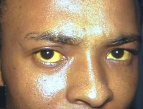
Scleral Icterus9
Why Check: Pregnancy adds a few important conditions to the Differential Diagnosis of jaundice. For instance, HELLP syndrome can present with jaundice (due to hemolysis and liver injury) and RUQ/Epigastric pain (liver capsule pain) along with proteinuria, nausea/vomiting, headache and visual changes. There is also acute fatty liver disease of pregnancy, presenting with jaundice, RUQ pain, N/V, and ascites. Alternatively, cholestatic disease of pregnancy presents with jaundice and pruritis (generally on palms and soles, worse at night) diagnosed with serum bile acid. Patients can still have acute cholecystitis, cholangitis and other liver diseases (e.g. Hepatitis B, Hepatitis C, Alcohol-related liver disease) occur during pregnancy as well.
Palpation
Symphysis-Fundal Height (SFH)
Say: I will use a tape measure to measure the symphysis-fundal height to get a sense of baby’s growth.
Do: Using a tape measure, palpate for the symphysis pubis, as well as the top of the uterine fundus. Measure this with your tape measure.
What is Normal: Emerges from pelvis at 12 weeks, 20 cm at 20 weeks (roughly at level of maternal umbilicus), + 1cm/week until labour when symphysis fundal height ↓ with fetal engagement in labour
Report: Symphysis-fundal height is _____ cm, which is normal for the gestational age of ___ weeks.
Why Check: Important to note growth of fetus to track during gestation. Abnormal symphysis-fundal height (i.e. not in keeping with expected size) can warrant further investigations such as U/S.
Leopold’s Maneuvers (Obstetrics)
Say: I will palpate the abdomen to assess the orientation of the fetus.
Do: 4 maneuvers to assess fetal positioning (see diagram). Four questions: (1) what is the presenting part?; (2) where is the back and where are the small parts?; (3) what is in the fundus?; and (4) where is the cephalic prominence? Is the head engaged? Do the examiner’s hands converge or diverge? During your palpation, fetal head and spine should feel firm as helpful landmarks.
What is Normal: Easiest to fully assess position >30 weeks (fetal head firm). Normal is a cephalic (head down) presentation.
Report: Fetus’ position is (breech/cephalic, or transverse lie); fetus is facing (left/right, opposite side of spine on palpation)
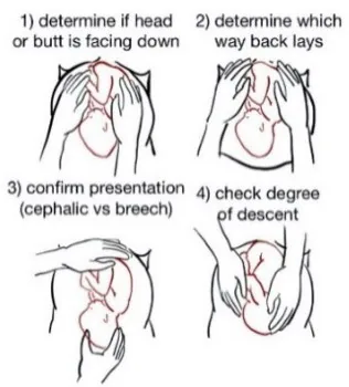
Why Check: Leopold’s maneuvers are commonly performed in the 3rd trimester, in the context that fetal positioning may change spontaneously before 34 weeks gestation. Within a prenatal visit, knowing fetal position helps determine Doppler placement for fetal heart rate assessment, where the doppler probe is commonly placed along fetal spine, closer to the head. The presentation of the fetus is important in preparing for delivery, for example determining whether fetus is cephalic or breech, which impacts risks of delivery, where the patient may be eligible for External Cephalic Version (attempt to rotate fetus into cephalic presentation). Presentations include: Complete Breech (Hips, Knees Flexed); Frank Breech (Hips flexed, Knees Extended); Footling/Incomplete Breech (Hips Extended, Foot/Buttocks presenting).
General Light Abdominal Palpation
AFMC level Pre-clerkship 3 · Clerkship 4
(see Ch 13 – Abdo exam)
Say: I will now palpate the abdomen in 9 regions using light palpation Are you experiencing any pain right now? If so, I will palpate that area last.
Do: Using the palm standing at patient’s side, palpate the abdomen (should be good surface contact), and progress along in a systematic way. Should be good amount of pressure, it is palpated using the fingers. Assess for masses, tenderness, distension, and hardness/rigidity (involuntary guarding). While doing this, look at patients’ face for signs of pain.
Report: No evidence of guarding, facial discomfort, or masses on light palpation in 9 abdominal regions
Why Check: Can identify tenderness, masses, hernias, and peritonitis. Relevantly, the likelihood of suspected ectopic pregnancy is mildly increased with positive peritoneal findings (LR+ 4.2-4.5, LR- 0.78-0.81) and with an adnexal mass (LR+2.4). The likelihood of suspected peritonitis increases slightly with rigidity (LR+ 3.9) and guarding (LR+ 2.6, LR- 0.6), and decreases with positive abdominal wall tenderness test (LR+ 0.1). Additionally, HELLP syndrome can present with RUQ pain (representing liver capsule pain). In terms of non-obstetrical findings, the typical presentations can change due to the presence of the fetus and enlarged uterus causing shifting of abdominal organs. For example, appendicitis may present with RUQ pain rather than RLQ pain in the later stages of pregnancy. Acute cholecystitis may still present with RUQ tenderness – however if the patient presents with jaundice, keep HELLP syndrome, acute fatty liver disease of pregnancy and cholestatic disease of pregnancy on the DDx.
Sensitive Pelvic Exams
Cervical Check and Modified Bishop Score (Similar to bimanual exam)
AFMC level Pre-clerkship 2 · Clerkship 3
Say: I will palpate the cervix to assess labour progress. Is it okay if I perform this exam first, and then the staff doctor double-checks my findings afterwards?
Setup: Patient should be in Dorsal Lithotomy Position. Lubricate one hand with gloves. Ensure chaperone present. Ask patient to breathe in, insert 2 fingers (index and middle) into vagina when breathes out.
Do: Assess the cervix for dilation, effacement/length, consistency, position, and station to complete a Modified Bishops Score (score each category and sum to total score). Station is determined by palpating the Ischial Spines and determining position versus the leading edge of the fetal skull.
- Bishop Score10 – Predicts likelihood of successful vaginal delivery and favourability of cervix
| Score | Dilation (cm) | Length (cm) (% Effacement) | Consistency | Position | Station |
|---|---|---|---|---|---|
| 0 | Closed | >4 cm (0-30%) | Firm | Posterior | -3 |
| 1 | 1-2 cm | 3-4 cm (40-50%) | Medium | Mid-position | -2 |
| 2 | 3-4 cm | 1-2 cm (60-70%) | Soft | Anterior | -1, 0 |
| 3 | 5+ cm | 0 cm (80-100%) | ----- | ---- | +1, +2 |
Report: The cervix is 4cm dilated, 3cm long, medium consistency, posterior, with station is -2. The Bishop Score is 5, and cervix is unfavourable. We may consider induction.
Why Check: The Cervical Exam is critical for monitoring stages of labour (remember that labour is defined as regular contractions + evidence of cervical change), including:
- Diagnosing transition from latent to active 1st stage of labour at 4cm dilatation → influences decisions to admit the patient (active labour) vs. watchful waiting (latent labour)
- The Bishop Score determines whether the cervix is favourable. For clinical decisions around induction/augmentation of labour, per SOGC guidelines11:
- Bishop Score of ≥ 6 is considered a favourable cervix, where we would consider oxytocin and may also perform artificial rupture of membranes.
- Bishop Score of <6 is considered an unfavourable cervix – consider first cervical ripening interventions (e.g. misoprostol, dinoprostone, balloon dilation of cervix), and once cervix becomes favourable, may consider oxytocin and may perform artificial rupture of membranes.
- Most important elements of Bishop Score: Dilatation most useful; Cervical consistency least useful
- Monitoring for dystocia (slowed or arrested progression of labour)
- As the cervix dilates and fetal head engages, assessing fetal head presentation by feeling suture lines (re: Occiput Anterior is easier to deliver with baby’s head facing floor vs. Occiput Posterior)
- In-office, useful to document pre-intervention for cervical stretch & sweeps offered beginning at 38 weeks (3 passes within the external cervical os with intent to ↑ local prostaglandin production).
- Why NOT Check:
- If you are concerned for a Placenta Previa (classically a painless vaginal bleed, versus Placental Abruption as a painful vaginal bleed) – do NOT do any cervical checks or utilize any vaginal instrumentation until this is safely assessed with an ultrasound.
- If you are concerned for Prelabour Rupture of Membranes (PROM) or Preterm Prelabour Rupture of Membranes (PPROM) digital exam for cervical check should be avoided to ↓ risk of infection until labour has begun or induction of labour is initiated (examine using sterile speculum exam – see below).
- Bimanual exam (in early pregnancy): The likelihood of suspected ectopic pregnancy is increased with positive cervical motion tenderness (LR+ 4.9, LR- 0.62) and with an adnexal mass (LR+ 2.4). The absence of adnexal tenderness decreases the likelihood of suspected ectopic pregnancy (LR- 0.57).
Sterile Speculum Exam for Rupture of Membranes (ROM)
AFMC level Pre-clerkship 2 · Clerkship 3
Say: I will perform a sterile speculum exam to assess for ruptured membranes.
Setup: Explain what speculum is, why doing it. Do NOT lubricate as this can give false positive results. Additionally, keep the speculum sterile (including not touching it against patient’s thigh to demonstrate temperature), as there is risk of infection if membranes have been ruptured. Prepare a slide for Ferning and have Nitrazine paper test ready.
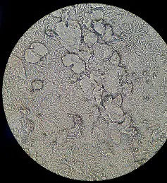
Insertion: Hold with 2 fingers over top of speculum, thumb in between speculum screw (speculum closed); angle towards sacral hollow. DO NOT TOUCH CLITORIS while inserting (painful). Ask patient to breathe in; as patient breathes out, insert speculum. Open speculum until you can see cervix. Tighten screw to keep in place.
Inspect Cervix – Look for pooling of fluid in posterior fornix, free flow from cervix, and cord prolapse.
Sample Collection: (1) Nitrazine Test Strip (Look for turning blue as a positive sign of ROM), and (2) Run Swab over microscope slide, dry slide for 10 minutes then inspect for Ferning (see Picture to right)
Removal: Unscrew the speculum and remove carefully
Report: Posterior pooling was seen on speculum with nitrazine test positive and Ferning noted on the slide under the microscope.
Why Check: Helps to confirm Rupture of Membranes (e.g. by patient report of a gush of fluid). On their prenatal chart, pay attention to any note of placenta previa, oligohydramnios/ polyhydramnios, and GBS Status. These can influence management of a premature ROM (PROM) or preterm premature ROM (PPROM), including the decision to administer antibiotics.
Why NOT Check: If you are concerned for a Placenta Previa (classically painless vaginal bleed, versus Placental Abruption as a painful vaginal bleed) – do NOT do any cervical checks or utilize any vaginal instrumentation until this is safely assessed with an ultrasound.
Part 4: Relevant Likelihood Ratios – from Rational Clinical Examination JAMA series
Clinical Question 1: Is this pregnant patient in a relationship with concerns for Intimate Partner Violence?7
| Screening Test (Reference Standard) – Population | Detail | Sensitivity (95% CI) | Specificity (95% CI) |
|---|---|---|---|
| AAS (CTS2) – 748 women post-partum, maternity ward | Minor violence | 0.32 (0.25-0.40) | 0.99 (0.98-0.996) |
| Severe violence | 0.61 (0.48-0.74) | 0.98 (0.96-0.990) | |
| Both | 0.32 (0.24-0.40) | 0.99 (0.98-0.997) |
Clinical Question 2: Does this woman have an Ectopic Pregnancy?12
| Procedure | Sensitivity | Specificity | LR+ (95% CI) | LR- (95% CI) |
|---|---|---|---|---|
| Physical Exam – Cervical Motion Tenderness | 0.45 (0.33-0.57 | 0.91 (0.80-0.96) | 4.9 (1.7-14) (about +15%) | 0.62 (0.47-0.83) (about −15%) |
| Physical Exam – Peritoneal findings | 0.23-0.27 | 0.94-0.95 | 4.2-4.5 (about +15%) | 0.78-0.81 (about −15%) |
| Physical Exam – Adnexal mass | 0.09 (0.02-0.27) | 0.96 (0.92-0.98) | 2.4 (1.6-3.7) (about +15%) | 0.94 (0.87-1.0) (no change: 95% CI includes 1) |
| Physical Exam – Adnexal tenderness | 0.61 (0.52-0.69) | 0.65 (0.42-0.83) | 1.9 (1.0-3.5) (no change: 95% CI includes 1) | 0.57 (0.48-0.67) (about −15%) |
| Imaging – Transvaginal Sonography | 0.88 (0.52-0.98) | 0.99 (0.96-1.0) | 111 (12-1028) (about +45%) | 0.12 (0.03-0.55) (about −30%) |
References
-
Paladine HL, et al. Postpartum care: an approach to the fourth trimester. Am Fam Physician. 2019;100(8):485-491. ↩
-
Perinatal Services BC. Accessed June 21, 2022. http://www.perinatalservicesbc.ca/Documents/Resources/Checklists/PSBC_Postpartum_Checklist.pdf ↩
-
Breastfeeding Resources Ontario. Accessed June 21, 2022. http://breastfeedingresourcesontario.ca/sites/default/files/pdf/Res_Strategy_IDM_Toolkit_EN.pdf ↩
-
Shakil A, Donald S, Sinacore M, Krepcho M. Validation of the HITS domestic violence screening tool with males. Fam Med. 2005;37(3):193-198. ↩ ↩2
-
Government of Canada. Accessed June 21, 2022. https://www.canada.ca/en/health-canada/services/food-nutrition/healthy-eating/healthy-weights/prenatal-guidelines-professionals-gestational-weight-gain.html#a5 ↩
-
Recommendations. Choosing Wisely Canada. Accessed August 1, 2022. https://choosingwiselycanada.org/recommendations/#list ↩
-
Nelson H, Bougatsos C, Blazina I. Screening women for intimate partner violence: a systematic review to update the US Preventive Task Force recommendation. Ann Intern Med. 2012;156:796-808. ↩ ↩2
-
Canada’s guidance on alcohol and health. Accessed February 25, 2023. https://ccsa.ca/canadas-guidance-alcohol-and-health ↩
-
Centers for Disease Control and Prevention. Accessed February 21, 2023. https://phil.cdc.gov/Details.aspx?pid=2860 ↩ ↩2 ↩3
-
Dore S, Ehman W. No. 396 – Fetal health surveillance: intrapartum consensus guideline. J Obstet Gynaecol Can. 2020;42(3):316-348. ↩
-
Leduc D, et al. No. 296 – Induction of labour. J Obstet Gynaecol Can. 2013;35(9):840-857. ↩
-
Crochet JR, Bastian LA, Chireau MV. Does this woman have an ectopic pregnancy? JAMA. 2013;309(16):1722-1729. ↩