Medical Foundations 4
Chapter 25Hand and Wrist Examination
Part 1: Focused History – Based on Chief Complaint (Begin with Open-Ended Questions)
HPI
Pain Assessment
- Onset:
- Acute onset of pain, disability immediate with injury (e.g. acute trauma, fracture) (see Mechanism of Injury below)
- Delayed onset of pain (e.g. ligamentous injury)
- Acute onset with no associated injury (e.g. septic arthritis)
- Insidious onset gradually worsening over time (e.g. overuse injury such as tenosynovitis)
- Pediatric patients: Is the patient in an active growth spurt → increased risk of overuse injuries
- Provoking:
- Aggravating postures: Wrist flexion/extension, radial/ulnar deviation; forearm pronation/supination
- Aggravating activities: Repeated activity (may suggest tenosynovitis); weightbearing (suggests bone or joint involvement); gripping (requires wrist stabilization); pinching; lifting – distinguish everyday lifting activity (e.g. picking up a cup) vs. heavy activity (e.g. repeated weightlifting)
- Palliating:
- Relieving postures/activities: Relieved with rest; wrist positions with ↓ pain
- Previous treatment: POLICE (Protect, Optimally Load, Ice, Compress, Elevate)? Physio? Analgesics?
- Rehab: Posture changes to activity; manual therapy; exercise (e.g. resistance, ROM); brace, cast, splint
- Note on Hand/Finger Injuries: Immobilization can exacerbate edema and stiffness – focus on protected stabilization with active motion splints (including e.g. “buddy taping” injured digit to uninjured digit to stabilize injury and encourage range of motion)
- Investigations: Type (x-rays, Ultrasound, MRI, etc.) and results (if available)
- Quality: Deep ache, sharp vs. dull? – Ask patient what words they would use
- Neuropathic Pain Descriptors: Pins/needles; hot/burning/stinging; numb; electrical shocks; sensitivity to touch (e.g. clothing, bed sheets)
- Region: Poorly localized (e.g.) vs. Well-localized (e.g.)
- Wrist Pain: Radial vs. Central vs. Ulnar – helps organize differential diagnosis
- Radiation: For example, radiation along a peripheral nerve (e.g. Carpal Tunnel Syndrome)
- Severity: Immediately after injury, vs. subsequently. Has it improved over time with rest?
- Was patient able to continue their activity (e.g. after the sport completed, noticed pain got worse)?
- Timing: Constant vs. Intermittent
- Night pain: Characteristic e.g. in Carpal Tunnel Syndrome
- Recurrence: Assess episodes and pattern of recurrence of pain
- Associated Symptoms:
- Stiffness after period of rest: May suggest inflammatory condition (e.g. Tenosynovitis)
- Lack of active hand motion: Warning sign of significant injury
- Joint click: e.g. carpal instability, Triangular Fibrocartilage Tear, Extensor Carpi Ulnaris Subluxation.
- Recurrent wrist swelling: ↑ suspicion for wrist instability
- Sensory neuropathy e.g. paresthesias or numbness – assess distribution e.g. Peripheral nerve, Dermatomal or Spinal level (See Ch 31 – Screening Neuro Exam) – e.g. impaired sensation over median nerve distribution (D1-D3 + radial side of D4) in Carpal Tunnel Syndrome vs. ulnar nerve distribution with Ulnar Nerve Compression at Level of Wrist (D4 and D5 of hand).
- Weakness: Weakness in specific motion may indicate a muscle tear; if concern for a neurologic cause (e.g. difficulty opening jar lids, dropping objects, difficulty turning keys or buttoning clothes) may need to do a screening neuro exam (See Ch 31 – Screening Neuro Exam).
Red Flags
- Pain: Pain at night/rest/unrelated to activity; Pain with passive movement; Poor Response to Treatment
- Constitutional Symptoms: Fever, night sweats, weight loss, anorexia
- Acute Compartment Syndrome (Can occur minutes-hours after acute injury, particularly closed fracture with compartments intact) → Surgical emergency (fasciotomy relieves compartment pressure)
- 6 Ps of compartment syndrome: pain, poikilothermia, paresthesia, paralysis, pulselessness, pallor.
- Pain: Out of proportion; not improving with analgesia; at rest; with passive muscle stretching
- LATE Findings of acute compartment syndrome: Pallor, paresthesia, paralysis, and pulselessness
- High Risk Facture of upper limb: Supracondylar Fracture (Pediatrics)
- Fullness or distension of involved compartment
- Inflammatory Arthritis:
- Intra-articular features: New onset joint swelling/effusion; morning stiffness >30 mins (vs. <30 mins in osteoarthritis); improves during day and with activity (vs. worsens in osteoarthritis); distribution (Monoarthritis = 1, Oligoarthritis = 2-4, Polyarthritis = 5+ joints, symmetrical vs. asymmetrical)
- Extraarticular features: Psoriasis, Inflammatory Bowel Disease; Uveitis/Iritis; Photosensitivity rash; Raynaud’s Phenomenon; Urethritis; Diarrheal Illness
- Septic Arthritis: Acute atraumatic pain. Look for adjacent skin infection (vs. hematogenous seeding e.g. in a patient with sepsis), fevers and chills with a warm, erythematous, swollen joint and painful restricted range of motion in all directions.
- Neurovascular Damage: High-velocity injury; no pulses; hand weakness.
- Non-Accidental Injury (Pediatrics): Delayed seeking medical attention. History not fitting with injury presentation. Unwitnessed injury. Bruises over soft tissue areas. Prior history of injuries or neglect. Types of fracture (e.g. metaphyseal)
Mechanism of Injury
- If injury occurred during an activity, ask open-ended questions about mechanism of injury e.g. “run me through what happened, play-by-play”. Try to visualize what happened. Assess low energy vs. high energy force injury and the forces involved (e.g. direct vs. indirect forces and their direction)
- Wrist Injury mechanism:
- Fall forward onto outstretched hand (FOOSH) → ↑ risk of Distal Radius Fracture, Scaphoid fracture, Scapholunate dissociation, etc.
- Falling backward → ↑ risk of triquetral fracture, lunate fracture, Lunotriquetral Dissociation
- Hand Injury mechanism: Direct, severe blow to fingers → Fracture; vs. Blow to point of finger → Interphalangeal dislocation, Joint sprain, or long flexor/extensor tendon avulsion
- Wrist Injury mechanism:
- If assessing an athlete, helpful to know/learn about their sport to understand common injuries and the demands of the position they play in the sport.
- Ask about possible overuse, what the patient does for work, etc. re: carpal tunnel syndrome.
Radial Wrist Pain
- Common Illness Presentations:
- De Quervain’s Tenosynovitis (Inflammation of Abductor Pollicis Longus and Extensor Pollicis Brevis at radial styloid): Gradual onset pain of dorsal radial wrist with localized swelling and tenderness that may radiate along the tendon lines, worse with pinch grasping or thumb and wrist movement.
- Carpometacarpal (CMC) Osteoarthritis: Presents with gradual onset of an aching, deep pain with joint stiffness worse with prolonged activity (e.g. painting, yard work), especially grasping and pinching, or forceful use of thumb (e.g. turning a key). May be associated with crepitus and loss of pincer strength.
- Can’t Miss Diagnosis Illness Presentations:
- Distal Radius Fracture: Acute onset pain of dorsal radial wrist following a FOOSH. May occur with a concurrent ulnar styloid fracture (see below), with associated swelling, deformity, and palpable bony step-off. May occur in young active patients, and in patients with osteoporosis.
- Colle’s Fracture: Common type of distal radius fracture following fall on palm of hand with upward tilt of radius, associated with a dinner fork sign (distal radius posteriorly displaced on X-ray)
- Smith’s Fracture: Distal radius fracture following fall on back of hand with downward tilt of radius
- Scaphoid Fracture (most common carpal fracture): Acute onset pain of dorsal radial wrist following a FOOSH, with tenderness of anatomical snuffbox. Pain may settle post-fall, where the patient presents late after injury. May be accompanied by loss of grip strength and swelling. The scaphoid has precarious blood supply → ↑ risk of Avascular Necrosis with chronic wrist pain.
- Trapezium Fracture: Acute onset pain of dorsal radial wrist following injury with axial loading of thumb or from direct blow. Associated with localized swelling. May be associated with dislocation or fracture of neighboring carpal bones (can’t miss diagnosis – see below), and weakness with pinching (e.g. cannot make “OK sign”). Long-term risk of ↓ CMC ROM with painful and/or weak pinch and grip.
- Distal Radius Fracture: Acute onset pain of dorsal radial wrist following a FOOSH. May occur with a concurrent ulnar styloid fracture (see below), with associated swelling, deformity, and palpable bony step-off. May occur in young active patients, and in patients with osteoporosis.
Central Wrist Pain
- Common Illness Presentations:
- Ganglion Cyst (Synovial cyst communicating with joint space): Often a relatively painless intermittent swelling, with intermittent wrist pain and reduced wrist range of motion. Ganglion cysts of wrist are common (about 2 per wrist in healthy populations – commonly in the dorsum of the wrist) and benign, requiring treatment if symptomatic.
- Carpal Tunnel Syndrome: Gradual onset burning volar central wrist pain, often worse at night. May present with paresthesias/numbness in distribution of median nerve (D1-D3 + radial side of D4 – LR+ 3.1 for hypoalgesia), where pain may radiate to forearm/elbow/shoulder.
- Do not diagnose new bilateral carpal tunnel syndrome until C-spine injury is excluded.
- Can’t Miss Diagnosis Illness Presentations:
- Lunate Fracture: Acute onset central dorsal wrist pain often occurring after wrist hyperextension injury or fall onto extended wrist, with pain worse with wrist motion or gripping.
- Scapholunate Dissociation/Instability: Acute onset central dorsal wrist pain localized to 2cm distal to Lister’s tubercle (radial side of Lunate). Injury following fall on extended wrist. May be associated with swelling of dorso-radial wrist, ↓ grip strength, and ↓ wrist ROM. Can’t miss diagnosis due to risk of ↓ grip strength, and osteoarthritis long-term.
- Avascular Necrosis of Lunate (Kienbock’s Disease – with progressive collapse of lunate): Gradual onset central dorsal wrist pain with swelling and tenderness over lunate, with limited wrist ROM. May occur due to previous lunate fracture, repetitive trauma. Can’t miss diagnosis re. risk of ↓ grip strength.
Ulnar Wrist Pain
- Common Illness Presentations:
- Triangular Fibrocartilage Complex tear (TFCC – complex between ulna and carpus, major stabilizer of distal radioulnar joint = DRUJ): Acute onset, intermittent dorsal ulnar wrist pain with dorsal wrist swelling. Pain worse with wrist dorsiflexion and ulnar deviation. Occurs following FOOSH or rotational stress to distal radio-ulnar joint, forced ulnar deviation and rotation. Look for TFCC Tear following distal radius fracture (above), ulnar styloid fracture (below) or DRUJ Instability. May also be an insidious onset of pain in athletes with repetitive wrist loading in extension (e.g. gymnasts).
- Extensor Carpi Ulnaris Tenosynovitis (stabilizes Distal Radioulnar Joint = DRUJ): Gradual onset of intermittent, dorsal ulnar wrist pain over area of ulnar styloid worse with active contraction or tendon stretch. Tendon may sublux with clicking or popping with wrist supination and extension.
- Ulnar Nerve Compression at Wrist (Guyon’s Canal Syndrome – vs. Cubital Tunnel Syndrome re. Ulnar Nerve compression at elbow): Diffuse Pain with paresthesias/numbness along ulnar nerve distribution in fingers (Ulnar side of D4 and D5). May occur due to acute trauma (e.g. karate), repetitive trauma (e.g. baseball catcher) or nerve compression (e.g. long-distance cycling supporting body weight).
- Can’t Miss Diagnosis Illness Presentations:
- Lunotriquetral Dissociation: Acute onset pain of dorsal ulnar wrist with pain at end-range pronation and supination following backwards fall. Does not commonly present with selling or bruising. Can’t Miss Diagnosis due to risk of carpal instability.
- Ulnar Styloid Fracture: Acute onset, constant pain of dorsal ulnar wrist following a FOOSH. May occur with a concurrent distal radius fracture (see above) and Triangular fibrocartilage complex tear. Concern if developing distal radial-ulnar joint (DRUJ) instability – refer to orthopedics.
- Hook of Hamate Fracture: Acute onset, constant pain of dorsal ulnar wrist following a FOOSH or following a swinging motion (e.g. golf club strikes ground instead of ball – forces transmit at golf club handle to hook of hamate of hand on top of golf club). May present with ulnar neuropathy (due to compression injury) including decreased grip strength. Swelling is usually minimal.
- Triquetral Fracture: Acute onset, constant pain of dorsal ulnar wrist following a fall on dorsiflexed and ulnar deviated hand (e.g. falling backward). Presents with point tenderness and painful wrist flexion. Look for comorbid peri-lunate fracture dislocation.
- Pisiform Fracture: Acute onset, constant pain of dorsal ulnar wrist following a FOOSH with wrist in extension, or direct force onto heel of hands (e.g. when striking object directly), presenting with pain and swelling at palmar and ulnar aspects of wrist.
Hand and Finger Pain
- Fractures:
- Base of 1st Metacarpal Fracture: Acute, constant deep pain following fall on abducted thumb, or punch of hard object. Requires urgent surgical referral.
- Fractures of 2nd-5th Metacarpals: Acute, constant deep pain following direct hits (e.g. hockey, baseball, cricket), punch of hard object, or contact with ball and ground. May see flexion deformity of distal fragment, which can impact grip strength. Look for rotated fractures requiring urgent surgical intervention – can cause crossover of fingers if left untreated.
- Proximal Phalanx Fracture: Acute, constant deep pain which can create functional impairment where extensor and flexor tendons contact callus and exposed bone, with potential Z-deformities through PIP and DIP joints due to imbalance of flexor and extensor tendons. Look for rotational deformities (e.g. better visualized with PIPs and DIPs flexed) which require urgent surgical intervention.
- Middle Phalanx Fracture: Acute, constant localized pain. Fractures distal to flexor tendon attachment show flexion of proximal fragment, and extension of distal fragment. A volar plate avulsion fracture at PIP joint can occur following a hyperextension injury – where this can cause impaired grip strength if untreated. Also look for rotated fractures requiring urgent surgical intervention – can cause stiff PIP joint if left untreated.
- Distal Phalanx Fracture: Acute, constant deep pain. Commonly occurs with a crush injury (e.g. finger jammed in door, or between fast-moving ball and stick or bat). May present with a subungual hematoma of associated nail bed.
- Dislocation Injuries:
- Carpo-Metacarpal Joint Dislocation: Occurs with hitting or impact on ground. May occur with carpal bone fracture (see above). CMC subluxation and dislocation require urgent surgical intervention to maintain strength and power in hand.
- Proximal Interphalangeal (PIP) Joint Dislocation: Occurs with hyperextension injury with longitudinal compression (e.g. ball sports). Look for proximal/middle phalanx fracture (e.g. volar plate avulsion fracture of middle phalanx – See Above).
- Distal Interphalangeal (DIP) Joint Dislocation: Occurs with hyperextension injury e.g. ball hitting tip of finger, with potential volar skin laceration. Important to test flexor tendon since avulsion may occur, or less commonly may damage extensor tendon with avulsion causing a Mallet finger (see below).
- Ligament and Tendon Injuries:
- Sprain of Ulnar Collateral Ligament of 1st MCP Joint (“Skier’s Thumb”): Acute onset, intermittent pain. Presents with laxity in radial direction and may present with soft end-feel in complete tear. Functionally impacts hand strength and precision, e.g. pinching. A complete tear requires urgent surgical referral.
- Sprain of Radial Collateral Ligament of 1st MCP Joint: Acute onset, intermittent pain with pain on grip and associated stiffness. Injury occurs with ulnar directed force on thumb (e.g. from a ball, or a punch).
- Capsular Sprain of 1st MCP Joint: Acute onset, intermittent pain following hyperextension injury. This injury is prone to recurrence.
- PIP Joint Sprains: Acute onset, intermittent pain following injury from sideways force. Look for instability with lateral stress as an indicator of a complete tear and may present with volar plate avulsion fracture (see above).
- Mallet Finger: Acute onset, intermittent pain over dorsal aspect of distal phalanx with loss of full extension of DIP joint. Pain may be relatively mild in comparison to the obvious flexion deformity. This occurs due to extensor tendon avulsion or rupture of extensor tendon proximal to insertion at distal phalanx, leaving unopposed action of flexor digitorum profundis tendon. Occurs with direct force (e.g. ball striking extended fingertip to catch ball). Requires urgent surgical referral with risk of chronic changes (e.g. Chronic Swan Neck Deformity) limiting hand function if left untreated.
- Chronic Swan Neck Deformity (PIP hyperextension, DIP flexion): Can be a sequela to Mallet Finger injury (see above) or volar plate injuries (see above). Chronic cases may present with PIP instability with visible snapping and discomfort; can impact fine motor activities with locking finger.
- Boutonniere Deformity (PIP flexion, DIP hyperextension): Acute onset, intermittent pain with injury to extensor tendon just proximal to insertion on middle phalanx (vs. extensor tendon injury at insertion proximal to distal phalanx in mallet finger) leaving unopposed action of flexor digitorum profundis tendon. Over time, can limit range of motion with ligament contraction. Occurs with direct force to dorsum of PIP, or PIP joint hyperflexion injury. May present with significant swelling and inability to actively extend PIP joint.
- Flexor Digitorum Profundis Avulsion (“Jersey Finger” – most common to D4): Acute onset, intermittent pain where patient hears a “snap”, with finger relatively extended at rest vs. other fingers, and inability to actively flex the DIP joint. Typically occurs with grabbing opponents clothing (e.g. in contact sports), with distal phalanx forced extension while athlete is flexing. May occur with avulsion fracture of distal phalanx, with a palpable lump proximally in finger (palpating the avulsed tendon) – Can’t miss diagnosis requiring urgent surgical intervention, limiting grip, hand strength, precision if left untreated.
- Trigger Finger (Stenosing flexor tenosynovitis): Gradual onset, intermittent pain in palm with painful clicking and locking of one or more fingers with active flexion of affected digit. Patient may also identify pain at the PIP joint.
- Dupuytren’s Contracture (Progressive fibrosis of palmar fascia – draws fingers into flexion at MCP and/or PIP joints): Gradual onset, intermittent episodic pain with progressive loss of full extension. Patient may notice nodules on palmar fascia, especially at D4 and D5 with puckering/tethering of skin.
Past Medical History (PMHx)
- Previous hand or wrist injuries
- ↑ Risk Carpal Tunnel Syndrome: Diabetes Mellitus; Arthritis (re. anatomic compression of carpal tunnel); Hypothyroidism (due to ↑ tissue edema); Pregnancy (re. accumulation of fluid); Acromegaly
- ↑ Risk Dupuytren’s Contractures: Diabetes Mellitus; Liver Disease/Cirrhosis
- Osteoporosis Risk Factors: History of Fragility # (e.g. fall from standing height or less); Vertebral Compression #; Inflammatory Arthritis (e.g. Rheumatoid Arthritis); Malabsorption Syndromes (e.g. Celiac disease, Inflammatory bowel disease); Endocrine Disorders (e.g. Hyperparathyroidism, Hyperthyroidism, Cushing’s, Hypogonadism)
- Risk Factors for Septic Arthritis: Immunosuppression (including diabetes mellitus and HIV); Pre-existing joint disease (e.g. Rheumatoid arthritis: LR+ 2.5, LR- 0.45); Advanced Age; Skin infection (LR+ 2.8, LR- 0.76)
- Lab Markers for Septic Arthritis:
- Peripheral – CRP >100 mg/L (LR+ 1.6, LR- 0.44), WBC >10 (LR+ 1.4), ESR >30mm/h (LR+ 1.3)
- Synovial fluid – WBC >100 (LR+ 28, LR- 0.71), WBC >50 (LR+ 7.7, LR- 0.42), WBC >25 (LR+ 2.9, LR- 0.32), PMN cells ≥90% (LR+ 3.4, LR- 0.34)
- Lab Markers for Septic Arthritis:
- Bleeding Disorder History: ↑ risk of serious pathology on injury.
- Risk Factors for Osteoarthritis: Age (>50 years); Female Gender; Obesity; Heavy manual labour; History of joint injury
- Psoriasis: Risk factor for Psoriatic Arthritis.
- Autoimmune disorders (↑ risk for Inflammatory Arthritis) – particularly Spondyloarthropathies e.g. Psoriatic arthritis, Uveitis, Ankylosing Spondylitis, Inflammatory Bowel Disease, Reactive Arthritis
Relevant Clinical Scores:
- CTS-6 – validated diagnostic scoring tool for Carpal Tunnel Syndrome (max score 26 points) where a score of ≥12 points is indicative of an 80% probability of Carpal Tunnel Syndrome.
Past Surgical History (PS Hx)
- Arthroscopic surgery; Open-Reduction-Internal-Fixation, Pinning
- Median nerve decompression e.g. for Carpal Tunnel Syndrome
- Palliative Radiation (e.g. for Painful Bony Metastasis in cancer)
- Risk Factors for Septic Arthritis: Recent joint surgery (LR+ 6.9, LR- 0.78)
Family Medical History (FMHx)
- Autoimmune disorders (↑ risk for Inflammatory Arthritis) – particularly Spondyloarthropathies e.g. Psoriasis, Uveitis, Ankylosing Spondylitis, Inflammatory Bowel Disease, Reactive Arthritis
- Gout
Social Hx
Substance Use
- Smoking: Risk factor for Osteoporosis, Avascular Necrosis, Dupuytren’s Contracture
- Alcohol: Risk factor for Gout; Osteoporosis (>3 units/day); Avascular Necrosis, Dupuytren’s Contracture
- IV Drug Use: Risk factor for Septic Arthritis
Occupational Hx
- Are you working right now? What do you do for work? What other kinds of work have you done in the past? Do you have benefits through work such as drug coverage (e.g. access to Physiotherapy coverage)?
- If injury occurred at work → separate Workplace Safety and Insurance Board (WSIB) process.
- Occupations ↑ Risk Wrist Injury: Office/admin, Manual Labour, Food Service Industry, Gardening
- Carpal Tunnel Syndrome ↑ Risk: Tasks include Repetitive hand/wrist use; Forceful hand/wrist use; Sustained wrist or palm pressure; Work with vibrating tools; Prolonged wrist extension/flexion; Use of hands in cold temperature – example occupations include manufacturing, office/admin support, food service industry.
- Dupuytren’s Contracture ↑ Risk: Repetitive handling of vibrating tools
- Musician History and Risk Assessment: Number of years playing, hours of practice per week, Recent changes (e.g. change in playing, complex piece, etc. – re. risk of overuse injury)
Sporting Hx
- History of Sport Played and Select ↑ Risk:
- ↑ Risk Fall on Outstretched Hand (risk of Scaphoid Fracture, Distal Radius Fracture, Triangular Fibrocartilage Complex Tear, Intercarpal Ligament Injury, etc.): Snowboarding, Rollerblading, Riding Sports (e.g. horseback riding, cycling)
- ↑ Risk Hook of Hamate Fracture: Swinging activity (e.g. Golf club, tennis racquet, baseball bat)
- ↑ Risk De Quervain’s Tenosynovitis: Volleyball, Racquet sports (Tennis, squash, badminton), Ten-pin bowling, Rowing, Canoeing, Golfing (especially of non-dominant hand)
- ↑ Risk Scapholunate Ligament Sprain: American Football
- ↑ Risk Extensor Carpi Ulnaris Tenosynovitis: Golf, Tennis, Weightlifters
- ↑ Risk Triquetral Fracture: Boxing
- ↑ Risk Avascular Necrosis of Lunate (Kienbock’s Disease): Particularly Golf and Tennis
- ↑ Risk Triangular Fibrocartilage Complex (TFCC) Tear: Stick sports (e.g. hockey), Racquet sports; and Repetitive Ulnar Compressive Loading (e.g. Gymnastics, weightlifting, boxing, surfing)
- ↑ Risk Ulnar Nerve Compression: Cyclists (prolonged support body weight), karate, baseball catchers
- ↑ Risk Metacarpal Fracture or carpometacarpal joint instability: Punching (e.g. boxing, ice hockey)
- ↑ Risk Carpo-Metacarpal (CMC) Joint Dislocation: Hitting (e.g. martial arts)
- ↑ Risk Flexor Digitorum Profundis Tendon Avulsion (“Jersey Finger”): Tackle sport (Football, Rugby)
- ↑ Risk Sprain of Ulnar Collateral Ligament of 1st MCP Joint (“Skier’s Thumb”): Ball-handling sports (e.g. basketball, football, volleyball), stick-holding sports (e.g. hockey)
- ↑ Risk Capsular sprain of 1st MCP Joint: Ball-handling sports
- ↑ Risk Mallet Finger: Catching (e.g. baseball, American football receiver, cricketer, basketball)
- ↑ Risk Boutonniere Deformity: Ball-handling sports (e.g. basketball, football, volleyball)
- Current level of sport played (e.g. recreational, competitive, league level) and desired future sporting activity level; Sports position played
- Current training history (e.g. for patients with overuse injuries) – quantity and quality of training; any recent changes to routines (e.g. training days per week, intensity, duration of training sessions)?
- Recent change of equipment (e.g. racquet size, grip size, string tension change in tennis)
- Overtraining Syndrome–excess fatigue, recurrent illness, ↓ motivation, persistent soreness/stiffness
Dietary Hx
- Inadequate nutrition for an athlete can contribute to overtraining syndrome
ADLs and IADLs
- Common activities impacted by wrist and hand pain: Gripping; fine motor movements (e.g. pinching); personal hygiene (e.g. brushing hair or teeth); eating; getting dressed (as screened for in the GALS exam)
- Use of Mobility Aids: e.g. cane, walker, crutches – any recent changes?
- Patient Handedness: e.g. in a patient who is right-handed, a right wrist injury may have greater impact on patient function vs. a left wrist injury
Medications Hx, Allergies
Medications with ↑ Osteoporosis Risk
- Glucocorticoid Use (e.g. 3+ months in past year, prednisone >7.5mg/day or equivalent)
- Aromatase Inhibitors (e.g. anastrozole, letrozole, exemestane)
- Androgen deprivation therapy (e.g. Leuprolide in prostate cancer treatment)
- Long-term use of Medroxyprogesterone Acetate injection (e.g. for contraception)
- Anti-Epileptic Drugs (e.g. Carbamazepine, Phenytoin – impact Vitamin D metabolism)
- Psychiatric Medication (e.g. SSRIs, Typical Antipsychotics e.g. Chlorpromazine, Haldol)
- Proton Pump Inhibitors (PPIs, e.g. Pantoprazole, Lansoprazole – re. ↓ Ca absorption)
- Loop Diuretic (e.g. Furosemide re. ↓ Ca reabsorption in Loop of Henle – conflicting data)
Osteoporosis Treatments
- Calcium Supplementation (Goal 1200mg/day from all sources including dietary)
- Vitamin D Supplementation (e.g. 1000-2000 IU PO Daily; 400 IU PO Daily for infants)
- Bisphosphonates (e.g. Alendronate, Risedronate, Zoledronic Acid)
- Denosumab (RANK Ligand monoclonal antibody)
- Selective Estrogen Receptor Modulator (SERMS – e.g. Raloxifene)
- Hormone Therapy (e.g. Estrogen ± Progesterone)
- Teriparatide (Endogenous Parathyroid Hormone, PTH)
- Calcitonin (↓ pain of acute vertebral fractures)
Inflammatory Arthritis
- Gout:
- Acute Flare: NSAIDs; Colchicine
- Urate-Lowering Therapy: Allopurinol, Febuxostat (safe in renal disease)
- Inflammatory Arthritis (e.g. Rheumatoid Arthritis, Spondyloarthropathies):
- Disease-Modifying Antirheumatic Drugs (DMARDs): Methotrexate, Sulfasalazine, Hydroxychloroquine, Leflunomide, Azathioprine
- Biologic DMARDs:
- TNF-α inhibitors: Etanercept, adalimumab, infliximab, golimumab, etc.
- Others: Abatacept, rituximab, tocilizumab, sarilumab, anakinra
- Acute Flare: Glucocorticoids (e.g. Prednisone)
Other Medications
- Fluoroquinolones (e.g. Ciprofloxacin) - ↑ risk of Tendinopathy or Tendon rupture
- Aromatase Inhibitors (e.g. anastrozole, letrozole, exemestane) - ↑ risk Carpal Tunnel Syndrome
Analgesics
- PO: NSAIDs (concern re. GI bleed, Hypertension, Renal impairment), Acetaminophen, Muscle Relaxant (e.g. Baclofen), Opioids – ask pain severity before versus after med, frequency and dosing (e.g. concern for Acetaminophen overdose >4g/day in healthy patient, >3g/day in elderly patient, >2g/day in liver disease)
- Topical: NSAIDs (E.g. Diclofenac), Lidocaine, etc.
- Joint Injections: Intra-articular corticosteroid injections, Diagnostic injections with lidocaine – ask about pain relief immediately after (effect of lidocaine) vs. sustained pain response (effect of corticosteroid) – Hand/Wrist Injections (e.g. for Trigger Finger) may be done via surface landmarking or with ultrasound guidance
Part 2: Physical Examination of the Hand and Wrist Joints:
- Note: If hand/wrist exam is unremarkable, consider examining the elbow. If suspecting radiculopathic pain, may need to examine neck and perform screening neurological exam (Chapter 31 – Screening Neuro Exam).
General Assessment:
Inspection (SEADS)
AFMC level Pre-clerkship 3 · Clerkship 4
- Approach: Offer a pillow for patient to rest their hands on during the exam. Ensure adequate exposure, at least to the level of the elbow.
- Swelling
- Erythema: Palmar erythema → associated with Portal hypertension, chronic liver disease, Pregnancy, Rheumatoid arthritis, Thyrotoxicosis, etc.
- Atrophy:
-
Thenar muscle wasting → Suggests Median nerve injury e.g. in severe carpal tunnel syndrome
-
Hypothenar muscle wasting → Suggest Ulnar nerve injury
-
Interossei muscle wasting → Suggests Disuse atrophy (e.g. severe arthritis) and/or Ulnar nerve injury
-
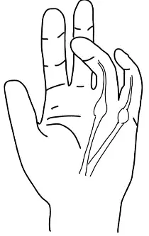
Dupuytren’s Contracture (See Diagram to right): Caused by thickening and puckering of palmar fascia) → non-specific sign often idiopathic, can be associated with Diabetes, Alcohol Use, Liver disease, or Liver cirrhosis.
-
- Deformities:
-
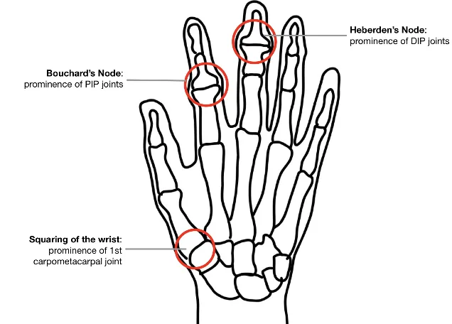
Osteoarthritis (OA) – look for Osteophyte development at specific locations:
- Squaring of the wrist – Bony prominence of 1st Carpometacarpal (CMC) Joint – an increased square wrist ratio increases the likelihood of suspected carpal tunnel syndrome (LR+ 2.7, LR- 0.5)
- Bouchard’s Nodes – Bony enlargement of Proximal Interphalangeal (PIP) Joints
- Heberden’s Nodes – Bony enlargement of Distal Interphalangeal (DIP) Joints
-
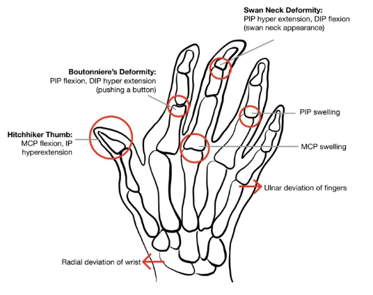
Rheumatoid Arthritis (RA):
- Joint swelling – Metacarpo-phalangeal (MCPs) and PIPs; DIPs are spared in Rheumatoid Arthritis
- Prominent ulnar head – dorsal subluxation if radioulnar ligament is disrupted
- Radial deviation of wrist
- Ulnar deviation of fingers and subluxation at the MCPs
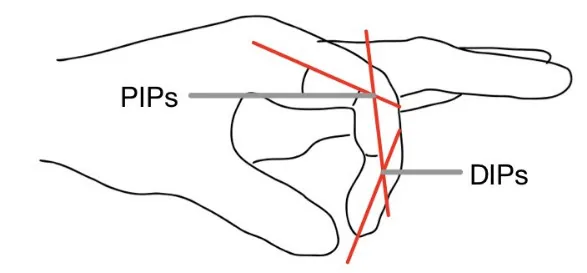
- Swan Neck Deformities (PIP hyperextension, DIP flexion – looks like a swan): Screen for Swan Neck Deformities with Prayer Sign where the patient put their palms together with wrists extended
- Boutonniere’s Deformities (PIP flexion, DIP hyperextension – looks like pushing on a button)
- Hitchhiker Thumb (MCP flexion, IP hyperextension of thumb)
-
Psoriatic Arthritis:
- Joint swelling – Any joint can be involved, including the DIPs
- Dactylitis (sausage digits) can be seen, as well as characteristic skin changes below.
-
- Scars and Skin Changes:
- Psoriatic: Psoriasis; Psoriasis Nail Changes (Pitting; Onycholysis = lifting off nail bed; Nail fold hemorrhages which can be asymmetrical)
- Gout: Gouty tophi
- Vasculitis: Splinter hemorrhages (look at nail beds), Peri-ungal infarcts
- Scars: For example, from a prior median nerve release procedure (for Carpal Tunnel Syndrome)
Palpation, including palpation for effusions at wrist, MCPs and IPs
AFMC level Pre-clerkship 3 · Clerkship 4
-
Commenting on: TESTCA – Tenderness, Effusions, Swelling, Temperature, Crepitus, & Atrophy
-
Check for warmth: Sweep with back of hand down forearm to the fingers.
- Warmth: Consider Inflammation such as septic arthritis and crystalline arthritis (e.g. Gout, Pseudo-gout), or other Inflammatory Arthritis causes
-
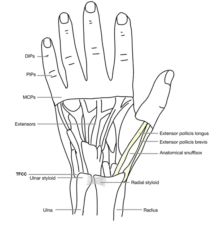
Palpate wrist:
- Ulnar head, ulnar styloid
- Triangular Fibrocartilaginous Complex (TFCC) found between ulnar head and carpal bone
- Distal radius, radial styloid
- Wrist Effusion: Palpate radiocarpal joint space using your thumbs.
- Press in with your thumbs to check for effusions (tense or boggy sensation)
- Slide thumbs across radial joint line to check for synovitis or effusion.
- Try to ballot fluid between your thumbs.
-
Palpate hand:
-
Anatomical Snuffbox:
- Floor of Snuffbox: Scaphoid bone → tenderness may indicate a scaphoid fracture (concern for Avascular Necrosis here due to precarious blood supply)
- Tendons of Snuffbox: Abductor pollicis longus and extensor pollicis brevis → can be swollen and tender in DeQuervain’s tenosynovitis.
-
Carpal Bones (Left hand shown):
- Palpate proximal carpal bones moving from hallux to D5:
- Scaphoid (Hallux) → Lunate → Triquetrum → Pisiform
- Palpate distal carpal bones moving from hallux to D5
- Trapezium (Hallux, distal row) → Trapezoid (distal) → Capitate (distal) → Hamate (distal)
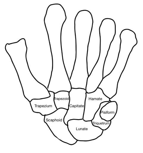
- Palpate proximal carpal bones moving from hallux to D5:
-
Flexor Tendonitis:
- Ask patient to supinate hand. Palpate flexor tendons in patient’s palm for tenderness and nodules.
- Clinician lines their fingertips along flexor tendon in palm and thumb under the patient’s hand. Apply pressure with fingertips on the flexor tendon, while passively flexing and extending each patient finger. Check for tenderness, crepitus and triggering.
-
-
Palpate fingers:
- Metacarpals:
- Screen for MCP synovitis with MCP squeeze: Grasp across MCPs with one hand and squeeze, and ask patient if this is painful
- MCP Effusion:
- Have patient flex fingers at MCPs, where joint line is 1cm distal to tip of knuckle.
- Use 4-finger technique to palpate – clinician’s middle fingers support underside of joint, tips of thumbs palpate joint line on dorsal side (roughly at 10 o’clock and 2 o’clock)
- Check for tenderness or fullness, and try to ballot fluid between your thumbs.
- Slide thumbs across the joint line to check for synovitis or effusion (indistinct on palpation)
- PIP and DIP Effusion:
- Palpate using 4 finger technique – clinician’s thumb and index of one hand on dorsal side (12 o’clock) and volar side (6 o’clock). Clinician uses thumb and index of their other hand to palpate the sides of the joint (at 9 o’clock and 3 o’clock).
- Check for tenderness or fullness and try to ballot fluid between your fingers.
- Metacarpals:
Why Check: Palpation can be very helpful to determine source of pain. Palpate for tenderness and swelling in a systematic manner, to assess for potential fractures and other abnormalities. In particular, note that tenderness on palpation of the anatomical snuffbox may suggest scaphoid fracture. These fractures commonly undetectable on initial X-ray, and thus may be missed. If initial X-ray is negative, immobilize the wrist and repeat X-ray in 14-21 days.
Range of Motion (ROM)
AFMC level Pre-clerkship 3 · Clerkship 4
-
Quick Screen: Make a fist with thumb out, then extend all fingers
-
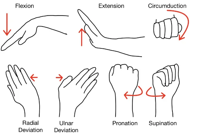
Wrist Range of Motion:
- Flexion (70-90°) and Extension (70-90°)
- Radial Deviation (20°) and Ulnar Deviation (30°)
- Supination (Palms up) and Pronation (Palms down)
- Circumduction (circular movement – combination of flexion, extension, abduction and adduction)
- Tuck Sign: During wrist or finger extension, see dorsal wrist bulge → suggests Extensor tenosynovitis.
-
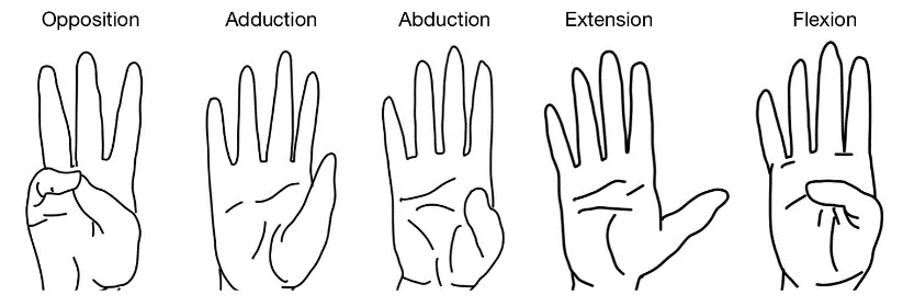
Thumb Range of Motion:
- Opposition (touching thumb to tip of each finger)
- Adduction and Abduction (perpendicular to plane of hand)
- Flexion and Extension (thumb movement in plane of hand)
-
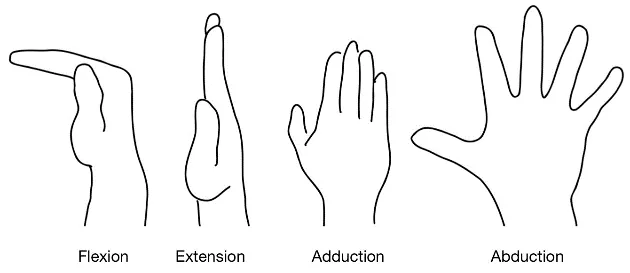
MCP Range of Motion: Extension (10-20°), Flexion (90-100°), Abduction and Adduction
-
PIPs (100-120°) and DIPs (0-80°) Flexion → These perform finger tuck.
Power Assessment
AFMC level Pre-clerkship 3 · Clerkship 4
- Approach: Resisted Isometric Testing i.e. patient resists force applied by clinician
- Grip Strength: Assess power across MCPs, PIPs and DIPs by squeezing your fingers
- Wrist Extension (Radial Nerve): Patient adopts elbow flexion with pronation; brace elbow, and apply downward force on dorsal surface of hand; patient resists motion.
- Wrist Flexion (Median Nerve): Patient adopts elbow flexion with pronation; brace elbow, apply upward force on palmar surface of hand; patient resists motion.
- Flexion at MCPs (D2 + D3 – Median Nerve; D4 + D5 – Ulnar Nerve): Patient adopts elbow flexion with pronation; brace wrist and apply a downward force on the DIPs grouped together; patient resists motion.
- Dorsal Interossei (Ulnar Nerve): Abduct fingers, patient resists motion.
- Palmar Interossei (Ulnar Nerve): Adduct fingers, patient resists motion.
- Thumb – Thumb Abduction, Flexion, Opposition (Median Nerve); Thumb Extension (Radial Nerve)
- The likelihood of suspected carpal tunnel syndrome is mildly increased by weakened thumb abduction (LR+ 1.8, LR- 0.5).
- Reporting: For each muscle group tested, assess per the Medical Research Council Power Grading. If it is all 5/5, you can say “Power was 5/5 bilaterally in both upper and lower limbs”, and report specific deficits. NOTE: This scale is detailed in Chapter 31 – Screening Neurological Examination
Neurological & Vascular Assessment
Sensory, Motor – [3, 4] Pulses– [1, 2] Cap Refill Time - [4, 4]
- Reflexes: Biceps (C5 & C6), Triceps (C7 & C8), Brachioradialis (C5 & C6)
- Sensory and Motor Assessment:
- Median nerve: Motor (“Okay” sign) and Sensory (Autonomous zone = palmar pad D2 distal phalanx)
- Radial nerve: Motor (“Thumbs Up”) and Sensory (Autonomous zone = dorsal 1st web space)
- Ulnar nerve: Motor (Finger abduction) and Sensory (Autonomous zone = palmar pad D5 distal phalanx)
- Vascular Assessment – includes Peripheral Pulses and Capillary Refill Time (see Chapter 8 – Peripheral Vascular Exam – MF1)
Special Tests:
Wrist Stability
Say: I will assess the wrist for stability.
Do: Stabilize the forearm and grasp the hand; Gently try to sublux the wrist up and down.
Report: No subluxation of wrist evident with gentle force.
Why Check: If wrist subluxes up and down, this is indicative of wrist instability
Piano Key Sign
Say: I will continue to assess the wrist for stability.
Do: Hold patient’s hand and press and release the ulnar head. Look for ulnar head to depress, then come back up (like a piano key).
Report: Piano key sign is negative.
Why Check: Positive piano key sign indicates disruption of the distal radioulnar ligament – this can occur in rheumatoid arthritis due to chronic inflammation.
Tinel’s Sign
AFMC level Pre-clerkship 3 · Clerkship 4
Say: I will examine the Carpal Tunnel in the wrist by gently tapping on an area of the wrist for 30 seconds. Please let me know if you feel any burning or pins and needles (paresthesias) during the test, and if so, where.
Do:
- Patient’s wrist in neutral position.
- Tap over the median nerve just proximal to the wrist on the palmar surface for 30 seconds.
- Assess for signs of burning or paresthesias over D1-D3 or radial half of D4 (median nerve distribution)
Report: No burning or paresthesias elicited – Tinel’s Sign is negative for Carpal Tunnel Syndrome.
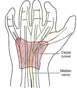
Why Check: A positive Tinel’s Sign mildly increases the likelihood of Carpal Tunnel Syndrome (LR+ 1.5, LR- 0.82). The Carpal tunnel is formed by the Flexor Retinaculum and the Carpal bones of the wrist. 9 tendons (4 for each of D2-D5, 1 for the Thumb) run in the Carpal tunnel along with the median nerve. The ulnar nerve and ulnar artery run separately along the palmar carpal ligament. Carpal Tunnel syndrome is caused by pathology ↑ pressure within the Carpal tunnel (e.g. from arthritis, effusion, fasciitis, etc.)
Phalen’s Sign
AFMC level Pre-clerkship 3 · Clerkship 4
Say: I will continue to examine the Carpal Tunnel in the wrist by bringing the hands together with palms down for 60 seconds. Please let me know if you feel any burning or pins and needles (paresthesias) during the test, and if so, where.
Do:
- Patient holds wrists in complete flexion by pushing their dorsal surfaces of hands together for 60 seconds.
- Assess for signs of burning or paresthesias over D1-D3 or radial half of D4 (median nerve distribution)
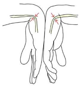
Report: No burning or paresthesias elicited – Phalen’s Sign is negative for Carpal Tunnel Syndrome.
Why Check: A positive Phalen’s Sign mildly increases the likelihood of Carpal Tunnel Syndrome (LR+ 1.3, LR- 0.74).
Finkelstein’s Test
Say: I will assess for DeQuervain’s Tenosynovitis. Please let me know if you feel any pain during the test, and if so, where.
Do:
- Patient makes a fist with their thumb in.
- Patient places their forearm on a table, with hand just off to the edge.
- Patient actively ulnar deviates the hand.
- Assess for signs of sharp pain along radial aspect of wrist (a +ve test result)
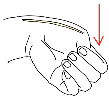
Report: No sharp pain elicited along radial aspect of the wrist – Finkelstein’s Test is negative for DeQuervain’s Tenosynovitis.
Why Check:
- A positive Finkelstein’s Test is suggestive of DeQuervain’s Tenosynovitis. This is caused by inflammation of the tendon sheaths of the abductor pollicis longus and extensor pollicis brevis (which are components of the anatomical snuffbox).
- The Finkelstein’s Test involves the patient actively deviating their hand. An alternate test, called Eichhoff’s Test, is similar with the clinician passively deviating the hand instead of active deviation by the patient.
Part 3: Select Special Tests – Not Part of Routine Shoulder Exam But Seen in Practice:
Metacarpophalangeal (MCP) Joint Stability
Say: I will assess the hand for stability.
Do:
- Ask patient to flex their knuckles (MCPs)
- Try to sublux the proximal phalanx anteriorly and posteriorly (like knee anterior and posterior drawer test)
- Look for significant movement (small amount of movement normal)
- Next, apply a varus and valgus stress to move each finger to the sides (should be little to no movement)
Report: MCP joints are stable.
Why Check: Significant movement with anterior/posterior force, any movement with valgus/varus force, indicates MCP Joint instability.
Interphalangeal (IP) Joint Stability
Say: I will continue to assess the hand for stability.
Do:
- Stabilize phalanx proximal to the joint (e.g. proximal phalanx if testing PIP)
- Grasp phalanx distal to joint (e.g. intermediate phalanx if testing PIP)
- Apply varus and valgus stresses to the joint, assess for collateral stability.
Report: IP joints are stable.
Why Check: If significant movement with varus or valgus forces, this indicates IP Joint instability.
Helpful Videos to Review:
- Video 1: Wrist/Hand Exam – McMaster MSK Examination Series (Link: HAND AND WRIST EXAMINATION – McMASTER UNIVERSITY)
- Video 2: Stethopedia Hand and Wrist Examination Video (Link: https://videopress.com/v/k5u1NYyl)
- Video 3 and 4: Bates’ Visual Guide to the Physical Exam – Anatomy Review and Examining the Wrists and Hands (Link: https://www.medportal.ca/ug/educational-resources/bates/lists/bates/musculoskeletal)
Part 4: Relevant Likelihood Ratios – from Rational Clinical Examination JAMA series
Clinical Question 1: Does This Patient have Carpal Tunnel Syndrome?2
| Procedure | LR+ (95% CI) | LR- (95% CI) |
|---|---|---|
| Classic or Probable Katz Hand Diagram | 2.4 (1.6-3.5) (about +15%) | 0.5 (0.3-0.7) (about −15%) |
| Age > 40 years old | 1.3 (1.0-1.7) (no change: 95% CI includes 1) | 0.5 (0.3-1.0) (no change: 95% CI includes 1) |
| Nocturnal Paresthesias (Pooled Results) | 1.2 (1.0-1.4) (no change: 95% CI includes 1) | 0.7 (0.5-0.9) (about −15%) |
| Bilateral Symptoms | 1.4 (1.0-2.1) (no change: 95% CI includes 1) | 0.7 (0.4-1.0) (no change: 95% CI includes 1) |
| Weak Thumb Adduction (Pooled Results) | 1.8 (1.4-2.3) (about +15%) | 0.5 (0.4-0.7) (about −15%) |
| Thenar Atrophy (Pooled Results) | 1.6 (0.9-2.8) (no change: 95% CI includes 1) | 1.0 (0.9-1.0) (no change: 95% CI includes 1) |
| Hypalgesia in median nerve territory (Pooled Results) | 3.1 (2.0-5.1) (about +15%) | 0.7 (0.5-1.1) (no change: 95% CI includes 1) |
| 2-Point Discrimination (Pooled Results) | 1.3 (0.6-2.7) (no change: 95% CI includes 1) | 1.0 (0.9-1.1) (no change: 95% CI includes 1) |
| Abnormal Vibration (Pooled Results) | 1.6 (0.8-3.0) (no change: 95% CI includes 1) | 0.8 (0.4-1.3) (no change: 95% CI includes 1) |
| Abnormal Monofilament | 1.5 (1.1-2.0) (about +15%) | 0.7 (0.5-0.9) (about −15%) |
| Square Wrist Sign (Pooled Results – Further evidence necessary) | 2.7 (2.2-3.4) (about +15%) | 0.5 (0.4-0.8) (about −15%) |
| Closed Fist Sign (Further evidence necessary) | 7.3 (1.1-49.1) (about +30%) | 0.4 (0.2-0.7) (about −30%) |
| Tinel Sign* (2009 Update) | 1.5 (1.2-2.1) (about +15%) | 0.82 (0.72-0.93) (about −15%) |
| Phalen Sign* (2009 Update) | 1.3 (1.2-1.5) (about +15%) | 0.74 (0.62-0.87) (about −15%) |
| Flick Sign* (2009 Update) | 1.4 (0.80-2.4) (no change: 95% CI includes 1) | 0.85 (0.68-1.1) (no change: 95% CI includes 1) |
| Pressure Provocation Test (Pooled Results) | 1.0 (0.8-1.3) (no change: 95% CI includes 1) | 1.0 (0.9-1.1) (no change: 95% CI includes 1) |
| Tourniquet Test (Pooled Results) | 1.0 (0.5-1.9) (no change: 95% CI includes 1) | 1.0 (0.7-1.5) (no change: 95% CI includes 1) |
*Note: Date presented for Tinel Sign, Phalen Sign and Flick Sign from 2009 Update in JAMA Rational Clinical Examination Series. Square Wrist Sign, Closed-Fist Sign and Flick Sign are described as less commonly used maneuvers requiring additional data.
Note on Katz Hand Diagram2:
- Procedure: Patient identifies on a diagram of a hand (or can trace own hand) and draws where symptoms are located on the diagram.
- Classic pattern: Affects ≥2 digits of D1-D3, without symptoms on palm or dorsum of hand. Allowed to have symptoms in D4-D5, so long as they are distal to metacarpals, as well as wrist pain and radiation of pain proximal to the wrist.
- Probable pattern: Affects ≥2 digits of D1-D3. Can involve palmar symptoms unless confined solely to ulnar aspect of hand around D4-D5.
- Possible pattern: Affects 1 digit of D1-D3. Can involve palmar symptoms unless confined solely to the ulnar aspect of hand around D4-D5.
- Unlikely pattern: No symptoms present in D1-D3.
References
-
Recommendations. Choosing Wisely Canada. Accessed July 22, 2022. https://choosingwiselycanada.org/recommendations/#list ↩ ↩2
-
D’Arcy CA, McGee S. Does this patient have carpal tunnel syndrome? JAMA. 2000;283(23):3110-3117. ↩ ↩2