Medical Foundations 4
Chapter 29Back Examination
Part 1: Focused History – Based on Chief Complaint (Begin with Open-Ended Questions)
HPI
Pain Assessment
- Onset:
- Acute onset of pain, disability immediate with injury (e.g. acute trauma, concern for fracture) (see Mechanism of Injury below)
- Neck Pain with trauma/accident/fall → Consider Canadian C-Spine Rule or NEXUS Rule (Pediatrics)
- Delayed onset of pain (e.g. ligamentous injury)
- Acute onset with no associated injury (e.g. septic arthritis)
- Insidious onset gradually worsening over time (e.g. overuse injury, osteoarthritis)
- Pediatric patients: Is the patient in an active growth spurt → increased risk of overuse injuries
- Acute onset of pain, disability immediate with injury (e.g. acute trauma, concern for fracture) (see Mechanism of Injury below)
- Provoking:
- Aggravating postures:
- C-Spine: Flexion, Extension, Rotation (unilateral vs. bilateral), Lateral flexion (unilateral vs. bilateral)
- Thoracic Spine: Primarily rotation
- Lumbar Spine: Flexion, extension, lateral flexion, rotation
- Aggravating activities: Standing; walking; lifting (re. spinal loading); throwing (e.g. cocking phase in throwing a ball); heading (e.g. in soccer); serving (e.g. in tennis); coughing/sneezing
- Aggravating postures:
- Palliating:
- Relieving postures/activities: Sleep; sitting; lying down; Flexion, Extension, Rotation, Lateral Flexion
- Previous treatment (including for previous episodes of pain e.g. in recurrent low back pain):
- Rehabilitation: Posture changes to activity; manual therapy; exercise (e.g. resistance, ROM)
- Scoliosis Treatments: e.g. Supportive back brace
- Investigations: Type (x-rays, Ultrasound, MRI, etc.) and results (if available)
- Quality: Deep ache, sharp vs. dull? – Ask patient what words they would use
- Neuropathic Pain Descriptors: Pins/needles; hot/burning/stinging; numb; electrical shocks; sensitivity to touch (e.g. clothing, bed sheets)
- Region: Central (along spine) vs. Unilateral vs. Bilateral
- C-Spine Pain: Where is pain most intense – Neck Dominant (e.g. sub-occipital, trapezius, parascapular, shoulder paraspinal) vs. Arm Dominant (shoulder, deltoid, upper arm, forearm, hand)
- C0-4 neck pain → Upper cervical or head pain
- C4-5 neck pain → Mid-lower neck pain
- C5-7 neck pain → Lower neck, upper thoracic, or shoulder/arm pain
- C-Spine Pain: Where is pain most intense – Neck Dominant (e.g. sub-occipital, trapezius, parascapular, shoulder paraspinal) vs. Arm Dominant (shoulder, deltoid, upper arm, forearm, hand)
- Radiation:
- Assess distribution e.g. unilateral vs. bilateral; dermatomal pattern (e.g. radiculopathic pain – See Chapter 31 – Screening Neuro Exam)
- Sciatica – radiation of pain from back down the leg in dermatomal pattern (commonly along L4-S1 dermatomes) has a Sensitivity of 95% and a Specificity of 88% for herniated disk.
- Assess distribution e.g. unilateral vs. bilateral; dermatomal pattern (e.g. radiculopathic pain – See Chapter 31 – Screening Neuro Exam)
- Severity: Immediately after injury, vs. subsequently. Has it improved over time with rest?
- Was patient able to continue their activity, e.g. after the sport completed, noticed pain got worse?
- Timing: Pain at rest (e.g. waking patient up at night); Constant vs. Intermittent
- Constant Pain → particularly important to rule out red flags (see below)
- Recurrence: Assess episodes and pattern of recurrence of pain (e.g. recurrent neck pain)
- Associated Symptoms:
- Sensory neuropathy e.g. paresthesias or numbness – assess distribution e.g. Unilateral vs. bilateral; Peripheral nerve, Glove and Stocking, Dermatomal or Spinal level (See Ch 31 – Screening Neuro Exam)
- Weakness: Assess distribution e.g. Unilateral vs. bilateral; Proximal vs. distal muscle weakness; Specific peripheral nerve vs. spinal level vs. brachial/lumbosacral plexopathy (See Chapter 31 – Screening Neuro Exam)
- Sleep Disturbance: Indicator of pain severity e.g. in neck pain
Red Flags (NIFTI mnemonic – concern for serious pathologies)1
- Neurological: Diffuse motor/sensory loss, progressive neurologic deficit, cauda-equina syndrome
- Cauda Equina Syndrome (surgical emergency): Have you had any unexpected accidents with your bowel or bladder function since this episode of your back pain started?
- Urinary retention followed by insensible urinary overflow
- Unrecognized fecal incontinence
- Distinct loss of saddle/perineal sensation
- Cauda Equina Syndrome (surgical emergency): Have you had any unexpected accidents with your bowel or bladder function since this episode of your back pain started?
- Infection: Fever, IV drug use (re: spinal epidural abscess or vertebral osteomyelitis), Immunosuppression
- Fracture: Trauma, Osteoporosis risk/fragility fracture
- Tumor: History of cancer; unexplained weight loss; significant unexplained night pain; severe fatigue
- In a patient with low back pain, Age >50 y or history of cancer or unexplained weight loss or failure of conservative therapy has a Sensitivity of 100% and Specificity of 60% for cancer.
- Inflammatory: Chronic back pain > 3 months; Onset < 45 years old; Morning stiffness > 30 minutes; Improving with exercise; Disproportionate night pain (e.g. with nocturnal awakening)
- Re: Ankylosing spondylitis – 4 of 5 Positive responses: Onset of back discomfort <40 years old or Problem began slowly or Pain duration 3+ months or Morning stiffness or Improved by exercise → has a Sensitivity of 23% and a Specificity of 82% for Ankylosing Spondylitis.
Yellow Flags (Psychological risk factors for developing chronicity)1
- Is there anything you cannot do now that you could do before the onset of your pain? → If yes, ask:
- Do you think your pain will improve or become worse? – listen for catastrophizing
- Do you think you would benefit from activity, movement or exercise? – listen for fear/avoidance
- How are you emotionally coping with your neck pain? – listen for social withdrawal, and low mood
- What treatments or activities do you think will help you recover? – listen for unrealistic expectations of treatments, expecting passive treatments rather than active participation will help
Mechanism of Injury
- If injury occurred during an activity, ask open-ended questions about mechanism of injury e.g. “run me through what happened, play-by-play”. Try to visualize what happened. Assess low energy vs. high energy force injury and the forces involved (e.g. direct vs. indirect forces and their direction)
- If assessing an athlete, helpful to know/learn about their sport to understand common injuries and the demands of the position they play in the sport.
- Trauma – motor vehicle accidents, falls, etc.
- Compression fractures may occur due to axial load injuries.
- C-spine injuries can occur in whiplash (cervical strain injury).
Canadian C-Spine Rules
- Population: Alert (GCS = 15) and stable trauma patients where Cervical spine injury is a concern
- Not Applicable if: Non-trauma case; Glasgow Coma Scale GCS <15; Unstable Vital Signs; Age <16 years Old; Acute Paralysis; Known Vertebral Disease; Previous C-Spine Surgery; Pregnant
- The Canadian C-Spine Rules have a Sensitivity of 90-100% and a Specificity of 1-77% for identifying clinically important cervical spine injury (i.e. any fracture, dislocation or ligamentous instability detectable by diagnostic imaging and requiring surgical or specialist follow-up).
- Part 1 – PRESENCE OF ANY HIGH-RISK FACTORS (YES → Image; NO → Proceed to Part 2)
- Age ≥ 65 years old
- Dangerous Mechanism: Fall from height ≥ 3 feet or 5 stairs; Axial Load to Head (e.g. Diving); Motor Vehicle Collision: High speed (>100km/hr), Rollover, Ejection; Motorized Recreational Vehicles; Bicycle Struck or Collision
- Paresthesias in Extremities
- Part 2 – ABSENCE OF ANY LOW RISK FACTORS ALLOWING SAFE RANGE OF MOTION ASSESSMENT (NO → Image; YES → Proceed to Part 3)
- Simple Rear-End Motor Vehicle Collision (excludes: Pushed into oncoming traffic; hit by bus or large truck; Rollover; or Hit by High-speed vehicle >100km/hr)
- Sitting position in Emergency Department
- Ambulatory at any time
- Delayed onset (not immediate) onset of neck pain
- Absence of midline C-spine tenderness
- Part 3 – ABLE TO ACTIVELY ROTATE NECK 45° LEFT AND RIGHT (ABLE → No Imaging; UNABLE → Image)
- Calculator2: https://www.mdcalc.com/canadian-c-spine-rule#next-steps
NEXUS C-Spine Criteria
- Population: Alert, stable trauma patients – can use in Pediatrics (vs. Canadian C-spine rules not applicable for patients < 16 years old)
- The NEXUS C-Spine Criteria have a Sensitivity of 83-100% and a Specificity of 2-46% for identifying clinically important cervical spine injury (i.e. any fracture, dislocation or ligamentous instability detectable by diagnostic imaging and requiring surgical or specialist follow-up).
- Imaging Not Required If All 5 Criteria Absent:
- Focal Neurological Deficit
- Midline Spinal Tenderness
- Altered Level of Consciousness
- Intoxication
- Distracting Injury
- Calculator3: https://www.mdcalc.com/nexus-criteria-c-spine-imaging#next-steps
Neck Pain (designed for movement)
- Common Illness Presentations:
- Cervicogenic Headache (results from physiological convergence of cervical and trigeminal afferents in trigeminocervical nucleus): Typically unilateral, side-consistent headache associated with neck pain, where pain begins in neck and later develops into a headache. May be associated with cervical movement dysfunction, muscle function impairments, and upper cervical joint dysfunction.
- Brachial Plexus Injury: Acute neck pain with upper extremity neuropathic pain (e.g., burning/stinging) that may be accompanied by paresthesias and/or weakness of upper extremity. Can occur with traction injury (e.g., downward shoulder displacement with contralateral side flexion of neck), cervical extension + ipsilateral side flexion, or direct blow to brachial plexus.
- Whiplash (Cervical Strain): Acute neck pain and/or stiffness following acceleration/deceleration injury (e.g., in Motor Vehicle Collision) causing sudden neck extension and flexion. Look for ↓ range of motion and neuromotor dysfunction (e.g., weakness, numbness)
- Can’t Miss Diagnosis Illness Presentations:
- Concussion: A form of mild Traumatic Brain Injury that affects brain function. For more information on assessment of Concussion and Traumatic Brain Injury, see Chapter 31 – Screening Neuro Exam.
- Helpful Resource: Peds Concussion Living Guideline for Pediatric Concussion Care
- Helpful Resource: Adult Concussion Living Guideline for Concussion and Prolonged Symptoms
- Cervical Myelopathy (Compression of cervical spinal cord): Presenting with gait disturbance, incoordination (e.g., clumsiness, imbalance), and loss of neuromotor function (e.g., doing up small buttons, new onset tingling/numbness in arms/hands).
- Cervical Arterial Dissection: Acute neck pain worsening over hours-to-days, with neurological or ischemic signs. Can occur in setting of recent infection or with minor trauma, or intense sneezing/coughing. Can’t miss diagnosis as an important cause of stroke e.g., in young patients.
- Cranio-Cervical Ligament Injury: Suspect in a patient with head and neck trauma (e.g., motor vehicle collision), presenting with acute neck pain with perioral paresthesia, tinnitus, “lump in throat” or nausea. Can’t miss diagnosis as it can lead to translational instability of C1-C2 vertebrae.
- Concussion: A form of mild Traumatic Brain Injury that affects brain function. For more information on assessment of Concussion and Traumatic Brain Injury, see Chapter 31 – Screening Neuro Exam.
Back Pain
- Pattern 1 Pain – Disc Pain (Acute Disc Prolapse, L5-S1 disc most common): Constant back/buttock dominant pain usually after forward bend with repeated flexion loading. Pain often worse with sitting, forward bend, lifting, cough/sneeze, and improved with lying down. May be worse with extension too.
- Pattern 2 Pain – Facet Joint Pain:
- Spondylolysis (Stress fracture of pars interarticularis): Intermittent, often gradual onset back/buttock dominant pain, worse with spinal side flexion, rotation and hyperextension. Pain may radiate to buttock and posterior thigh.
- Spondylolisthesis (Anterior displacement of vertebra– most common L5 vertebrae slips forward on S1 vertebrae): Intermittent, often gradual onset back/buttock dominant pain, worse with extension activities and postures. Concern (red flag) if occurs with progressively worsening neurologic signs.
- Pattern 3 Pain– Compressed Nerve Pain (e.g. Acute Disc Prolapse with Nerve Root Compression): Constant leg dominant pain usually after forward bending movement with repeated flexion loading, with radicular pain on affected side. Pain often worse with all movements and improved with lying down particularly on asymptomatic side.
- Pattern 4 Pain – Symptomatic Spinal Stenosis (Neurogenic Claudication): Intermittent leg-dominant diffuse pain, aggravated by standing and walking, improved with sitting, lying and lumbar flexion.
- Can’t Miss Diagnosis Illness Presentations (assessed for by Red Flags, above) – NIFTI Acronym:
- Neurological: e.g. Spinal Cord Compression, Cauda-Equina Syndrome
- Infection: e.g. Spinal Epidural Abscess; Vertebral Osteomyelitis; Discitis
- Fracture: e.g. Vertebral Compression Fracture
- Tumor: e.g. Metastatic Cancer (e.g. breast, prostate, lung, thyroid, kidney); Multiple Myeloma
- Inflammatory: e.g. Ankylosing Spondylitis
Past Medical History (PMHx)
- Previous back or neck injuries
- Osteoporosis Risk Factors: History of Fragility # (e.g. fall from standing height or less); Vertebral Compression #; Inflammatory Arthritis (e.g. Rheumatoid Arthritis); Malabsorption Syndromes (e.g. Celiac disease, Inflammatory bowel disease); Endocrine Disorders (e.g. Hyperparathyroidism, Hyperthyroidism, Cushing’s, Hypogonadism).
- Risk Factors for Septic Arthritis: Immunosuppression (including diabetes mellitus and HIV); Pre-existing joint disease (e.g. Rheumatoid arthritis: LR+ 2.5, LR- 0.45); Advanced Age (>80 years old – LR+ 3.5); Skin infection (LR+ 2.8, LR- 0.76)
- Lab Markers for Septic Arthritis:
- Peripheral – CRP >100 mg/L (LR+ 1.6, LR- 0.44), WBC >10 (LR+ 1.4), ESR >30mm/h (LR+ 1.3)
- Synovial fluid – WBC >100 (LR+ 28, LR- 0.71), WBC >50 (LR+ 7.7, LR- 0.42), WBC >25 (LR+ 2.9, LR- 0.32), PMN cells ≥90% (LR+ 3.4, LR- 0.34)
- Lab Markers for Septic Arthritis:
- Bleeding Disorder History: ↑ risk of serious pathology on injury.
- Risk Factors for Osteoarthritis: Age (>50 years); Female Gender; Obesity; Heavy manual labour; History of joint injury
- Psoriasis: Risk factor for Psoriatic Arthritis.
- Autoimmune disorders (↑ risk for Inflammatory Arthritis) – particularly Spondyloarthropathies e.g. Psoriatic arthritis, Uveitis, Ankylosing Spondylitis, Inflammatory Bowel Disease, Reactive Arthritis.
Relevant Clinical Scores:
- Canadian C-Spine Rule – Rules out need for C-spine imaging (more sensitive/specific than NEXUS; applicable to patients ≥ 16 years old)
- NEXUS C-Spine Criteria – Rules out need for C-spine imaging (less sensitive/specific than Canadian C-Spine Rule; applicable to pediatric and adult populations)
- Spinal instability neoplastic score (SINS) – Helps to assess tumor-related instability of the vertebral column, helping to guide management
- PEG Scale – Chronic pain assessment in primary care patients to monitor treatment response over time
Past Surgical History (PS Hx)
- Scoliosis Treatments: Posterior Spinal Fusion, Anterior Spinal Fusion, Instrumentation, Bone Grafting
- Osteoporotic Vertebral Compression Fracture Treatments: Vertebroplasty (bone cement injected in fracture site); Kyphoplasty (balloon inflation in fracture site followed by bone cement injection)
- Spinal Cord Compression Treatments: Surgical decompression (e.g. Laminectomy); Palliative Radiation (e.g. Stereotactic Body Radiation Therapy for Malignant Spinal Cord Compression), Vertebroplasty or Kyphoplasty (as above)
- Other Spinal Surgery: Spinal Fusion (may be seen e.g. in Ankylosing Spondylitis), Artificial Disc Replacement
- Risk Factors for Septic Arthritis: Recent joint surgery (LR+ 6.9, LR- 0.78)
Family Medical History (FMHx)
- Parental History of Hip Fracture → ↑ risk for Osteoporosis
- Autoimmune disorders (↑ risk for Inflammatory Arthritis) – particularly Spondyloarthropathies e.g. Psoriasis, Uveitis, Ankylosing Spondylitis, Inflammatory Bowel Disease, Reactive Arthritis
Social Hx
Substance Use
- Smoking: Risk factor for Osteoporosis, Avascular Necrosis
- Alcohol: Risk factor for Gout; and for Osteoporosis (>3 units/day), Avascular Necrosis
- IV Drug Use: Risk factor for Septic Arthritis
Occupational Hx
- Are you working right now? What do you do for work? What other kinds of work have you done in the past? Do you have benefits through work such as drug coverage (e.g. access to Physiotherapy coverage)?
- If injury occurred at work → separate Workplace Safety and Insurance Board (WSIB) process.
Sports History
- History of sports played and select ↑ risk:
- Thoracolumbar Junction (T12-L1) Injury ↑ Risk → Overloading injury (e.g. skiing, horseback riding)
- Sternoclavicular Joint Injury ↑ Risk: Direct/indirect blow to shoulder (e.g. gymnastics)
- Spondylolysis (Stress fracture of pars interarticularis) and Spondylolisthesis (Anterior displacement of vertebra) ↑ Risk: Repeated spinal loading and bending (e.g. tennis, weightlifting, throwing sports, dance, gymnastics, diving); for pediatrics, increased risk during adolescent growth phase.
- Disc Prolapse ↑ risk: Repeat spinal loading with flexion + rotation (e.g. cycling, field hockey, rowing)
- Current level of sport played (e.g. recreational, competitive, league level) and desired future sporting activity level; Sports position played (some positions may place unique stresses on back)
- Current training history (e.g. for patients with overuse injuries) – quantity and quality of training; any recent changes to routines (e.g. training days per week, intensity, duration of training sessions)?
- Recent change of equipment (e.g. footwear for running – note shoe type, age, wear pattern)
- Overtraining Syndrome–excess fatigue, recurrent illness, ↓ motivation, persistent soreness/stiffness
Dietary Hx
- Particularly dietary calcium (1200mg/day) and Vitamin D (e.g. 1000-2000 IU/day; 400/day for infants)
ADLs and IADLs
- Common activities impacted by back pain: Running; walking; jumping; looking around (e.g. neck pain); lifting; tying up shoes (e.g. bending); going up and down stairs (as screened for in the GALS exam)
- Use of Mobility Aids: e.g. cane, walker, crutches – any recent changes?
Medications Hx, Allergies
Medications with ↑ Osteoporosis Risk
- Glucocorticoid Use (e.g. 3+ months in past year, prednisone >7.5mg/day or equivalent)
- Aromatase Inhibitors (e.g. anastrozole, letrozole, exemestane)
- Androgen deprivation therapy (e.g. Leuprolide in prostate cancer treatment)
- Long-term use of Medroxyprogesterone Acetate injection (e.g. for contraception)
- Anti-Epileptic Drugs (e.g. Carbamazepine, Phenytoin – impact Vitamin D metabolism)
- Psychiatric Medication (e.g. SSRIs, Typical Antipsychotics e.g. Chlorpromazine, Haldol)
- Proton Pump Inhibitors (PPIs, e.g. Pantoprazole, Lansoprazole – re. ↓ Ca absorption)
- Loop Diuretic (e.g. Furosemide re. ↓ Ca reabsorption in Loop of Henle – conflicting data)
Inflammatory Arthritis
- Inflammatory Arthritis (e.g. Rheumatoid Arthritis, Spondyloarthropathies):
- Disease-Modifying Antirheumatic Drugs (DMARDs): Methotrexate, Sulfasalazine, Hydroxychloroquine, Leflunomide, Azathioprine
- Biologic DMARDs:
- TNF-α inhibitors: Etanercept, adalimumab, infliximab, golimumab, etc.
- Others: Abatacept, rituximab, tocilizumab, sarilumab, anakinra
- Acute Flare: Glucocorticoids (e.g. Prednisone)
Osteoporosis Treatments
- Calcium Supplementation (Goal 1200mg/day from all sources including dietary)
- Vitamin D Supplementation (e.g. 1000-2000 IU PO Daily; 400 IU PO Daily for infants)
- Bisphosphonates (e.g. Alendronate, Risedronate, Zoledronic Acid)
- Denosumab (RANK Ligand monoclonal antibody)
- Selective Estrogen Receptor Modulator (SERMS – e.g. Raloxifene)
- Hormone Therapy (e.g. Estrogen ± Progesterone)
- Teriparatide (Endogenous Parathyroid Hormone, PTH)
- Calcitonin (↓ pain of acute vertebral fractures)
Analgesics
- PO: NSAIDs, Acetaminophen, Muscle Relaxant (e.g. Baclofen), Opioids – ask pain severity before versus after med, frequency and dosing.
- Topical: NSAIDs (E.g. Diclofenac), Lidocaine, etc.
- Joint Injections: Intra-articular corticosteroid injection; Diagnostic injection (e.g. lidocaine) – ask about pain relief immediately after (effect of lidocaine) vs. sustained pain response (effect of corticosteroid) – Back injections (e.g. facet joint, epidural injection) under image guidance (e.g. CT-guided fluoroscopy); Trigger Point Injections with surface landmarking.
Part 2: Physical Examination of the Back:
General Assessment:
Gait Assessment
Gen - [3, 4] Trendelenburg [2, 4]
- Components of Gait Cycle:
- Stance phase (60% of gait cycle): initial contact (heel strike) → mid-stance → pre-swing (toe-off)
- Swing phase (40% of gait cycle): initial swing → mid-swing → terminal swing
- Note: Former terminology in brackets re. heel-strike and toe-off.
- Quality of Gait: Smoothness of movement (legs, arm swing, pelvic tilt), symmetry, normal stride length, ability to turn quickly, symmetry.
- Abnormal Gaits to Assess for: Antalgic Gait (shortened stance phase on affected side, typically indicates pain with weight-bearing); and Trendelenburg Gait (weak hip abductors); bilateral weak hip abductors produce a Waddling Gait).
- Transition from Sitting to Standing: Ask patient to rise from a chair; helpful to identify source of pain
Inspection (SEADS)
AFMC level Pre-clerkship 3 · Clerkship 4
- Approach: Ensure adequate but respectful exposure during your examination
- Swelling
- Erythema
- Atrophy: Sternocleidomastoid, Shoulder Girdle, Chest Wall, Intercostals, Paraspinals, Gluteal, Calf Muscles – likelihood of lumbosacral radiculopathy in patients with sciatica is increased with ipsilateral calf muscle wasting (LR+ 5.2, LR- 0.8)
- Deformities:
- Normal Curvatures of the Spine:
- Cervical Lordosis
- Thoracic Kyphosis – ↑ in Ankylosing Spondylitis (AS) and in vertebral fractures (e.g. osteoporosis)
- Lumbar Lordosis – ↑ in flexion contracture of hip; Flattened e.g. in Ankylosing Spondylitis (AS)
- Sacrococcygeal Kyphosis
- Scoliosis: Look for asymmetric shoulder height, abnormal curvature in frontal plane
- Adam’s Forward Bend Test (see Special Tests below)
- Normal Curvatures of the Spine:
- Scars or Skin Changes: Inspect for sinus or hair tufts → Suggests concern for Spina Bifida
Palpation
AFMC level Pre-clerkship 3 · Clerkship 4
- Approach: Palpate bones and soft-tissue structures for tenderness – helpful to determine source of pain. Palpate systematically e.g. begin at Occiput, palpate inferiorly and stabilize the patient.
- Canadian C-Spine Rule: Palpate for midline C-Spine tenderness.
- NEXUS C-Spine Criteria: Palpate for midline spinal tenderness.
- Commenting on: TESTCA – Tenderness, Effusions, Swelling, Temperature, Crepitus, & Atrophy
- Palpate:
- Muscles – can be tender with Myofascial Pain
- Spinous processes and Interspinous ligaments – can be tender with enthesitis
- Bony Landmarks:
- C-Spine: C7: Bony prominence at Base of Neck
- T-Spine: T3: Spine of scapula; T7: Angle of scapula
- L3-L4: Iliac Crests; Palpate space from ribs to iliac crests (Normal 4 finger breadths, ↓ in kyphosis)
- S2: Posterior Superior Iliac Spine (PSIS) – see Ch 27 – Hip Exam
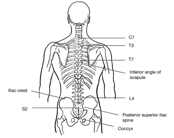
Why Check: Palpation can be very helpful to determine source of pain. Palpate for tenderness and swelling in a systematic manner, to assess for potential fractures and other abnormalities. Particularly, when palpating the spine, observe for midline point-tenderness.
Range of Motion (ROM)
AFMC level Pre-clerkship 3 · Clerkship 4
- Approach: Usually performed as active ROM (patient does it). Clinician may assist for Thoracolumbar rotation by holding both shoulders and rotating beyond patient active ROM.
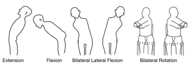
- Cervical Spine (Among regions of spine, the Cervical spine has greatest range and variety of movement):
- Flexion (Look Down): Normal 80-90°; Chin should touch chest
- Extension (Look Up): Normal 70°; Chin should pass the level of the ears
- Lateral Flexion: Normal 20-45°; Ear should touch the shoulder (be sure patient isn’t elevating shoulders to do so)
- Rotation (Look Side to Side): Normal is up to 90° to each side –likelihood of cervical radiculopathy in patients with arm/neck pain is increased with <60° rotation of neck to involved side (LR+ 1.7, LR- 0.2).

- Thoracolumbar Spine (Thoracic Spine primarily Rotatory movements, restricting flexion, extension, and lateral flexion; Lumbar Spine primarily Flexion, Extension, and Lateral Flexion, with restricted rotation):
- Chest Expansion: See Bilateral Chest Expansion below
- Flexion: Ask patient to stand and keep their knees straight and end forward to touch the floor by sliding their hands down the anterior surfaces of each leg. Normally expect to see lumbar lordosis transition to kyphosis with Thoracolumbar flexion.
- Measurement – Modified Schober’s Test: See Modified Schober’s Test below
- Extension: Ask patient to stand and keep legs straight with lower body supported against a stable structure behind (e.g. exam table) and lean backwards without bending knees.
- In a Facet Arthropathy, Extension becomes limited
- Lateral Flexion: Ask patient to stand with back against the wall and with their hands at their sides with fingers straight. Ask patient to bend to either side, sliding their fingers down the side of their leg.
- Rotation: Ask patient to sit (so pelvis is anchored) and give themselves a hug (arms crossed across chest). Ask patient to twist to either side. Clinician may apply over-pressure to fully assess range, while observing for pain.
Power Assessment
AFMC level Pre-clerkship 3 · Clerkship 4
- Approach: The back-examination’s power assessment is considered to be part of the neurological examination. When examining the back, you’ll be asked to examine the power of the major flexors and extensors of the lower limb –Hip, Knee, and Ankle/Foot, NOT the posterior muscles of the back. This is to discern whether there is any spinal cord compression, etc. going on along with a chief complaint such as back pain which would trigger you to perform the back MSK examination.
Why Check: In the upper limbs, likelihood of suspected cervical radiculopathy in patients with neck/arm pain is increased by weakness of any arm muscle (LR+ 1.9, LR- 0.4). Specifically, weakness of elbow flexion increases the likelihood of C5 radiculopathy (LR+ 5.3); weakness of wrist extension increases the likelihood of C6 radiculopathy (LR+ 2.3); weakness of elbow extension increases the likelihood of C7 radiculopathy (LR+ 4.0, LR- 0.4); and weakness of finger flexion increases the likelihood of C8 radiculopathy (LR+ 3.8).
In the lower limbs, the likelihood of suspected lumbosacral radiculopathy in patients with sciatica is increased by weak ankle dorsiflexion (LR+ 4.9, LR- 0.5). Specifically, weakness of knee extension increases the likelihood of L3-L4 radiculopathy (LR+ 3.7, LR- 0.7); weakness of hallux extension increases the likelihood of L5 radiculopathy (LR+ 1.6); and normal strength of ankle plantarflexion decreases the likelihood of S1 radiculopathy (LR- 0.7).
Neurological & Vascular Assessment
Tone, Power, Deep Tendon Reflexes – [3, 4] Peripheral Pulses – [1, 2] Capillary Refill Time - [4, 4]
- How Do I Perform This Assessment? See Screening Neurological Examination for full details in Chapter 31. Brief overview is included here for reference.
- Inspection: Inspection begins in waiting room or as patient enters the exam room. Observe their gait and posture, abnormal limb position, weakness, muscle atrophy, fasciculations, involuntary movements, and lack of coordination.
- Assess Tone:
- With patient relaxed and supine, support and passively move the limb across joints (Hip, Knee, Ankle)
- Assess for hypotonia (↓ tone in lower motor neuron lesions e.g. nerve root, peripheral nerve)
- Assess for hypertonia (↑ tone in upper motor neuron lesions)
- Spasticity: Variable resistance, often worse at extremes of ROM e.g. in Cerebral Palsy or Stroke
- Cogwheeling Rigidity: Intermittent ↑ resistance and tremor e.g. in Parkinson’s Disease
- Lead-Pipe Rigidity: (↑ resistance throughout range of motion e.g. in Parkinson’s Disease, or Neuroleptic Malignant Syndrome (NMS)
- Assess for ankle clonus (upper motor neuron lesion)
- Assess Power: Assess with resisted isometric force. Report with MRC Power Grading Scale (out of 5)
- Hip Flexion (L1-L3)
- Hip Extension (S1-S2)
- Knee Extension (L2-L4)
- Knee Flexion (L5-S2)
- Ankle Dorsiflexion (L4-L5)
- Ankle Plantarflexion (S1-S2)
- Great Toe Dorsiflexion (L5-S1)
| MRC | Description |
|---|---|
| 5 | Movement of body part against gravity with full resistance |
| 4 | Movement of body part against gravity with some resistance • 4+ and 4-: Graduations of 4/5 strength re how much examiner had to lower resistance |
| 3 | Movement of body part against gravity only |
| 2 | Movement of body part with gravity eliminated |
| 1 | Muscle contraction, but with no joint movement |
| 0 | No muscle contraction |
- Assess Reflexes (Diminished/Absent → suggests Lower Motor Neuron Lesion; Hyperactive/Sustained → suggests Upper Motor Neuron Lesion)
- Patellar Reflex (L2-L4 – Mainly L4)
- Achilles Reflex (S1-S2 – Mainly S1)
- Plantar Response (If plantars upgoing, this indicates an Upper Motor Neuron lesion).
| Reflex Grade | Description |
|---|---|
| 0 | Absent |
| 1+ | Diminished (reinforcement needed to elicit e.g. patient clenches teeth, locks fingers) |
| 2+ | Normal |
| 3+ | Hyperactive without clonus |
| 4+ | Hyperactive with clonus |
- Assess Dermatomes: Assess with both Posterior Columns (Cotton Ball) and Spinothalamic Tract (Sharp/Dull pin testing differentiation). Map out area of altered sensation to determine spinal level. The following are particular sites to examine for a back exam (i.e. testing Lumbar and Sacral spinal levels:
- Groin (L1); Just Inferior to Groin (L2)
- Medial Aspect of Knee (L3); Lateral Aspect of Knee (L4)
- Dorsum of Foot (L5); Lateral Border of Foot (S1)
- Vascular Assessment – includes Peripheral Pulses and Capillary Refill Time (see Chapter 8 – Peripheral Vascular Exam – MF1)
Why Check: In the upper limbs, the likelihood of suspected cervical radiculopathy in patients with neck/arm pain is increased by weakness of any arm muscle (LR+ 1.9, LR- 0.4), reduced biceps reflex (LR+ 9.1), or any reduced arm reflex (biceps, triceps or brachioradialis reflexes – LR+ 3.6, LR- 0.8). Specifically, the likelihood of a C5 radiculopathy is increased with weakness of elbow flexion (LR+ 5.3). The likelihood of a C6 radiculopathy is increased with weakness of wrist extension (LR+ 2.3), reduced biceps or brachioradialis reflex (LR+ 14.2, LR- 0.5), and sensory loss of the thumb (LR+ 8.5). The likelihood of a C7 radiculopathy is increased with weakness of elbow extension (LR+ 4.0, LR- 0.4), and diminished triceps reflex (LR+ 3.0). The likelihood of a C8 radiculopathy is increased with weakness of finger flexion (LR+ 3.8) and sensory loss of D5 (LR+ 41.4).
In the lower limbs, the likelihood of suspected lumbosacral radiculopathy in patients with sciatica is increased by weak ankle dorsiflexion (LR+ 4.9, LR- 0.5), abnormal ankle jerk reflex (LR+ 2.7) and ipsilateral calf muscle wasting (LR+ 5.2, LR- 0.8). Specifically, the likelihood of an L3-L4 radiculopathy is increased with weakness of knee extension (LR+ 3.7, LR- 0.7) and asymmetric quadriceps reflexes (LR+ 8.7, LR- 0.6). The likelihood of an L5 radiculopathy is increased with weakness of hallux extension (LR+ 1.6) and L5 distribution sensory loss (LR+ 3.1, LR- 0.8). The likelihood of an S1 radiculopathy is increased with ipsilateral calf muscle wasting (LR+ 2.4, LR- 0.7), asymmetric Achilles reflexes (LR+ 2.9, LR- 0.4) and S1 distribution sensory loss (LR+ 2.4, LR- 0.7), and decreased by normal strength of ankle plantarflexion (LR- 0.7).
Special Tests:
Adam’s Forward Bend Test
Peds - [1, 2]
Say: I will inspect for scoliosis. Please stand with feet together and knees straight. I will stand behind you and watch the back’s movements with bending.
Do:
- Patient stands with knees in full extension. Clinician stands behind patient.
- Patient bends forward keeping knees in full extension.
- Clinician observes for asymmetry with a prominent rib hump.
Report: I note no asymmetry on forward bending – Adam’s Forward Bend Test is negative.
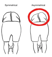
Why Check: Adam’s Forward Bend Test’s diagnostic value is to rule out thoracic spine curvature re Scoliosis. Note that a diagnosis of scoliosis is defined objectively by a Cobb Angle >10% on Anteroposterior (AP) Xray. A positive Adam’s Forward Bend Test has a Sensitivity of 92% and a Specificity of 60% for scoliosis.
Occiput-Wall Distance
AFMC level Pre-clerkship 2 · Clerkship 4
Say: I will assess for kyphosis in the neck using a measuring tape. Please stand with the back to the wall.
Do:
- Patient stands with back to wall, nose and ear at same level (controls for neck hyperextension)
- Observe distance from occiput to wall; in this position, distance should be 0 cm (Normal: 0-4cm)
Report: Occiput-Wall distance is less than 4.0 cm, with no abnormal cervical or thoracic kyphosis.
Why Check: Occiput-Wall distance > 4.0 cm’s diagnostic value is to rule in thoracic vertebral fracture. It is increased in abnormal cervical or thoracic kyphosis, for example in Ankylosing Spondylitis (tends to begin in the neck in females, vs the lower spine in males) or Thoracic vertebral compression fractures (e.g. in Osteoporosis). An abnormal occiput-wall distance has a Sensitivity of 41% and a Specificity of 92% for thoracic vertebral fracture.
Bilateral Chest Expansion
AFMC level Pre-clerkship 2 · Clerkship 4
(Also seen in Ch 5 – Resp Exam – MF1)
Say: If you can sit up and turn to the side of the exam table, I will palpate the back for chest expansion with breathing.
Do:
- Place hands at 10th rib, press together.
- Ask patient to breathe out fully, then position hands.
- Have patient take a deep breath.
- Hands should move equidistant on each side with ≥4cm deviation bilaterally
Alternative Method: Measure horizontally with measuring tape around patient at level of the xiphisternum. Note: takes longer, can comment that you would measure only if there was time in an OSCE scenario.
Report: The chest shows chest expansion of 4 cm with bilateral symmetry, a normal value.
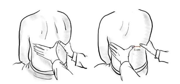
Why Check: Bilateral ↓ expansion can occur in asthma and COPD (Ch 5 – Resp Exam – MF1), as well as Thoracic Scoliosis and Ankylosing Spondylitis (from costovertebral fusion); unilateral ↓ expansion seen in pleural effusion, pneumothorax, etc. on the affected side.
Rib-Pelvis Distance
AFMC level Pre-clerkship 2 · Clerkship 4
Say: I will palpate the distance between the ribs and pelvis for signs of kyphosis in the lower back.
Do:
- With patient standing and clinician’s fingers outstretched, place your hand in patient’s side between lower rib and the iliac crest (located at mid-axillary line). The distance is usually about 4 fingerbreadths (some variation is expected as fingers vary)
- Abnormal if the distance is 2 finger breadths or less.
Report: The rib-pelvis distance is 4 finger breadths.
Why Check: A Rib-Pelvis distance of > 2 finger breadth’s diagnostic value is to rule out lumbar vertebral fractures. A Rib-Pelvis distance of ≤ 2 finger breadths has a Sensitivity of 87% and a Specificity of 47% for Lumbar Vertebral Fractures. The Rib-pelvis distance is decreased in lumbar kyphosis e.g. in Ankylosing Spondylitis or Lumbar vertebral compression fractures (e.g. in Osteoporosis)
Modified Schober’s Test
AFMC level Pre-clerkship 2 · Clerkship 4
Say: I will assess for reduced flexion in the low back using a measuring tape. Please stand and keep the knees straight.
Setup: With patient standing, using your measuring tape mark the midline between the Dimples of Venus, and mark 10 cm above and 5 cm below this point (total of 15 cm distance while standing).
Do: Have patient bend forward trying to touch the floor while keeping their knees straight. Measure the distance between your two points –should now be > 5cm longer (i.e. ≥20 cm).
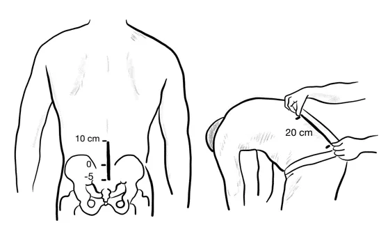
Report: There is an increase of greater than 5 cm in the distance between the two measured points – the Modified Schober’s Test is negative.
Why Check: An increase in the distance of < 5cm indicates a restricted lumbar flexion, which can occur in conditions such as ankylosing spondylitis (which tends to begin in the lumbar region in men).
Straight Leg Raise and Lasegue’s Sign
AFMC level Pre-clerkship 2 · Clerkship 4
Say: I will examine for sciatica nerve pain by moving the leg away from the exam table while it is relaxed. Let me know if you feel any pain during the test, and if so, where.
Do:
- Patient lying supine with knees extended. Clinician raises the SYMPTOMATIC straightened leg.
- Observe for pain radiating down the back of the raised leg into foot, (usually at 30-70° of leg raise). This suggests Sciatic Nerve Root Irritation. Pain that is only in the low back or hamstring is NOT a positive straight leg raise (could be due to Hamstring muscle strain e.g. due to tight musculature).
- If the pain on straight leg raise is consistent with sciatic nerve root irritation, slowly lower the leg until the pain stops and then dorsiflex the ankle. Return of pain is a Positive Lasegue’s Sign.
Report: No pain elicited on straight leg raise – Straight Leg Raise test is negative.
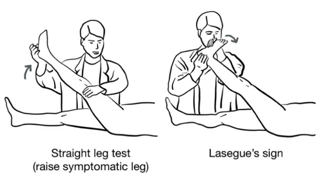
Why Check: Straight Leg Raise test’s diagnostic value is to rule out lumbar disc herniation, via testing for irritation of the Sciatic Nerve Roots (L4, L5, S1, S2) by manipulating the symptomatic leg i.e. Sciatica. A positive straight leg raise test has a Sensitivity of 91% and a Specificity of 32% for lumbar disc herniation (sciatica). Further, the likelihood of lumbosacral radiculopathy in patients with sciatica is moderately decreased with a negative straight leg raise test (LR+ 1.3, LR- 0.3).
Crossed Straight Leg Raise
AFMC level Pre-clerkship 2 · Clerkship 4
Say: I will continue to examine for sciatica nerve pain now by moving the opposite leg away from the exam table while it is relaxed. Let me know if you feel any pain during the test, and if so, where.
Do:
- Patient lying supine with knees extended. Clinician raises the ASYMPTOMATIC straightened leg.
- Observe for pain radiating down the back of the symptomatic leg into foot (symptomatic leg still on exam table) This suggests Sciatic Nerve Root Irritation.
Report: No pain elicited on crossed straight leg raise – Crossed Straight Leg Raise test is negative.
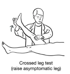
Why Check: Crossed Straight Leg Raise test’s diagnostic value is to rule in lumbar disc herniation. A positive crossed straight leg raise test has a Sensitivity of 32% and a Specificity of 98% for lumbar disc herniation (sciatica). Further, the likelihood of lumbosacral radiculopathy in patients with sciatica is mildly increased with a positive crossed straight leg raise test (LR+ 3.4, LR- 0.8).
Femoral Nerve Stretch
Say: I will examine for femoral nerve pain by moving the leg away from the exam table. I will ask you to lie on the stomach for this test with the legs relaxed. Let me know if you feel pain during the test, and if so, where.
Do:
- Patient lying prone with knees extended. Clinician passively flexes the knee and extend the hip lifting thigh off the exam table.
- Observe for pain radiating down the anterior thigh (Note: Femoral nerve is in the anterior compartment).
Report: No pain elicited on femoral nerve stretch – Femoral Nerve Stretch test is negative.
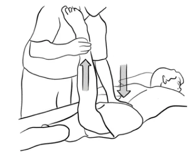
Why Check: Femoral Nerve Stretch test’s diagnostic value is to rule in mid-lumbar nerve impingement, via testing for irritation of the Femoral Nerve Roots (L2, L3, L4). A positive femoral nerve stretch test has a Sensitivity of 50% and a Specificity of 100% for mid-lumbar nerve impingement.
FABER Test
(same as in Hip Exam - Chapter 27)
Say: I will rest the foot on the opposite knee and apply pressure to the inside of the knee to look for signs of Hip or Sacroiliac Joint pain when moving the hip. Please let me know if you feel pain, and if so, where.
Do:
- Patient lying supine, Flex, Abduct, and Externally Rotate one hip (placing heel on opposite knee, making a figure-of-4).
- Push down on knee; press on opposite hemipelvis to stabilize.
- Observe for pain in lower back or buttock
Assess: No pain elicited with FABER test.
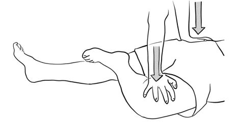
Why Check: FABER (Flexion, Abduction, External Rotation) test with inability to achieve FABER position or eliciting pain in the anterior/lateral of the hip or groin pain suggests a Hip Joint Pathology (can cause pain in healthy patients due to stretch); pain elicited in lower back/gluteal region suggests a Sacroiliac Joint Pathology.
Gaenslen’s Test
Say: I will continue to assess for Sacroiliac Joint pain when you bring one knee towards the chest while letting the other leg be relaxed off the exam table. Please let me know if you feel pain, and if so, where.
Do:
- Patient lying supine with one leg hanging off the exam table (the thigh must be off the exam table)
- Clinician asks patient to bring the other knee (still on the exam table) to their chest using their hands.
- Observe for pain in the lower back or buttock.
Report: No pain elicited on flexion of knee to chest using hands – Gaenslen’s test is negative.
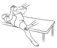
Why Check: Gaenslen’s test eliciting pain in the back or buttocks suggests Sacroiliac Joint pathology.
Part 3: Select Special Tests – Not Part of Screening Back Exam But Seen in Practice:
Spurling Test
Say: I will now perform a test for radicular pain in the neck by applying a pressure to the top of the head. Please tilt the head to one side and back. Please let me know if you feel pain or pins and needles during the test, and if so, where.
Do:
- Review any contraindications to performing Spurling Test (See When Not to Check)
- Patient sitting up, laterally flexes and then extends the neck to affected side. After each neck motion, ask if this causes pain or paresthesias (tingling).
- If patient neck in lateral flexion and neck extended without causing their symptoms, apply axial pressure on spine, and ask if this reproduces their pain or paresthesias.
Report: Spurling’s test is negative for cervical radiculopathy.
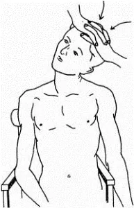
Why Check: The Spurling test is a provocative test, where a positive spurling test in patients with neck/arm pain increases the likelihood of suspected cervical radiculopathy (LR+ 4.5, LR- 0.6).
When Not to Check: Avoid in patients suspected of having cervical instability – this includes structural (e.g. trauma, cervical spondylosis with myelopathy), infectious (osteomyelitis or other infectious process), inflammatory (e.g. rheumatoid arthritis), or malignant (e.g. metastatic bone disease).
Helpful Videos to Review:
- Video 1: Back Exam – McMaster MSK Examination Series (Link: BACK EXAMINATION - McMASTER UNIVERSITY)
- Video 2 and 3: Bates’ Visual Guide to the Physical Exam – Anatomy Review and Examining the Spine (Link: https://www.medportal.ca/ug/educational-resources/bates/lists/bates/musculoskeletal)
- Video 4: Dr. Janice Harvey’s Joint Assessment Made Easy – Back Assessment (Link: https://www.medportal.ca/ug/educational-resources/joint-assessment-made-easy)
Part 4: Relevant Likelihood Ratios – from Rational Clinical Examination JAMA series
Clinical Question 1: Does this patient have a clinically important cervical spine injury?5
Note: Clinically significant defined as any fracture, dislocation or ligamentous instability detectable by diagnostic imaging and requiring surgical or specialist follow-up.
| Procedure | Sensitivity | Specificity |
|---|---|---|
| Canadian C-Spine Rules | 0.90-1.00 | 0.01-0.77 |
| NEXUS C-Spine Criteria | 0.83-1.00 | 0.02-0.46 |
Clinical Question 2: Does this patient have Scoliosis?6
| Procedure | Sensitivity | Specificity |
|---|---|---|
| Adam’s Forward Bend Test | 0.92 | 0.60 |
Clinical Question 3: Does this patient have Thoracic Vertebral Fractures?7
| Procedure | Sensitivity | Specificity |
|---|---|---|
| Occiput-Wall Distance (>4cm = Positive Result) | 0.41 | 0.92 |
Clinical Question 4: Does this patient have Lumbar Vertebral Fractures?8
| Procedure | Sensitivity | Specificity |
|---|---|---|
| Rib-Pelvis Distance (≤ 2 fingerbreadths = Positive Result) | 0.87 | 0.47 |
Clinical Question 5: Does this patient have Lumbar Disc Herniation (Sciatica)?9
| Procedure | Sensitivity | Specificity |
|---|---|---|
| Straight Leg Raise | 0.91 | 0.32 |
| Crossed Straight Leg Raise | 0.32 | 0.98 |
Clinical Question 6: Does this patient have Mid-Lumbar Nerve Impingement?10
| Procedure | Sensitivity | Specificity |
|---|---|---|
| Femoral Nerve Stretch | 0.50 | 1.00 |
Clinical Question 7: What can the medical history tell us about Low Back Pain?11
| Disease | Medical History | Sensitivity | Specificity |
|---|---|---|---|
| Cancer | Age >50 yo or cancer history or unexplained weight loss or failed conservative treatment | 1.0 | 0.60 |
| No relief with bed rest | >0.90 | 0.46 | |
| Age >50 y | 0.77 | 0.71 | |
| Duration of pain > 1 mo | 0.50 | 0.81 | |
| History of cancer | 0.31 | 0.98 | |
| Failure to improve with a month of therapy | 0.31 | 0.90 | |
| Unexplained weight loss | 0.15 | 0.94 | |
| Spinal Osteomyelitis | IV Drug Abuse, UTI, or Skin Infection | 0.40 | N/A |
| Compression Fracture | Age >50 y | 0.84 | 0.61 |
| Age >70 y | 0.22 | 0.96 | |
| Trauma | 0.30 | 0.85 | |
| Corticosteroid use | 0.06 | 0.995 | |
| Herniated Disk | Sciatica | 0.95 | 0.88 |
| Spinal Stenosis | Age >50 y | 0.90 | 0.70 |
| Pseudoclaudication | 0.60 | NA | |
| Ankylosing Spondylitis | Age at onset <40 y | 1.0 | 0.07 |
| Pain not relieved supine | 0.80 | 0.49 | |
| Morning back stiffness | 0.64 | 0.59 | |
| Pain duration 3+ months | 0.71 | 0.54 | |
| 4 of 5 Positive responses: Onset of back discomfort <40 y; Problem began slowly; Pain duration 3+ months; Morning stiffness; Improved by exercise | 0.23 | 0.82 |
Clinical Question 8: Does my patient with neck and/or arm pain have a cervical radiculopathy?12
| Procedure | LR+ (95% CI) | LR- |
|---|---|---|
| Motor: Weakness of any arm muscle | 1.9 (1.4-2.5) (about +15%) | 0.4 (0.3-0.6) (about −30%) |
| Sensory: Reduced vibration or pinprick sensation in arm | 0.7 (0.5-1.0) (no change: 95% CI includes 1) | 1.4 (1.0-1.8) (no change: 95% CI includes 1) |
| Reflex: Reduced Biceps reflex | 9.1 (1.2-69.4) (about +30%) | 0.9 (0.8-1.0) (no change: 95% CI includes 1) |
| Reflex: Reduced brachioradialis reflex | 7.3 (0.9-56.8) (no change: 95% CI includes 1) | 0.9 (0.9-1.0) (no change: 95% CI includes 1) |
| Reflex: Reduced triceps reflex | 2.3 (0.7-7.0) (no change: 95% CI includes 1) | 0.9 (0.9-1.0) (no change: 95% CI includes 1) |
| Reflex: Reduced Biceps, Brachioradialis, or triceps reflex | 3.6 (1.4-9.2) (about +15%) | 0.8 (0.7-0.9) (about −15%) |
| Spurling Test | 4.5 (3-6.8) (about +15%) | 0.6 (0.4-0.8) (about −15%) |
| ROM: Rotation of neck to involved side <60 degrees | 1.7 (1.3-2.3) (about +15%) | 0.2 (0.1-0.9) (about −30%) |
Clinical Question 9: Where does this patient’s Cervical Radiculopathy localize to?12
| Level | Procedure | LR+ (95% CI) | LR- (95% CI) |
|---|---|---|---|
| C5 Radiculopathy | Elbow Flexion – Weak | 5.3 (2.7-10.5) (about +30%) | 0.2 (0-2.5) (no change: 95% CI includes 1) |
| C6 Radiculopathy | Biceps or Brachioradialis Reflex – Diminished | 14.2 (4.3-46.7) (about +45%) | 0.5 (0.3-0.8) (about −15%) |
| Thumb – Sensory Loss | 8.5 (2.3-31.1) (about +30%) | 0.7 (0.5-1.0) (no change: 95% CI includes 1) | |
| Wrist Extension – Weak | 2.3 (1.1-5.0) (about +15%) | 0.8 (0.5-1.1) (no change: 95% CI includes 1) | |
| C7 Radiculopathy | Elbow Extension – Weak | 4.0 (1.8-9.2) (about +15%) | 0.4 (0.3-0.6) (about −30%) |
| Middle finger – Sensory Loss | 3.2 (0.2-60.1) (no change: 95% CI includes 1) | 1.0 (0.9-1.0) (no change: 95% CI includes 1) | |
| Triceps Reflex – Diminished | 3.0 (1.6-5.6) (about +15%) | 0.6 (0.3-1.4) (no change: 95% CI includes 1) | |
| C8 Radiculopathy | Little finger – Sensory Loss | 41.4 (2.1-807.3) (about +45%) | 0.8 (0.6-1.1) (no change: 95% CI includes 1) |
| Finger Flexion – Weak | 3.8 (1.7-8.5) (about +15%) | 0.6 (0.3-1.1) (no change: 95% CI includes 1) |
Clinical Question 10: Does this patient with Sciatica have a Lumbosacral Radiculopathy?12
| Procedure | LR+ (95% CI) | LR- (95% CI) |
|---|---|---|
| Ipsilateral calf wasting | 5.2 (1.3-20.8) (about +30%) | 0.8 (0.6-0.9) (about −15%) |
| Weak ankle dorsiflexion | 4.9 (1.9-12.5) (about +15%) | 0.5 (0.4-0.7) (about −15%) |
| Crossed Straight Leg Raise | 3.4 (1.8-6.4) (about +15%) | 0.8 (0.7-0.9) (about −15%) |
| Abnormal ankle jerk | 2.7 (1.6-4.5) (about +15%) | 0.8 (0.7-1.0) (no change: 95% CI includes 1) |
| Straight Leg Raise | 1.3 (1.1-1.6) (about +15%) | 0.3 (0.2-0.6) (about −30%) |
| Leg sensation abnormal | 1.0 (0.7-1.5) (no change: 95% CI includes 1) | 1.0 (0.8-1.1) (no change: 95% CI includes 1) |
Clinical Question 11: Where does this patient’s Lumbosacral Radiculopathy with Sciatica localize to?12
| Level | Procedure | LR+ (95% CI) | LR- (95% CI) |
|---|---|---|---|
| L3-L4 Radiculopathy | Quadriceps Reflex - Asymmetric | 8.7 (4.9-15.5) (about +30%) | 0.6 (0.5-0.8) (about −15%) |
| Knee Extension – Weak | 3.7 (1.9-7.6) (about +15%) | 0.7 (0.5-0.8) (about −15%) | |
| L5 Radiculopathy | L5 Distribution – Sensory Loss | 3.1 (1.8-5.6) (about +15%) | 0.8 (0.7-0.9) (about −15%) |
| Hallux Extension – Weak | 1.6 (1.3-2.0) (about +15%) | 0.8 (0.6-1.0) (no change: 95% CI includes 1) | |
| Ankle Dorsiflexion – Weak | 1.3 (0.9-1.8) (no change: 95% CI includes 1) | 0.8 (0.6-1.0) (no change: 95% CI includes 1) | |
| S1 Radiculopathy | Ankle Plantarflexion – Weak | 4.8 (0.4-60.4) (no change: 95% CI includes 1) | 0.7 (0.6-0.9) (about −15%) |
| Achilles Reflex – Asymmetric | 2.9 (1.8-4.7) (about +15%) | 0.4 (0.3-0.6) (about −30%) | |
| S1 Distribution – Sensory Loss | 2.4 (1.3-4.2) (about +15%) | 0.7 (0.6-0.9) (about −15%) | |
| Calf Muscles – Ipsilateral Wasting | 2.4 (1.2-4.7) (about +15%) | 0.7 (0.5-0.9) (about −15%) |
References
-
Centre for Effective Practice. CORE Back Tool. Published 2016. Accessed March 12, 2022. https://cep.health/clinical-products/low-back-pain/ ↩ ↩2
-
Canadian C-Spine Rule. MDCalc. Accessed March 12, 2022. https://www.mdcalc.com/canadian-c-spine-rule#next-steps ↩
-
NEXUS criteria for C-spine imaging. MDCalc. Accessed March 12, 2022. https://www.mdcalc.com/nexus-criteria-c-spine-imaging#next-steps ↩
-
Recommendations. Choosing Wisely Canada. Accessed July 22, 2022. https://choosingwiselycanada.org/recommendations/#list ↩ ↩2
-
Michaleff ZA, et al. Accuracy of the Canadian C-spine rule and NEXUS to screen for clinically important cervical spine injury in patients following blunt trauma: a systematic review. CMAJ. 2012;184(16):E867-E876. ↩
-
Cote P, Cassidy JD. A study of the diagnostic accuracy and reliability of the scoliometer and Adam’s forward bend test. Spine (Phila Pa 1976). 1999;24(22):2411-12. ↩
-
Siminoski K, Warshawski RS, Jen H, Lee KC. The accuracy of clinical kyphosis examination for detection of thoracic vertebral fractures: comparison of direct and indirect kyphosis measures. J Musculoskelet Neuronal Interact. 2011;11(3):249-256. ↩
-
Siminoski K, et al. Accuracy of physical examination using rib-pelvis distance for detection of lumbar vertebral fractures. Am J Med. 2003;115(3):233-236. ↩
-
Vroomen PC, et al. Diagnostic value of history and physical examination in patient suspected of sciatica due to disc herniation: a systematic review. J Neurol. 1999;246(10):899-906. ↩
-
Suri P, Rainville J, Katz KN, et al. The accuracy of the physical examination for the diagnosis of midlumbar and low lumbar nerve root impingement. Spine (Phila Pa 1976). 2011;36(1):63-73. ↩
-
What can the medical history tell us about low back pain. In: Rational Clinical Examination Series. Chapter 7. 2009:76. ↩
-
McGee S. Evidence Based Physical Diagnosis. Saunders; 2012:836-840. ↩ ↩2 ↩3 ↩4