Medical Foundations 4
Chapter 31“Complete” Screening Neurological Examination
Part 1: Focused History – Based on Chief Complaint (Begin with Open-Ended Questions)
Approach
Headache
- Onset (gradual vs. sudden; any typical time of day/week/month/year?)
- New onset headache after age 50 years old – Red Flag for secondary headache
- New onset in pregnancy or post-partum period – Red Flag for secondary headache (re: pre-eclampsia)
- New onset headache after trauma – Red Flag for secondary headache (re: intracranial hemorrhage)
- Frequency/Duration (both when headaches first started and currently; keeping a headache journal?)
- Duration 30mins-7 days re: Tension Headache; 4-72 hours (if untreated) re: Migraine Headache
- Cluster Headache: Episodes of 15 to 180-minute duration; occurring q2days up to 8 per day in cluster.
- Recurrence upon awakening – Red Flag for secondary headache
- Pattern change of headaches from patient baseline – Red Flag for secondary headache
- Location:
- Unilateral – consistent with migraine or cluster headache (orbital, supraorbital and/or temporal pain)
- Bilateral – consistent with tension-type headache
- Character (specifically, pounding/pulsating, pressure-like)
- Change in character of headache – Red Flag for secondary headache
- Intensity (rate out of 10; what are they usually? How bad do they get? How often are they this bad?)
- Thunderclap headache: Sudden severe headache reaching max intensity in few seconds or <1 minute – red flag, concerning for subarachnoid hemorrhage – “worst headache of my life” – can’t miss diagnosis
- Aggravating factors:
- Worse with normal activity e.g. walking, climbing stairs – associated with migraine
- Painful on direct pressure – concern for sinus headache
- Positional headache – Red Flag for secondary headache
- Precipitated by coughing, sneezing, Valsalva or exercise – Red Flag for secondary headache
- Alleviating Factors (what do you do to make the headache feel better?)
- Ask about headache medications – what have they tried? What dose? How often? Effectiveness?
- Medication Overuse Headache: Concern if using Acetaminophen or NSAIDs >15 days/month; or use of Ergots, Triptans, Combination analgesics or opioids >10 days/month
- Ask about headache medications – what have they tried? What dose? How often? Effectiveness?
- Associated symptoms
- Nausea/Vomiting – associated with migraine
- Note: N/V with papilledema, neuro deficit, headache → risk of Raised ICP – Can’t Miss diagnosis.
- Aura (symptoms preceding <60 minutes before headache with reversible neurologic symptoms e.g. visual, sensory, speech/language, motor, brainstem and/or retinal) – associated with migraine
- Photophobia re. sensitivity to light, phonophobia re. sensitivity to noise – associated with migraine
- Ipsilateral conjunctival injection and/or lacrimation; nasal congestion and/or rhinorrhea; eyelid edema; forehead/facial sweating; miosis and/or ptosis – associated with cluster headache
- Note: Acute/subacute neck pain or headache with Horner Syndrome (ptosis, miosis, anhidrosis) and/or neurologic deficit raises concern for Cervical Artery Dissection – can’t miss diagnosis.
- Restlessness or agitation – associated with cluster headache
- Systemic symptoms (e.g. fevers, chills, night sweats, weight loss) – Red Flag for secondary headache
- Neurologic deficit (e.g. altered mental status, vision loss, dysphagia, dysarthria, aphasia, weakness, paresthesias, pulsatile tinnitus) – Red Flag for secondary headache
- Neck Stiffness and fever – concern for meningitis
- Painful eye with autonomic features, Vision loss – Red Flag for secondary headache (e.g. Acute angle closure glaucoma – can’t miss diagnosis)
- Nausea/Vomiting – associated with migraine
- Assess for life-style contributors
- Sleep – how much sleep do they get on average? Do they wake refreshed? Do they snore?
- Diet – do they skip meals? Amount of caffeine in diet? Red wine? Old cheddar cheese?
- Activity level – sedentary vs. athlete?
- Stress level – any relaxation, CBT, biofeedback, pacing activities they’ve tried that help?
- Family history of headaches (especially if character is similar to those described by patient)
- Medical History:
- History of immunosuppression (e.g. HIV) – Red Flag for secondary headache
- History of cancer (solid tumor, or hematologic malignancy) – Red Flag for secondary headache
- New drug at onset of headache – Red Flag for secondary headache
- Assess the impact the headaches are having on the patient’s life (e.g., missed work/school, stops them from participating in desired activities)
Why Check: The International Classification of Headache Disorders (ICHD-3) divides headaches into primary and secondary headaches. The 3 main types of primary headache disorders to be aware of are:
Tension-Type Headache
- Duration 30 mins-7 days
- Bilateral, non-pulsating pain
- Mild-moderate intensity
- Not worsened by normal activity (e.g. walking, climbing stairs)
- No nausea/vomiting
- Not both of: Photophobia and phonophobia (may have 1 of 2)
Migraine Headache
- Duration 4-72hrs untreated
- Unilateral, pulsating pain
- Moderate-severe intensity
- Worsened by normal activities (e.g. walking, climbing stairs) – desire to rest/avoid activity
- Nausea/vomiting
- Photophobia and phonophobia
- May have aura: Symptoms precede headache (<60 mins usually) with reversible neurologic symptoms (e.g. visual, sensory, speech, language, motor, brainstem, retinal)
Cluster Headache
- Duration 15-180 mins untreated
- Unilateral orbital, supraorbital and/or temporal pain
- Severe-very severe intensity
- Sense of agitation/restlessness
- Associated with at least 1 of: Ipsilateral conjunctival injection and/or lacrimation; nasal congestion and/or rhinorrhea; eyelid edema; forehead/facial sweating; miosis and/or ptosis
- Cluster: Episodes every other day up to 8 episodes/day through min. 50% of time-course
Headache can be a symptom caused by underlying disorders, some of which may be serious or life-threatening. The mnemonic SNNOOP10 is a mnemonic for red flags of secondary headache to urgently investigate:
- a. Systemic Symptoms (e.g. Fever – e.g. concern for meningitis, encephalitis, tumor)
- b. Neoplasm History
- c. Neurological Deficit (including decreased level of consciousness – e.g. Horner’s syndrome with cervical artery dissection; CN VI palsy with raised intracranial pressure)
- d. Onset sudden or abrupt (e.g. thunderclap headache of subarachnoid hemorrhage)
- e. Older Age (onset after 50 years old – e.g. tumor)
- f. Pattern change or recent onset of new headache
- g. Positional headache (e.g. intracranial hypotension worse with upright posture relieved by recumbency)
- h. Precipitated by coughing, sneezing or exercise
- i. Papilledema (e.g. raised intracranial pressure, idiopathic intracranial hypertension)
- j. Progressive headache and atypical presentations
- k. Pregnancy or postpartum period (e.g. pre-eclampsia/eclampsia)
- l. Painful eye with autonomic features (e.g. Acute angle closure glaucoma, optic neuritis)
- m. Post-traumatic onset of headache (e.g. traumatic brain injury, intracerebral hemorrhage)
- n. Pathology of immune system (e.g. HIV)
- o. Painkiller (analgesic) overuse (e.g. medication overuse – Ergots, triptans, analgesics, codeine/opioids >10 days/month, or Acetaminophen/NSAIDS >15 days/month) or new drug at onset of headache
One helpful resource for an approach to headache is the Core Neck Tool and Headache Navigator from the Centre for Effective Practice (PDF).
Acute Stroke and Transient Ischemic Attack (TIA)
- If unable to establish symptom time onset, clarify last time patient seen “normal” (or at baseline)
- Is the patient pregnant? (relative contraindication to rtPA treatment)
- Cincinnati Pre-Hospital Stroke Scale:
- Assess for facial droop (ask patient to smile/show teeth)
- Assess for arm drift (test as pronator drift – see Coordination Exam below)
- Assess for abnormal speech (ask patient to repeat “you can’t teach an old dog new tricks”)
- Is the patient presenting with seizure <24 hours before stroke onset?
- Medication History: Particularly insulin/oral antihyperglycemic agents; anticoagulants (including warfarin, heparin, DOACS); antiplatelets (ASA, clopidogrel, ticagrelor); antihypertensives
- Neurological PMHx: Significant head trauma or prior stroke last 3 months; history of intracerebral hemorrhage (including neoplasm, arteriovenous malformation or aneurysm); recent intracranial or intraspinal surgery; history of seizures/epilepsy
- Bleeding Hx: Arterial puncture at non-compressible site last 7 days; active internal bleeding; acute bleeding diathesis (stat labs – Platelets, aPTT/INR); major surgery or trauma last 14 days; GI or urinary tract hemorrhage last 21 days; recent MI last 3 months
- STAT Investigations: Glucose, CBC, INR, aPTT, Lytes, Cr; 12-Lead EKG; EMERGENT CT Head (Non-Contrast CT head; or CT Angiography from arch-to-vertex depending on the hospital centre and available imaging)
Why Check: In acute stroke, time is brain (penumbra – at risk brain around ischemic area), where time last seen normal is vital to determine eligibility to receive rtPA or endovascular thrombectomy.
- To assess out-of-hospital patients for stroke, a score of ≥1 on Cincinnati Pre-Hospital Stroke Scale has LR+ of 5.5 (95% CI = 3.3-9.1)1. Once in ER, assess stroke severity using National Institute of Health Stroke Scale (NIHSS).
- Ischemic and hemorrhagic strokes have distinct, emergent management approaches distinguished by a rapid CT (target <25mins to CT, <45 minutes to read CT after ER arrival – a plain CT will light up if hemorrhagic stroke). If ischemic stroke is within window for rtPA (Less than 3-4.5hrs) use ACLS rtPA checklist. A stat glucose assesses for hypoglycemia as a stroke mimic – correct immediately (glucose <2.7 is an exclusion criteria in rtPA checklist).
- A transient ischemic attack (TIA) is on the spectrum of minor stroke, defined by the American Heart Association as a transient (traditional definition < 24 hours) episode of neurological dysfunction caused by focal brain, spinal cord, or retinal ischemia, without acute infarction2. TIAs should be risk stratified e.g. using the ABCD₂ score to determine risk of 2-day, 7-day and 90-day stroke risk and to guide management.
- The likelihood of unilateral cerebral hemispheric disease is greatly increased by a positive arm rolling test (LR+ 21.7), upgoing plantar response (LR+ 19.0, LR- 0.6), and pronator drift (LR+ 10.3, LR- 0.1). The likelihood of unilateral cerebral hemispheric disease is moderately increased by a positive finger tapping test (LR+ 6.6, LR- 0.3) and hyperreflexia (LR+ 5.8, LR- 0.4). Finally, the likelihood of unilateral cerebral hemispheric disease is slightly decreased by the absence of hemianopia (LR- 0.7) and absence of hemisensory disturbance (LR- 0.7).
- The likelihood of significant aspiration risk in patients with recent strokes is slightly increased by drowsiness (LR+ 3.4, LR- 0.5), abnormal water swallow test (LR+ 3.2, LR- 0.4), oxygen desaturation 0-2 min after swallowing (LR+ 3.1, LR- 0.3), absent pharyngeal sensation (LR+ 2.4, LR- 0.03 – i.e. normal pharyngeal sensation greatly reduces this likelihood), abnormal voluntary cough (LR+ 1.9, LR- 0.6), dysarthria (LR+ 1.6, LR- 0.5), abnormal gag reflex (LR+ 1.5, LR- 0.6), and dysphonia (LR+ 1.3, LR- 0.4). It is slightly decreased by absent tongue weakness (LR- 0.6) and normal gag reflex (LR- 0.6).
- Clinical Stroke Syndromes may be identified based on site of occlusion causing infarction distal to the occlusion and associated neurologic deficit – where the Circle of Willis is the last line of defense for collateral blood flow as a series of anastomoses (see diagram above)3.
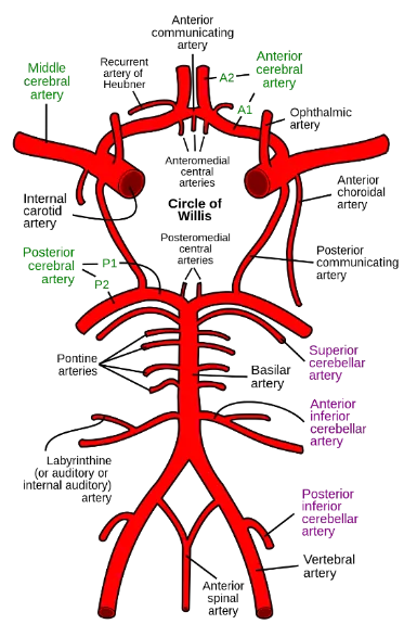
Stroke Syndrome (Arterial Occlusion Site)3: Clinical Manifestations
Anterior Cerebral Artery Occlusion (Anterior Cerebral Artery)
- Motor and Sensory Deficit: Contralateral upper and lower extremity motor weakness and sensory loss (lower > upper)
- Others: Apraxia; incontinence; slowed mentation
Middle Cerebral Artery Occlusion, Dominant Hemisphere (usually left)
- Motor and Sensory Deficit: Contralateral upper and lower extremity motor weakness and sensory loss (upper > lower); contralateral facial droop
- Aphasia: Expressive, receptive, and/or global aphasia
- Others: Homonymous hemianopsia; gaze deviation to side of lesion
Middle Cerebral Artery Occlusion, Non-Dominant Hemisphere (usually right)
- Motor and Sensory Deficit: Contralateral upper and lower extremity motor weakness and sensory loss (upper > lower); contralateral facial droop
- Others: Homonymous hemianopsia; gaze deviation to side of lesion; hemi-neglect
Lateral Superior Pontine Syndrome (Superior cerebellar artery)
- Ipsilateral: Limb ataxia; loss of facial sensation; Horner’s Syndrome
- Contralateral: Loss of pain and temperature sensation
- Others: Hiccup; Nausea/vomiting; Nystagmus; absence of specific cranial nerve signs
Lateral Mid-Pontine Syndrome (Short circumferential artery – a pontine artery)
- Ipsilateral: Limb ataxia; loss of facial sensation; Horner’s Syndrome
- Contralateral: Loss of pain and temperature sensation
- Others: Hiccup; Nausea/vomiting; Nystagmus; trigeminal nerve impairment (bilateral lesion = difficulty chewing; unilateral = ipsilateral jaw deviation on mouth opening)
Lateral Inferior Pontine Syndrome (Anterior inferior cerebellar artery)
- Ipsilateral: Limb ataxia; loss of facial sensation; Horner’s Syndrome; facial paralysis; gaze paralysis
- Contralateral: Loss of pain and temperature sensation
- Others: Hiccup; Nausea/vomiting; Nystagmus; deafness and tinnitus
Lateral Medullary Syndrome = Wallenberg Syndrome (Posterior inferior cerebellar artery)
- Ipsilateral: Limb ataxia; loss of facial sensation; Horner’s Syndrome; ipsilateral vocal cord paralysis → hoarseness of voice; ipsilateral loss of pharyngeal reflex
- Contralateral: Loss of pain and temperature sensation
- Others: Hiccup; Nausea/vomiting; Nystagmus; dysphagia
Locked In Syndrome (Basilar artery occlusion → bilateral ventral pontine lesions)
- Complete quadriplegia; inability to speak and loss of all facial movements
- Normal level of consciousness; patient may communicate with eye or eyelid movements
Dorsal midbrain syndrome (compression by extra-axial lesion e.g. pinealoma)
- Paralysis of upward gaze
Central midbrain syndrome (Paramedian branches of basilar artery
- Ipsilateral: Oculomotor nerve palsy
- Contralateral: Loss of cutaneous sensation and proprioception; hemichorea of limbs
Ventral midbrain syndrome = Weber syndrome (Paramedian branches of basilar artery
- Ipsilateral: Oculomotor nerve palsy
- Contralateral: Hemiparesis; supranuclear facial paresis
Medial superior pontine syndrome (Paramedian branches of basilar artery
- Ipsilateral: Limb ataxia
- Contralateral: Hemiparesis; loss of proprioception/vibration sensation
- Others: Internuclear ophthalmoplegia; palatal myoclonus
Medial inferior pontine syndrome (Paramedian branches of basilar artery
- Ipsilateral: Limb ataxia; gaze paralysis; lateral rectus paralysis
- Contralateral: Hemiparesis; loss of proprioception/vibration sensation
- Others: Gaze-evoked nystagmus
Medial medullary syndrome (Paramedian branches of basilar artery
- Ipsilateral: Limb ataxia; Tongue weakness
- Contralateral: Hemiparesis; loss of proprioception/vibration sensation
Weakness
- Pattern of weakness:
- Proximal vs. distal? Diffuse vs. focal? Symmetrical vs. asymmetrical? Upper vs. lower extremities?
- Facial involvement? Ocular involvement? Bulbar weakness?
- Time course of the condition (acute, sub-acute, chronic, relapsing, progressively worsening)
- Patient response to exercise – easy fatigue vs. warm up phenomenon vs. static weakness
- Assess for associated symptoms:
- Muscle pain? Cramping? Contractures or other musculoskeletal deformities?
- Tea/coke coloured urine? Fasciculations? Sensory fibers involved: Paresthesias or numbness?
- Confusion, seizures, or other symptoms of brain dysfunction? Cardiorespiratory symptoms?
- Antecedent Event: Recent infection? Or, in Guillain Barre Syndrome (GBS), potentially increased risk with recent influenza vaccination (note that influenza infection is a greater risk for GBS).
- Medication History:
- Medication-induced neuropathies: Chemotherapy (e.g. platinum-based, taxanes, vincristine); antimicrobials (e.g. isoniazid, fluoroquinolones); antiretrovirals, TNF-inhibitors (e.g. infliximab); immune checkpoint inhibitors (e.g. ipilimumab); antiseizure drugs (e.g. phenytoin); colchicine, etc.
- Medication-induced myopathies: Glucocorticoid; statins; antimalarials (e.g. chloroquine); antipsychotics (re. neuroleptic malignant syndrome); colchicine; antiretrovirals (e.g. zidovudine); alcohol; cocaine, etc.
- Family History: Early use of assistive devices for mobility? Early cardiac disease (non-atherosclerotic)? Early dementia, deafness, short stature?
Approach: Delineate whether it is a Neural, Myopathic, or NMJ issue:
- Neural: Fine motor muscles most impacted
- Glove and Stocking Distribution: Suggests length dependent process – motor neurons in LL are longer, such that in a length-dependent process (e.g. Guillain Barre Syndrome) expect to find neurological deficits ascend to level of knees before symptoms begin in fingers.
- Myopathic: Larger, proximal muscles most impacted due to forces required to contract these muscles (e.g. Duchenne Muscular Dystrophy, Becker Muscular Dystrophy, steroid-induced myopathy, myositis)
- NMJ: Expect frequently used, fatigable muscles (for example, in the eyes) to be affected first
- The likelihood of Myasthenia Gravis is greatly increased by provocative testing including Sleep Test (LR+ 53, LR- 0.01), Ice Test (LR+ 24, LR- 0.16), Peek Sign (LR+ 30), and Anticholinesterase Test (LR+ 15, LR- 0.11 – not commonly performed due to risk of bradycardia, asystole, bronchoconstriction); and moderately increased by unintelligible speech after prolonged speaking (LR+ 4.5, LR- 0.61)
- In the upper limbs, likelihood of suspected cervical radiculopathy in patients with neck/arm pain is increased by weakness of any arm muscle (LR+ 1.9, LR- 0.4). Specifically, weakness of elbow flexion increases the likelihood of C5 radiculopathy (LR+ 5.3); weakness of wrist extension increases the likelihood of C6 radiculopathy (LR+ 2.3); weakness of elbow extension increases the likelihood of C7 radiculopathy (LR+ 4.0, LR- 0.4); and weakness of finger flexion increases the likelihood of C8 radiculopathy (LR+ 3.8).
- In the lower limbs, the likelihood of suspected lumbosacral radiculopathy in patients with sciatica is increased by weak ankle dorsiflexion (LR+ 4.9, LR- 0.5). Specifically, weakness of knee extension increases the likelihood of L3-L4 radiculopathy (LR+ 3.7, LR- 0.7); weakness of hallux extension increases the likelihood of L5 radiculopathy (LR+ 1.6); and normal strength of ankle plantarflexion decreases the likelihood of S1 radiculopathy (LR- 0.7).
- Check for reversible causes: metabolic (electrolytes, acidosis, etc.), endocrine (TSH, glucose, etc.), infectious.
Concussion and Traumatic Brain Injury (TBI) Assessment
- Patient Age:
- Age ≥ 16 years old: Use Canadian CT Head Injury/Trauma Rule for patients with mild TBI (GCS 13-15 at 2 hours post-injury with 1 of: loss of consciousness, amnesia re. event, and/or witnessed disorientation) to rule out intracranial injuries needing neurosurgical intervention without CT imaging.
- Age <16 years old: Use CATCH2 for patients with minor head injury (GCS 13-15) to identify patients requiring a CT head.
- Alternative: Use PECARN for patients with mild TBI to rule out clinically important TBI without CT head.
- Age ≥ 65 years old: ↑ risk intracranial injury; CT indicated unless asymptomatic and no other risk factors1.
- Patient Medical Profile4:
- Multisystem Trauma: Beware patient with unstable vital signs or distracting injuries or receiving analgesia/anesthesia – where significant head injury may be missed due to confounding injuries.
- Known coagulopathy or bleeding disorder: Strong indication for early CT scan and to check the patient’s INR
- Known Neurosurgery/Neurological Impairment: e.g. Hydrocephalus with shunt, AVM, tumor, mild cognitive impairment/dementia → clinical assessment less reliable, may ↑ risk of intracranial injury
- Establish Plausible Injury Mechanism: Ask open-ended questions about mechanism of injury e.g. “run me through what happened, play-by-play.” Try to visualize what happened and to distinguish patient’s personal memories from facts they inferred or learned from other observers afterwards. Assess low energy vs. high energy force injury and the forces involved (e.g. direct vs. indirect forces and their direction).
- Dangerous Mechanism ≥16 years old: Pedestrian struck by motor vehicle; occupant ejected from motor vehicle; fall > 3 feet or > 5 stairs; others – falls from horse/bicycle; focal blunt trauma e.g. bat/ball/club4.
- Dangerous Mechanism <16 years old (CATCH2): Motor vehicle crash; fall ≥ 3 feet or ≥ 5 stairs; fall from bicycle without helmet.
- Sport-Related Concussion: If assessing an athlete, helpful to know/learn about their sport to understand common injuries and the demands of the position they play in sport – sports with ↑ risk of concussion include rugby, ice hockey, American football, lacrosse, boxing and soccer.
- Ask about signs and symptoms of altered mental status immediately after impact4:
- Do you remember the impact and moments just after?
- Did anyone see you lay still and unresponsive right after the accident?
- Were you confused or unsure about where you were and what was happening?
- Were you able to think clearly about what to do after the accident?
- Were you able to answer questions appropriately and follow instructions from people at the scene?
- Did anyone tell you that your speech was incoherent or not making sense?
- Rule out Confounding Factors:
- Substance Use: Were you drinking alcohol or using substances just before the accident?
- Acute Psychological Stress: Did you see the impact coming? Did you think that you or others would be seriously injured or killed? Did you feel panicked or scared?
- Other Injuries: Did you injure other parts of your body? Were you in severe pain?
- Concussion – Common and Red Flag Symptoms:
Physical
- Headache
- Nausea/Vomiting
- Visual issues (blurred vision, seeing stars/light)
- Balance problems
- Dizziness
- Tinnitus
- Photophobia
- Phonophobia
Red Flag Symptoms
- Worsening/severe headache
- Neck pain
- Double vision
- Focal neurologic symptoms/signs
- Seizure/convulsions
- Persistent vomiting
Cognitive
- “Slowed down/In a fog”
- Difficult to concentrate/ remember
Red Flag Symptoms
- Altered Mental Status e.g. agitation, somnolence, repetitive questioning, slow response to verbal communication.
Sleep
- Excessive sleep difficulty
- Difficulty falling asleep (insomnia)
Red Flag Symptoms
- N/A
Emotional/Behavioural
- Drowsiness
- Fatigue/Lethargy
- Irritability
- Emotional lability
- Depression
- Anxiety
Red Flag Symptoms
-
In pediatrics patients; Inconsolable; unwilling to eat/drink/nurse.
-
Physical Examination – Including but not limited to:
- Vital Signs (e.g. q1hour) and ABCDEs (Airway, Breathing, Circulation, Disability, Environment)
- Head and Neck Exam
- Look for signs of basilar skull fracture: Peri-orbital ecchymosis (racoon eyes), retro-auricular ecchymosis (Battle’s sign), CSF leakage (clear rhinorrhea or otorrhea); hemotympanum on otoscopy
- Look for large, boggy scalp hematoma, or open/depressed skull fracture (<16 years old – CATCH2 rule)
- Cervical Spine Exam – palpate cervical spine, and posterior and anterior paraspinal musculature for tenderness or reactive muscle guarding; assess cervical spine range of motion
- Screening Neurological Exam – looking for focal neurological deficits – including papilledema (sign of raised ICP) on direct fundoscopy; cranial nerves; motor; sensory; reflex; cerebellar; gait and balance
- Mental Status and Cognition – assess level of consciousness (re. Glasgow Coma Scale, GCS – serially assess e.g. q1hour) and assess for confusion, disorientation, amnesia, concentration, etc.
Why Check456: Traumatic brain injury (TBI) is a common diagnosis – almost 50% of patients sustain 1 TBI in their lifetime. Falls are the most common cause of TBI, particularly in children and older adults. Other common causes include sports-related injuries and motor vehicle collisions, as well as non-accidental injury in pediatrics. While the majority of TBIs are classified as mild, we assess for red flags to rule out neurosurgical emergencies.
- Traumatic Brain Injury (TBI): TBI is a disruption of normal function or structure of the brain caused by traumatic biomechanical forces – either by direct blow (e.g. blunt trauma, penetrating injury) or indirectly (e.g. acceleration/deceleration of brain). TBI severity is classified according to Glasgow Coma Scale (See Below), where a mild TBI has GCS 13-15; a moderate TBI has GCS 9-12, and a severe TBI has GCS ≤ 8.
- Primary TBIs include both skull fractures and intracranial lesions, as follows:
-
Skull Fracture – Linear: May be defined as open or closed – concerning particularly if crossing middle meningeal groove in temporal bone, or venous dural sinuses with risk of significant extra-axial bleed.
-
Skull Fracture – Depressed: Displacement of skull below level of adjacent skull with ↑ risk of associated intracranial lesions, CNS infection and seizure. May be classified as open (overlying/adjacent scalp laceration) or closed (no overlying/adjacent scalp laceration).
-
Skull Fracture – Basilar (Neurosurgical emergency): Fracture of bones of skull base. Basilar skull fractures are at high risk of epidural hematoma (re. temporal bone is relatively weak versus other skull bones with close proximity to middle meningeal artery).
-
Skull Fracture – Elevated: One fracture fragment elevated above level of adjacent skull – uncommon, due to tangential force impact on skull)
-
Skull Fracture – Penetrating: For example, due to gunshot, stab wound, or blast injury.
-
Intracranial Lesions – Focal:
- Epidural Hemorrhage (EDH): Bleed between skull and dura mater – hemorrhage does not cross sutural margins but does cross dural attachments – lens-shaped appearance on CT)
- Subdural Hemorrhage (SDH): Bleed between dura and arachnoid mater, hemorrhage does cross sutural margins but does not cross dural attachments – crescent-shaped appearance on CT)
- Subarachnoid Hemorrhage (SAH): Bleed between arachnoid mater and pia mater
- Intracerebral Hemorrhage (ICH): Brain parenchymal bleed
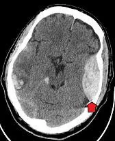
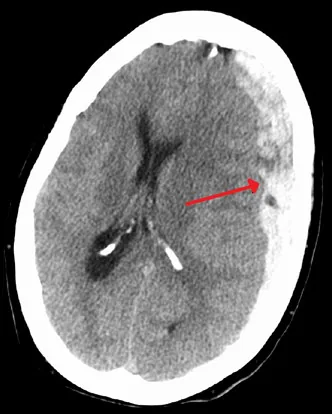
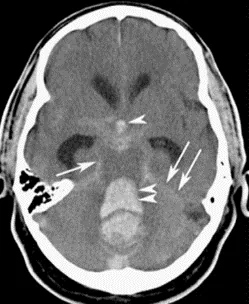
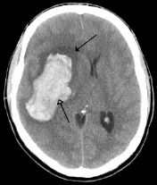
Epidural Hemorrhage (EDH)7; Subdural Hemorrhage (SDH)8; Subarachnoid Hemorrhage (SAH)9; Intracerebral Hemorrhage (ICH) (with surrounding edema)10
-
Intracranial Lesions – Diffuse:
- Cerebral Contusion: Due to direct impact, commonly occur in basal frontal/temporal areas.
- Diffuse Axonal Injury: Due to shearing force on white matter tracts (commonly at grey-white matter junction, corpus collosum, and brainstem in severe cases) – ↑ risk poor outcome.
-
- Secondary Brain Injury (sequelae from primary injury): Includes hypoxic-ischemic damage and swelling
- Concussion: A form of mild TBI that affects brain function in absence of structural injury on imaging. Once diagnosis is made, core principles include11:
- Early recognition and directing to care: If a concussion is suspected, remove from activity immediately for assessment and to avoid another injury – “if in doubt, sit them out.”
- Close follow-up (in 1-2 weeks) to monitor symptoms including development of persistent post-concussion symptoms (>10-14 days in adults, >4 weeks in children)
- Graded return to activity after a brief period of physical and cognitive rest (24-48 hours) – tailored to patient, encouraging return to cognitive activity and low-risk physical activity as soon as tolerated while staying below symptom-exacerbation thresholds. Return to contact sport or high-risk activities avoided until (1) post-concussion symptoms subside; (2) return to full school activities without accommodations related to post-concussion symptoms; and (3) medically cleared following return-to-sport protocol.
- Helpful Resource: Peds Concussion Living Guideline for Pediatric Concussion Care
- Helpful Resource: Adult Concussion Living Guideline for Concussion and Prolonged Symptoms
| Assessment Tool | Description |
|---|---|
| Concussion Recognition Tool 6 | To help identify concussion in children, adolescents and adults (can be used by non-Healthcare Providers) |
| SCAT6 | Sport Concussion Assessment Tool for adolescents (13+) and adults in the acute phase (ideally within 72 hours of injury) |
| SCOAT6 | Sport Concussion Office Assessment Tool for adolescents (13+) and adults in a controlled office environment (typically from 72 hours after injury) |
| Child SCAT6 | Sport Concussion Assessment Tool for children ages 8-12 years in the acute phase (ideally within 72 hours of injury) |
| Child SCOAT6 | Sport Concussion Office Assessment Tool for children ages 8-12 years in a controlled office environment (typically from 72 hours after injury) |
Seizure
- Ideally obtain collateral history from a witness of the episode, with a video if available.
- Assess if presenting with one or multiple seizure types. For each type, determine:
- Onset: When did the seizures begin?
- Timing: How long did the episode last? How often are they occurring? How long can patient go without a seizure? What is longest seizure duration? When do the seizures occur (e.g. wake, sleep)?
- Aura: Do they know when it is going to occur?
- Characterization/Localization: What do seizures look like? Part of body vs. entire body? Rhythmic vs. arrhythmic? Stiff vs. limp? Impaired consciousness? Gaze deviation or eye rolling? Repetitive movements (e.g., lip smacking, tongue thrusting)? Any video of event? Associated incontinence?
- Post-ictal: Confusion, drowsiness, agitation, fatigue, amnesia, paralysis, weakness? How long?
- Medical Profile:
- Hx of CNS lesions (e.g., injury, stroke, tumours)? Dysfunction (global developmental delay, autism)?
- Are they on any anticonvulsants (both maintenance and rescue)? Are they typically adherent?
- Is there a family history of epilepsy?
- Find out about burden of the illness (e.g., limitations, missed work/school).
- If patient is older than 16 years old, do they drive?
- Does the patient drink alcohol, benzos, or use any other substances? (re: withdrawal seizures)
- Any history of diabetes or low blood sugars? (hypoglycemia is the most common cause of seizures, and the quickest to reverse)
- Any known triggers of seizures (e.g. flashing lights, hyperventilation)?
Approach:
- For a first episode of seizure, important to consider differential diagnoses including syncope, TIA, migraine, movement disorder, conversion disorder, panic disorder. rule out organic causes:
- Neuro: Brain tumor (primary or metastatic – especially low-grade tumors); stroke/TIA; Intracerebral Hemorrhage; Trauma; Radiation-Induced Edema/Necrosis; Pre-Existing Epilepsy
- Metabolic: Hypoxia; Hypo/hyper-natremia, hypo/hyper-glycemia, hypercalcemia; hypomagnesemia; uremia; hepatic failure; renal failure; hyperthyroidism
- Drug Toxicity: Opioid-induced neurotoxicity; amphetamines, cocaine
- Drug Withdrawal: EtOH, Barbiturates, Benzodiazepines, Opioids
- Infection: Including meningitis, encephalitis
- Medications Lowering Seizure Threshold: Including antipsychotics, antidepressants, stimulants, opioids, and select immunosuppressants
- Hypoglycemia: Seizures due to hypoglycemia will often quickly resolve after administration of glucose.
- Alcohol Withdrawal: Patients can be placed on CIWA protocol if experiencing alcohol withdrawal. As these patients are at risk of seizures, can provide benzodiazepines (as per the CIWA protocol), which raise the seizure threshold.
- Epilepsy: Defined as 2 unprovoked seizures > 24 hrs apart. Seizure semiology can help determine your approach to control seizure activity. Seizure semiologies include:
- Generalized Seizures (arising within and rapidly engaging bilateral distributed networks) → Tonic (“stiffening”), Clonic (“jerking”), Tonic-Clonic, Absence (impaired consciousness in brief episodes, pediatrics especially), Atonic (“drop attacks”), Myoclonic
- Focal Seizures (identify altered level of consciousness/awareness; localization; and progression) – including Focal; Focal Aware; Focal Impaired Awareness; and Focal to bilateral tonic-clonic
- Status epilepticus: Failure of mechanisms responsible for seizure termination
- Prolonged seizures – Tonic-clonic: ≥5min; Focal with impaired consciousness: ≥10min; Absence: ≥10min
- ≥2 seizures in 30 minutes without return to baseline neurological functioning in between
- Risk of neuronal death/injury (after 30mins in tonic-clonic, >60mins in focal with impaired consciousness)
- Driving and Epilepsy: In general, patients who experience an unprovoked seizure have to be reported to the Ministry of Transportation of Ontario (e.g. a diagnosis of epilepsy would be required for mandatory reporting under the CPSO Mandatory and Permissive Reporting Policy). As per the CMA Driving Guide, private drivers presenting with their first unprovoked seizure should not drive for at least 3 months and not before a complete neurologic evaluation to determine the cause. If a diagnosis of epilepsy is made, private drivers may drive if seizure-free on medication for 6 months. For commercial drivers, an even greater certainty is needed before resuming driving that another seizure will not occur while driving than private drivers.
Simple Febrile Seizure (Pediatric)
- What makes a febrile seizure simple? Brief (<15 minute) generalized seizures occurring once during a 24-hour period in a febrile child who does not have an intracranial infection, metabolic disturbance, or history of afebrile seizures; commonly occur in children 6 months – 6 years of age.
- Simple febrile seizure is a clinical diagnosis – NOT a diagnosis of exclusion. Be sure to assess for Red Flags:
- Meningeal or Encephalitis Signs
- Focal onset of seizure
- Altered Level of Consciousness (should return to full alertness within 1 hour)
- Unvaccinated child (raises suspicions of bacterial meningitis)
- Partially healed infections (especially sinusitis or otitis media)
Key Ideas for Simple Febrile Seizures:
- Role of Fever: Fever does not cause seizure; it ↓ seizure threshold. There is no correlation between risk of having a simple febrile seizure and the absolute temperature or its rate of increase. The real mediator is cytokines that increase neuronal excitability, not pyretic effects themselves.
- Role of Vaccines: Some risk of seizures from vaccines (highest in Measles and Pertussis vaccinations). Keep in mind significant consequences of disease (including neuro problems) vs. seizure risk of vaccine.
- Absent Red Flags, risk of progression to epilepsy in a child with a single febrile seizure is low: 0.5-1% of general population, vs. 1-2% with children who have had a simple febrile seizure.
- Recurrence Risk: 30% of children will have a recurrence; 75% of these will have a recurrence in the 1st year, while almost all who are going to have a recurrence will do so in the first 2 years.
- Treatment: Otherwise healthy children with simple febrile seizures do not need to be on anti-epileptics, which have significant side effects and do not prevent epilepsy (which most of these children will not have). If child has significant comorbidities (e.g. cystic fibrosis), consider giving anti-epileptics.
Aphasia
- Assess Naming: Ask patient to name their hand, finger, fingernail, common objects (e.g. pencil)
- Fluency: Ask “how do you get from your house to the hospital?
- Commands: Ask patient a simple 3-step command across the midline, e.g. with your right hand, touch your left shoulder, then point to the ceiling, then close your eyes.
- Repetition: Ask patient to repeat: “no ifs, ands or buts”; or “you can’t teach an old dog new tricks”
Approach: Ascertaining deficits in fluency (effort, rate, and prosody = rhythm, stress and intonation); comprehension (e.g. in response to commands); verbal repetition and naming can help differentiate aphasia subtypes, which can help with localization of the lesion. Common Aphasia Syndromes include:
| Aphasia | Speech Fluency | Comprehension | Verbal Repetition |
|---|---|---|---|
| Global | No | No | No |
| Mixed Transcortical | No | No | Yes |
| Broca’s (Expressive) | No | Yes | No |
| Transcortical Motor | No | Yes | Yes |
| Wernicke’s (Receptive) | Yes | No | No |
| Transcortical Sensory | Yes | No | Yes |
| Conduction | Yes | Yes | No |
| Anomic | Yes | Yes | Yes |
Supportive Communication Skills for Patients with Aphasia: Goal to ↑ communicative access to your service, as well as efficiency of your service with trained conversation partners. Key principles include:
- Acknowledge Competence (Help patients feel they’re being treated respectfully): you want patient to know we I still know they’re very smart people, problem is with getting the words out
- Reveal Competence (Make sure message goes IN and you get message OUT accurately): Can use paper (yes/no/maybe cards – to point); Gestures (up or down, hold pen in each hand and ask which one they use, directions for N/W/E/S, pay attention to their pointing when where); Facial Expressions; Write Key Words (black marker); Use pictures; see if they need glasses or hearing aids.
- Verify Info – sometimes doesn’t work at all, but it’s vital to put in the effort. Paraphrase and say you do not understand or repeat back if comprehension is issue.
Frailty
Definition12: A state of increased vulnerability from age-associated decline in reserve and function, resulting in reduced ability to cope with everyday or acute stressors. It is a confluence of medical and geriatric conditions and disabilities, as well as socioeconomic circumstances, which puts many elderly patients at greater risk of health destabilization.
Assessing Frailty Using the Clinical Frailty Scale: In assessing for Frailty, the Clinical Frailty Scale (CFS) (see Right)13 emerged as a valuable tool with an Inter-Rater Reliability of κ = 0.6412 between medical students and critical care physicians; guidelines such as the Canadian Diabetes Association 201814 have begun to incorporate Frailty Assessment (specifically Clinical Frailty Scale) in adjusting glycemic control targets. It is likely this will become more common in other guidelines for management of chronic diseases.
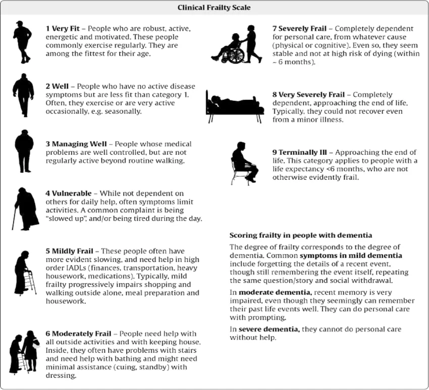
Importance of Recognizing Frailty in Clinical Practice12: Frailty is common, particularly in elderly persons with complex chronic conditions. Emerging evidence demonstrates importance of frailty in elderly patients as a predictor of adverse outcomes and mortality. Frailty is important to consider in discussing risks and benefits of treatments and can guide counseling and anticipatory preventive measures for patients with frailty, including management of comorbidities that can contribute to or be affected by frailty.
Behaviours that Challenge (BTC) in Adults with Intellectual and Developmental Disabilities (IDD)15
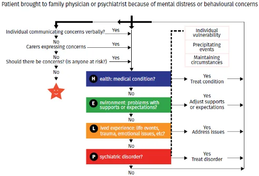
Why Check: Adults with IDD are often not able to communicate their needs. Behaviours that Challenge (BTC) are frequently a way in which patients with IDD communicate unmet needs. Unfortunately, when these behaviours present, a common response from many health care providers is to feel overwhelmed by the complexity of the patient’s diagnoses and the challenges in communicating with the patient. The focus, in this case, should be on treating the underlying cause of the behaviours of concern.
Past Medical History (PMHx)
- Risk Factors for Stroke:
- General Risk Factors: Hypertension; Dyslipidemia; Obstructive Sleep Apnea
- ↑ Risk of Embolic Ischemic Stroke: Atrial fibrillation; Recent myocardial infarction (<1 month); Cardiac Valvulopathy (e.g. Rheumatic mitral or aortic valve disease; infective endocarditis); Heart Failure with Reduced Ejection Fraction (e.g. LVEF <30%); Dilated cardiomyopathy
- ↑ Risk Thrombotic Ischemic Stroke: Vasculitis (e.g. Giant Cell Arteritis, Takayasu Arteritis)
- Coagulation/Bleeding Disorders (e.g. Antiphospholipid syndrome, inherited thrombophilia, cancer-related hypercoagulability, Factor V Leiden mutation, Protein C or S deficiency, etc.) → ↑ ischemic, thrombotic stroke risk
- Risk Factors for Epilepsy:
- Diabetes Mellitus: Hypoglycemia and hyperglycemia can cause seizure (hypoglycemia most common)
- Hyperthyroidism: Can cause seizures, or exacerbate seizures in patients with epilepsy
- Risk Factors for Epilepsy in Older Adults: Intracranial tumors, vascular malformations, head trauma; Alzheimer disease; Subacute and Chronic Stroke
- Risk Factors and Protective Factors re. Infection:
- ↑ Risk of Guillain-Barre Syndrome: Recent URTI; Gastroenteritis; Campylobacter jejuni; Influenza A/B; CMV
- ↑ Risk of Bell’s Palsy: Recent Herpes Simplex Virus (HSV) Activation; other causes include CMV, Epstein-Barr Virus (EBV), Influenza, Mumps, Rubella, Adenovirus, Coxsackievirus, severe COVID-19
- ↑ Risk of Multiple Sclerosis: History of Epstein-Barr Virus infection (EBV – cause of infectious mononucleosis)
- Childhood Vaccinations: Vaccinations against Haemophilus influenzae B (HIB vaccination series), Streptococcus pneumoniae (Pneu-C-13 vaccination series), Neisseria meningitidis (Men-C-C and Men-C-ACYW vaccinations) protect against most common historical pathogens causing bacterial meningitis.
Relevant Clinical Scores
- Glasgow Coma Scale (GCS) – Coma severity based on Eye, Verbal and Motor Criteria
- Canadian CT Head Injury/Trauma Rule – Used for patients >16 years old with GCS 13-15 to rule out intracranial injuries that would require neurosurgical intervention without the need for CT imaging
- CATCH2 (Canadian Assessment of Tomography for Childhood Head Injury) Rule – Predicts clinically significant head injuries in children up to 16 years old with minor head injury and initial GCS at least 13
- PECARN Pediatric Head Injury/Trauma Algorithm – Used for pediatric patients with GCS of 14-15 to rule out clinically important TBI including those that would require neurosurgical intervention without need for CT imaging
- ABCD² Score for TIA – Estimates the risk of stroke after suspected Transient Ischemic Attack (TIA)
- Cincinnati Prehospital Stroke Severity Scale – Predicts large vessel occlusion and severe stroke in patients with stroke symptoms
- NIH Stroke Scale (NIHSS) – Quantifies stroke severity
- Pediatric NIH Stroke Scale – Quantifies stroke severity, child-specific version of NIH Stroke Scale
- tPA Contraindications for Ischemic Stroke – Provides inclusion/exclusion criteria when deciding to use tPA on a patient with acute ischemic stroke
- CHA₂DS₂-VASc Score – stroke risk in atrial fibrillation; used for initiation of anticoagulation, builds on CHA₂DS₂ Score
- HAS-BLED Score – Bleeding risk for patients on anti-coagulation
- SORCAN – For patients with acute ischemic stroke, estimates mortality at 30 days and 1 year, and 30-day functional outcomes (e.g. death/disability or death/institutionalization)
- Intracerebral Hemorrhage (ICH) Score – Estimates 30-day mortality for patients with ICH
- Functional Outcome in Patients with Primary ICH (FUNC) Score – Estimates functional independence at 90-days for patients with ICH
- Barthel Index for ADLs – Assesses functional independence, generally in stroke patients
- McDonald Criteria for Multiple Sclerosis (2017) – Diagnosis of multiple sclerosis
- Revised Amyotrophic Lateral Sclerosis Functional Rating Scale (ALSFRS-R) – Stratifies severity of ALS
- Modified Hoehn and Yahr Scale for Parkinson Disease – Grades severity of symptoms
Past Surgical History (PS Hx)
- Cerebrovascular Disease:
- Risk Factors for Embolic Ischemic Stroke: Coronary artery bypass graft surgery; Prosthetic/Mechanical Heart Valves
- Brain Aneurysm: Neurosurgical options include aneurysm repair with surgical clipping or endovascular coiling
- Acute Ischemic Stroke: Interventional option includes intra-arterial mechanical thrombectomy (clot retrieval – e.g. for a proximal large artery occlusion in the anterior circulation)
- CNS Tumors:
- History of Ionizing Radiation: Risk factor for primary CNS tumors
- Primary CNS Tumors – Non-Malignant: Meningiomas; Pituitary tumors; Nerve sheath tumors; and others
- Primary CNS Tumors – Malignant: Glioblastomas; Diffuse Astrocytoma; Oligodendrogliomas; Ependymal tumors; and others (e.g. anaplastic meningioma, malignant peripheral nerve sheath tumor, CNS lymphoma)
- Brain Metastasis: Most common – lung, breast, colon, renal cell, melanoma
- Neurosurgical Resection of CNS Tumors: e.g. Craniotomy (patient may be awake to facilitate functional cortical mapping for brain tumors close to cortex controlling motor, sensory or language function – to maximize tumor resection, and ↓ neurologic injury risk); transsphenoidal surgery (e.g. for pituitary adenomas, Sellar masses)
- Radiation (may be adjuvant intent e.g. post primary tumor resection with curative intent, or palliative intent e.g. for brain metastasis causing neurologic deficit, headache, seizures, etc.):
- Modalities include stereotactic radiosurgery (SRS – delivers high dose radiation to radiographically discrete treatment volume) or whole brain radiation therapy (WBRT)
- Deep Brain Stimulation: Placement of electrodes into target area of brain
- For Advanced Parkinson Disease: e.g. motor fluctuation, refractory tremor, inconsistent treatment response
- Other Neurologic Indications for Deep Brain Stimulation: Epilepsy; Essential Tremor; Dystonia
- Psychiatric Indications for Deep Brain Stimulation: Obsessive compulsive disorder; Refractory major depression
- Epilepsy (e.g. for medically refractory epilepsy that is drug-resistant):
- Neurosurgical Resection: e.g. for patients with refractory neocortical focal seizures – e.g. mesial temporal lobe epilepsy secondary to mesial temporal sclerosis; tumors or vascular malformations with focal-onset seizures
- Vagus Nerve Stimulation: e.g. for patients who are not surgical candidates/decline surgery, or persistent seizures after neurosurgical resection
- Others: Responsive Cortical Stimulation
- Hydrocephalus:
- CSF Shunt: Catheter connecting lateral ventricle to systemic circulation or peritoneum, to bypass mechanical or functional obstruction causing hydrocephalus
- Endoscopic Third Ventriculostomy (ETV): Creating opening in floor of 3rd ventricle to allow CSF to flow into prepontine cistern and subarachnoid space – ↓ risk of infection/malfunction vs. CSF shunt; generally ineffective for hydrocephalus due to impaired CSF absorption.
- ↑ Intracranial Pressure (ICP):
- Invasive ICP monitoring: Intraventricular, intraparenchymal, subarachnoid, epidural –Burr Hole placement
- Decompressive craniectomy (can significantly ↓ ICP) – risks include herniation through skull defect, CSF leak, wound infection, epidural/subdural hematoma
- FAILS Mnemonic re. red flags to perform CT before Lumbar Puncture (LP) in a patient with fever and headache:
- Focal Neurological Deficit (e.g. decreased level of consciousness, inability to answer questions and follow commands, gaze palsy, abnormal visual fields, facial palsy, abnormal motor function, or abnormal language)
- Age >60 years old
- Immunocompromised (e.g. HIV, on immunosuppressants, solid organ or hematopoietic stem cell transplant recipient)
- Lesion (history of central nervous system mass lesion)
- Seizures (new onset seizure within 1 week of presentation)
Family Medical History (FMHx)
- Epilepsy: Most recognized genetic epilepsies begin in childhood (e.g. childhood absence epilepsy, juvenile absence epilepsy, juvenile myoclonic epilepsy)
- Parkinson Disease: ↑ risk particularly if age at symptom onset <50 years old
- Huntington Disease: autosomal-dominant disease caused by trinucleotide repeat expansion in huntingtin (HTT) gene
- Multiple Sclerosis: Association with Class I and Class II alleles of major histocompatibility complex (MHC) e.g. HLA-DRB1
- Dystrophinopathies (e.g. Duchenne muscular dystrophy, Becker muscular dystrophy): X-linked recessive inheritance
- Migraine and Cluster Headaches: Tends to have familial association
- Amyotrophic Lateral Sclerosis (ALS): Family history ↑ risk of ALS
- Essential Tremor: ↑ risk if first-degree relative has Essential Tremor
Social Hx
ADLs and IADLs
- Driving and Mandatory Reporting: For many neurologic diseases (e.g. diseases causing cognitive impairment; sudden incapacitation as in seizures; motor, sensory or visual impairment as in cerebrovascular disease, movement disorders, etc.), physicians must report individuals to the Ministry of Transportation of Ontario and are advised to inform the patient in advance of doing so. For specific recommendations for given conditions, consider reviewing the CMA Driving Guide
- For many patients with neurologic illnesses, allied health supports are vital to support and enhance patient functioning and patient-centred quality of life. This could be delivered as an inpatient (e.g. on acute admission, inpatient rehab), outpatient (e.g. outpatient rehab, etc.), or in the home. Examples include, but are not limited to:
- Nursing: Focus on promotion of health, assessment, provision of care, treatment and evaluation of health conditions by supportive, preventive, therapeutic, palliative and rehabilitative means in order to attain or maintain optimal function.
- Speech and Language Pathologist (SLP): Focus on communication (4 key tasks – speaking, listening, reading and writing) and swallowing disorders. Example interventions include support for patients with aphasia, dysarthria, dysphagia, etc.
- Physiotherapy: Focus on improving patient-centred outcomes e.g. reducing pain, improving function and quality of life across the lifespan. Examples include strengthening/rehabilitation for patients with motor neurologic deficits; chest physiotherapy e.g. for patients with amyotrophic lateral sclerosis, etc.
- Occupational Therapy: Focus on assisting patients to overcome limitations in order to promote function and independence. Examples of interventions include, but are not limited to:
- Assistive Devices: e.g. canes, walker, crutches, wheelchairs, etc.
- Home Safety Equipment: e.g. stairlift, higher toilet seat, shower chair, grab bars, bathtub lifts
- Eating Supports: e.g. specialized eating utensils, grips, holders, etc.
- Sleeping Supports: e.g. pressure-relieving mattress to prevent pressure ulcer development, etc.
- Respiratory Therapy: Support e.g. in neuromuscular disorders such as ALS with non-invasive positive pressure ventilation, tracheostomy, etc. as life-sustaining treatment if within their goals of care
- Dietitian: Focus on providing expert nutrition and dietary advice, e.g. optimizing nutritional support, including artificial nutrition (e.g. PEG-tube feeding for patients with dysphagia as life-sustaining treatment e.g. in neuromuscular disorders, post-stroke patients, etc. if within their goals of care).
- Social Work: Focus on the pursuit of social justice, enhancement of quality of life, and to development of the full potential of each individual, group and community in society. Example interventions include systems navigation and access to services, case management, counseling supports, etc.
- Personal Support Workers: e.g. to assist with activities of daily living (e.g. bathing, toileting), etc.
- Home Care: To coordinate publicly funded services in the home, including those listed above.
Substance Use
- Smoking - ↑ risk: Cluster headaches; ischemic stroke; intracerebral hemorrhage; neurocognitive disorders (e.g. vascular dementia); may ↑ risk of MS and ALS; Note - ↓ risk Parkinson Disease
- Alcohol:
- Seizures may occur due to alcohol withdrawal (often generalized tonic-clonic seizures 6-48 hours after cessation of alcohol use – can also occur with benzodiazepine withdrawal).
- Heavy alcohol use (e.g. >10 drinks/week for females, >15 drinks/week for males) ↑ risk of ischemic stroke and intracerebral hemorrhage (ICH)
- Alcohol noted to ↓ essential tremor
- Recreational Drug Use:
- Stimulant intoxication (e.g. cocaine, amphetamines) ↑ risk of seizures, ischemic and hemorrhagic stroke
- IV drug use ↑ risk of infective endocarditis (where infective endocarditis also ↑ risk of stroke).
Occupational Hx
- Are you working right now? What do you do for work? What other kinds of work have you done in the past? Do you have benefits through work such as drug coverage (e.g. access to Physiotherapy coverage)?
- If injury occurred at work → separate Workplace Safety and Insurance Board (WSIB) process.
- Important component of neurological history given impact of neurologic deficits on functional status – e.g. may require workplace accommodations, physician advocacy for supports, etc.
Lifestyle History
- Ketogenic Diet (high fat, adequate protein, low-carb diet): Recommended for drug-resistant epilepsy
- DASH or Mediterranean Diet: Recommended for patients with history of stroke or TIA
- Low Sodium Diet (<2000 mg/day): Recommended for patients with history of stroke or TIA
- University students living in dormitory accommodations: ↑ risk of Neisseria meningitidis meningitis (9-23X higher than baseline population risk)
- Lifestyle Factors in Management of Headache: Regular meals (avoid skipped meals); sleep hygiene (avoid irregular or too little sleep); exercise; limit caffeine consumption (e.g. <100 mg/day – important to taper slowly, where caffeine withdrawal can also cause headaches); stress management
- Sun Exposure, UV Exposure: Inverse relationship with the risk of Multiple Sclerosis
- Exercise: Aerobic exercise/physical activity may be protective against development of Parkinson Disease and Neurocognitive disorders (e.g. Alzheimer disease); regular aerobic exercise (150 minutes/week) recommended for medically stable patients for secondary prevention of stroke.
Medication Hx, Allergies
Primary Headache Pharmacologic Management17
- Tension-Type Headache
- Acute Management: Non-Steroidal Anti-Inflammatory Drugs (NSAIDs – e.g. ibuprofen, naproxen); Acetaminophen; Acetylsalicylic Acid (ASA)
- Prophylactic Management: Tricyclic Antidepressants (TCAs – 1st line: e.g. amitriptyline, nortriptyline); Antidepressants (2nd line: Mirtazapine, Venlafaxine)
- Migraine Headache:
- Acute Management:
- 1st line: NSAIDs (e.g. Ibuprofen, Naproxen); Acetaminophen; Acetylsalicylic Acid (ASA)
- 2nd Line: Triptans (e.g. sumatriptan, rizatriptan, almotriptan, zolmitriptan, etc.)
- Prophylactic Management (e.g. if headache >3d/month where acute medication ineffective, headache > 8d/month, or disability despite acute medication management):
- 1st Line: Beta-blockers (e.g. propranolol, metoprolol, nadolol); Tricyclic Antidepressants TCAs (e.g. amitriptyline, nortriptyline)
- 2nd Line: Topiramate; Gabapentin; Candesartan
- Others: Divalproex; pizotifen; botulinum toxin injections; flunarizine; venlafaxine
- Over the Counter: Magnesium citrate; riboflavin; butterbur; coenzyme Q10
- Acute Management:
- Cluster Headache:
- Acute Management: Triptans (e.g. sumatriptan, zolmitriptan); 100% oxygen
- Prophylactic Management (e.g. if disability despite acute medication management):
- 1st Line: Verapamil (Non-dihydropyridine calcium channel blocker)
- 2nd Line: Lithium (Mood stabilizer)
- Others: Topiramate; Melatonin
↑ Intracranial Pressure (ICP) Management
- Osmotic Therapy and Diuresis: including Hypertonic Saline and Mannitol
- Glucocorticoids (e.g. Dexamethasone): May be considered when ↑ ICP due to brain tumor or CNS infection – avoid in traumatic brain injury; not helpful in intracranial hemorrhage
- Other Interventions: Hyperventilation (e.g. ↑ ICP complicating cerebral edema, tumor, intracranial hemorrhage – avoid in patients with traumatic brain injury or stroke)
Acute Ischemic Stroke Treatment18
- Acute Ischemic Stroke Treatment (after CT Scan rules out hemorrhagic stroke – if hemorrhagic stroke, care directed by neurosurgery e.g. managing bleeding, ↑ICP, seizure):
- rtPA (Alteplase –door-to-needle time <60 mins): Review exclusion criteria (tPA Contraindications for Ischemic Stroke) – given <3 hours from symptom onset (extended to 3-4.5 hours in select patients)
- Antiplatelet: If not receiving rtPA, given ASA 160mg x 1 after brain imaging excludes intracranial hemorrhage. If receiving rtPA, delay ASA for 24 hours (and confirm no secondary hemorrhage with CT prior to starting)
- Endovascular Thrombectomy Treatment: Highly selected patients with disabling stroke symptoms may benefit from endovascular thrombectomy up to 24 hours from time last known well (advanced neuro imaging needed if > 6 hrs from time last known well)
- BP Management:
- Eligible for rtPA: Target BP <185/110mmHg pre-rtPA, <180/105mmHg x 24 hrs post-rtPA
- Ineligible for rtPA: Generally permissive hypertension – treat if BP > 220/120 mmHg in 1st 24 hours
- Blood Glucose Management: All patients check glucose on arrival to ER – correct hypoglycemia immediately.
Stroke Secondary Prevention18
- Antiplatelet Therapy (for patients without atrial fibrillation):
- Short Term Dual Antiplatelet Therapy (DAPT): ASA + Clopidogrel x 21 days; or ASA + Ticagrelor x 30 days
- Long Term Antiplatelet Therapy: ASA, Clopidogrel, or combined ASA + Dipyridamole
- Anticoagulant Therapy for Atrial Fibrillation:
- For patients with ischemic stroke or TIA and atrial fibrillation, recommend oral anticoagulant over ASA or Dual Antiplatelet therapy – for example:
- Direct Oral Anticoagulants (DOACs): e.g. Dabigatran, Apixaban, Rivaroxaban, Edoxaban
- Vitamin K Antagonist: Warfarin (note: bridging with heparin until therapeutic INR 2-3 reached is not recommended; suggested to bridge with low-dose ASA)
- For patients with ischemic stroke or TIA and atrial fibrillation, recommend oral anticoagulant over ASA or Dual Antiplatelet therapy – for example:
- Blood Pressure Management:
- History of Ischemic Stroke or TIA: Target BP <140/90 mmHg (including patients with CKD)
- History of Small Subcortical Stroke (e.g. Lacunar): Target SBP < 130 mmHg
- History of Intracerebral Hemorrhage (ICH): Target BP < 130/80 mmHg
- History of Stroke and Diabetes: Target BP < 130/80 mmHg
- General Medication Class Recommendations:
- Angiotensin Converting Enzyme inhibitors (ACEi): End in “-pril” e.g. Perindopril, Ramipril
- Thiazide-Like Diuretic: Hydrochlorothiazide, Chlorthalidone, Metolazone, Indapamide
- Lipid Management:
- Recommend Statin Pharmacotherapy (e.g. Atorvastatin, Rosuvastatin) for patients with history of non-cardioembolic ischemic stroke or TIA – target LDL <1.8mmol/L
- Ezetimibe (Cholesterol absorption inhibitor) or PCSK9 inhibitor (e.g. Evolocumab, alirocumab) may be considered if LDL >1.8 despite maximal tolerated statin therapy
- Recommend against Statin Pharmacotherapy for secondary prevention of ICH
- Recommend Statin Pharmacotherapy (e.g. Atorvastatin, Rosuvastatin) for patients with history of non-cardioembolic ischemic stroke or TIA – target LDL <1.8mmol/L
- Diabetes Management:
- Glycemic Targets: A1c ≤7.0%; FBG 4-7 mmol/L; 2-hr post-prandial glucose 5-10 mmol/L
- Anticoagulation
- ↑ risk of ICH (particularly warfarin)
- Stroke and TPA ↑ risk of ICH
Myasthenia Gravis Management
- Symptomatic Treatment – Acetylcholinesterase Inhibitors: Pyridostigmine (↑ Ach at NMJ)
- Chronic Immunotherapies (e.g. for patients with symptoms on pyridostigmine, to target underlying immune dysregulation): e.g. Glucocorticoids (e.g. prednisone); glucocorticoid-sparing therapies (e.g. Azathioprine, mycophenolate mofetil)
- Rapid Short-Acting Immunomodulating Treatments: e.g. glucocorticoid therapy, Plasma exchange (PLEX, to remove acetylcholine receptor antibodies from circulation), Intravenous Immune globulin (IVIG) – e.g. in myasthenic crisis.
Amyotrophic Lateral Sclerosis (ALS) Management
- Disease-Modifying Treatment Options:
- Riluzole (Glutamate inhibitor): Impacts survival and slows progression to modest degree – particularly in patients with symptoms present for < 5 years
- Edaravone (Free radical scavenger): May be considered for patients living independently with disease duration <2 years
Multiple Sclerosis (MS) Management
- Disease-Modifying Therapy (e.g. in relapsing-remitting MS in adults):
- Monoclonal Antibodies: e.g. Natalizumab; Ocrelizumab; Rituximab; Ofatumumab; Alemtuzumab
- Ocrelizumab used in Primary Progressive MS with pts <55y or active disease on MRI
- Oral Therapies:
- Fumarates: e.g. Dimethyl fumarate; diroximel fumarate; monomethyl fumarate
- Sphingosine 1-phosphate (S1PR) modulators: e.g. Fingolimod, Siponimod, ozanimod, ponesimod
- Others: Teriflunomide; cladribine
- Platform Injection Therapies: e.g. Interferons; glatiramer acetate
- Monoclonal Antibodies: e.g. Natalizumab; Ocrelizumab; Rituximab; Ofatumumab; Alemtuzumab
- Acute Exacerbations of MS:
- High dose, short-term glucocorticoid therapy: IV methylprednisolone, oral prednisone
- Alternative – Adrenocorticotropic hormone (ACTH)
- Refractory Relapse: Plasma Exchange (PLEX – nonselective removal of plasma proteins); Immunoadsorption (selective binding and removal of circulating immunoglobulins)
Duchenne and Becker Muscular Dystrophy
- Glucocorticoid Treatment (improve motor function, strength, pulmonary function, ↓ risk of scoliosis, and possibly delaying cardiomyopathy onset): e.g. Prednisone
- Novel Disease-Modifying Treatments (e.g. for specific genetic mutations to ↑ dystrophin expression): Casimersin; Eteplirsen; Golodirsen; Viltolarsen; Alaluren → note that clinical benefit of these medications not yet established.
Antiseizure Drugs (ASDs)19
Goal of treatment: To provide best quality of life with no seizures and fewest adverse effects from treatment.
- First Line Antiseizure Drugs (ASDs):
- Adults with Focal Seizures: Carbamazepine; Eslicarbazepine; Lamotrigine; Levetiracetam; Oxcarbazepine; Phenytoin; Topiramate; Valproic Acid
- Adults with Generalized Tonic-Clonic Seizures: Clobazam; Lamotrigine; Levetiracetam; Perampanel; Valproic Acid
- Children with Focal Seizures: Carbamazepine; Clobazam; Oxcarbazepine; Phenobarbital; Topiramate; Valproic Acid
- Children with Generalized Tonic-Clonic Seizures: Carbamazepine; Lamotrigine; Levetiracetam; Phenobarbital; Topiramate; Valproic Acid
- Children with Absence Seizures: Ethosuximide; Valproic Acid; consider Lamotrigine
- Benign Epilepsy of Childhood with Centrotemporal Spikes: Carbamazepine; Valproic Acid
- Myoclonic (incl. Juvenile Myoclonic Epilepsy): Lamotrigine; Levetiracetam; Topiramate; Valproic Acid
- Infantile Spasms: Vigabatrin; Steroids (oral prednisolone/injection ACTH)
- Dravet Syndrome: Topiramate; Valproic Acid
- Lennox-Gastaut Syndrome: Rufinamide; Valproic Acid
- Convulsive Status Epilepticus:
- 1st Treatment – Benzodiazepines: e.g. diazepam, lorazepam, midazolam
- 2nd Treatment – Antiseizure Drug (ASD): e.g. Levetiracetam, Fosphenytoin, Valproic Acid
Medications Lowering Seizure Threshold
- Antipsychotics: e.g. clozapine has ↑ risk in particular
- Stimulants: e.g. methylphenidate
- Antidepressants: e.g. bupropion, tricyclic antidepressants (TCAs), SSRIs, SNRIs, buspirone; atomoxetine (used in Attention Deficit/Hyperactivity Disorder treatment)
- Lithium (treatment for Bipolar Affective Disorder)
- Immunosuppressants: e.g. azathioprine, cyclosporine, mycophenolate, tacrolimus
- Anticancer Drugs: e.g. doxorubicin, etoposide, fluorouracil, cisplatin, methotrexate, etc.
- Opioids: e.g. meperidine, tramadol
- Antibiotics: e.g. carbapenems, 4th gen cephalosporins, fluoroquinolone, isoniazid, Penicillins
- Pulmonary Drugs: e.g. theophylline, aminophylline
Bell’s Palsy
- Glucocorticoids (short course): e.g. Prednisone, Prednisolone x 1 week
- Antiviral therapy (for severe facial palsy): e.g. valacyclovir, acyclovir
- Eye Care (to prevent corneal injury)
Parkinson Disease Management
(Note: current medications help to alleviate symptoms and improve patient-centred quality of life, but do not impact disease progression)
- Common Medications:
- Dopamine Replacement Therapy: e.g. Levodopa-Carbidopa → most effective for treatment of motor symptoms; a clear and dramatic beneficial response to dopaminergic therapy is considered supportive criteria in diagnosis of Parkinson Disease
- Dopamine Agonists: e.g. Pramipexole, Ropinirole, Rotigotine → Limited use in patients >65 years old due to risk of adverse effects (e.g. somnolence, peripheral edema, nausea, dizziness, impulse control disorders)
- Anticholinergic Agents: e.g. Trihexyphenidyl, Benztropine → useful for patients ≤65 years old with tremor-dominant disease without significant bradykinesia or gait disturbance; limited use in patients >65 years old due to anticholinergic side-effects.
- Other Medications (e.g. advanced Parkinson Disease, with motor fluctuations re. “wearing off” of levodopa dosing, and dyskinesia due to overstimulation of dopamine receptors):
- Monoamine Oxidase (MAO)-B Inhibitors: e.g. Rasagiline, Safinamide, Selegiline → e.g. useful to reduce “wearing off” as an adjunctive therapy with levodopa
- Catechol-O-Methyl Transferase (COMT) Inhibitors: e.g. Entacapone, Opicapone, Tolcapone → e.g. useful to reduce “wearing off” as an adjunctive therapy with levodopa
- Amantadine: Useful for dyskinesia in advanced Parkinson Disease where other antiparkinsonian medications have already been optimized
- Apomorphine: Dopamine agonist useful as rescue medication re. sudden “off” periods
Other Tone and Movement Disorder Symptom Management
(To improve symptoms and quality of life)
- Essential Tremor Management:
- 1st Line: Beta-blockers (e.g. Propranolol); Antiseizure Drugs (specifically Primidone)
- 2nd Line: Gabapentin; Topiramate; Benzodiazepines (e.g. Alprazolam, Clonazepam – particularly for patients where tremor is aggravated by anxiety)
- Restless Leg Syndrome:
- Intermittent Symptoms: Dopamine replacement as needed (e.g. Carbidopa-levodopa)
- Chronic Persistent Symptoms:
- 1st Line: Gabapentinoids (e.g. Gabapentin, Pregabalin); or Dopamine Agonist (e.g. Pramipexole, Ropinirole, Rotigotine)
- Spasticity Management (e.g. in Cerebral Palsy, Amyotrophic Lateral Sclerosis): Options include Baclofen (GABA agonist); Benzodiazepines (e.g. clonazepam, lorazepam, diazepam); Dantrolene; Alpha-2 adrenergic agonists (e.g. Tizanidine); and Botulinum Toxin injection
- Chorea Management (may be hereditary e.g. Huntington’s Disease, or acquired e.g. post-stroke, Sydenham’s Chorea, etc.):
- Vesicular Monoamine Transporter Type 2 (VMAT2) Inhibitors: e.g. Tetrabenazine, Deutetrabenazine → dopamine-depleting agents, inhibit dopamine release at synapse.
- Second-Line and Adjunctive Treatments:
- Antipsychotics (re. D2 antagonist activity): e.g. 2nd generation antipsychotics (risperidone, olanzapine, aripiprazole) – 1st generation antipsychotics (e.g. Haldol) limited by side-effects e.g. parkinsonism, tardive dyskinesia
- Benzodiazepines (GABA inhibitor): e.g. clonazepam, lorazepam –e.g. in severe chorea
- Dystonia Management (e.g. in Cerebral Palsy, in Huntington’s Disease): Options include Anticholinergics (e.g. trihexyphenidyl); Benzodiazepines (e.g. clonazepam, lorazepam); Baclofen (GABA agonist); Botulinum Toxin injection (for focal dystonia)
- Myoclonus Management (may be physiologic, essential, epileptic, or symptomatic e.g. secondary to major neurocognitive disorder; basal ganglia degenerative diseases; stroke; neuroinflammation e.g. encephalitis; metabolic e.g. hyponatremia, hypoxia, hypoglycemia, renal failure, hepatic failure)
- Cortical myoclonus: Levetiracetam (1st line – Antiseizure drug); adjuvant Benzodiazepines (e.g. clonazepam), Valproic Acid (Antiseizure drug) or Perampanel (Antiseizure drug)
- Cortical-Subcortical Myoclonus: Valproic Acid (1st line – Antiseizure drug); Levetiracetam or Lamotrigine (2nd line – Antiseizure drugs)
- Subcortical-nonsegmental or spinal-segmental myoclonus: Benzodiazepines (e.g. clonazepam); may consider Botulinum Toxin injections e.g. in palatal myoclonus
- Peripheral: Consider Botulinum Toxin injection to lesion
Sialorrhea Symptom Management
- Sialorrhea (Drooling, e.g. in Amyotrophic Lateral Sclerosis, Tardive Dyskinesia):
- Anticholinergics – e.g. Atropine, Hyoscyamine, Glycopyrrolate, Scopolamine patch
- Tricyclic Antidepressants (TCAs – e.g. Amitriptyline – with anticholinergic effect)
- Botulinum toxin injection into salivary glands
Neuropathic Pain Symptom Management20
- 1st Line: Gabapentinoids (e.g. Pregabalin, Gabapentin); Tricyclic Antidepressants (TCAs, e.g. Amitriptyline, Nortriptyline, Desipramine); or Serotonin-Norepinephrine Reuptake Inhibitors (SNRIs, e.g. Venlafaxine, Duloxetine)
- 2nd Line: Tramadol (opioid agonist, SNRI activity); Opioids (e.g. Morphine, Hydromorphone)
- 3rd Line: Cannabinoids (e.g. Nabilone, Nabiximols)
- 4th Line: Topical lidocaine (Sodium channel blocker – 2nd line for post-herpetic neuralgia); Methadone (opioid agonist and NMDA-receptor antagonist activity); Tapentadol (opioid agonist, Norepinephrine reuptake inhibition activity); Antiseizure drugs (Lamotrigine, Lacosamide); Botulinum Toxin Injections
Part 2: Pertinent Notes about the Physical Exam
- Key Principle: Within a screening neuro exam, ask where is the lesion – and does this test help me with figuring that out? For particular patient presentations, it is important to establish a baseline neuro exam that can be tracked serially.
- Tools you will need for a screening neuro exam: Percussion hammer (Reflex Testing); Pocket Snellen Acuity Card (CN II); Flashlight (CN II); Ophthalmoscope (CN II); Pins (Spinothalamic Sensory Exam); Cotton Ball (Posterior Column Sensory Exam)
- Pediatric Neurologic Exam: Age-Appropriate Toys and Objects (especially for Cranial Nerve & Coordination Exams)
Part 3: Specific Maneuvers in the Physical Exam – Say, Setup, Do, Report
Notes on Vitals
Vitals – Always do HR, BP, RR, Temp, SpO2
In This Exam
- Blood Pressure:
- Hypertension – Risk factor for stroke/TIA, cerebral artery dissection and intracranial hemorrhage.
- Hypertensive Emergency – Severely elevated BP (e.g. >180/120 mmHg) with end-organ damage (e.g. hypertensive encephalopathy, acute aortic dissection, acute LV failure, acute coronary syndrome, acute kidney injury, intracranial hemorrhage, acute ischemic stroke, eclampsia in pregnancy).
- Hypertensive urgency: Severely elevated BP without evidence of end-organ damage.
- Elevated ICP – Look for Cushing’s Triad (bradycardia, widened pulse pressure, irregular respirations)
- In a patient with a coma, the likelihood of having an intracranial structural lesion is greatly increased with sBP >160 mmHg (LR+ 10.4), and greatly decreased with sBP <120 mmHg (LR+ 0.1).
- Oxygen Saturation: In patients with recent strokes, the likelihood of significant aspiration risk is greatly increased if patient experiences oxygen desaturation 0 to 2 min after swallowing (LR+ 3.1, LR- 0.3).
- Temperature: Classic triad for meningitis = fever, nuchal rigidity and altered mental status
General Assessment
Observations to make during history taking, as well as comment on during physical exam
Level of Consciousness – Glasgow Coma Scale
AFMC level Pre-clerkship 2 · Clerkship 4
- Consider assessing Glasgow Coma Scale (GCS) – Minimum Score of 3, Max of 15:
| Eyes (Max Score 4) | Verbal (Max Score 5) | Motor (Max Score 6) |
|---|---|---|
| 4 – Open eyes spontaneously | 5 – Oriented, converse normally | 6 – Obeys commands for movement |
| 3 – Open eyes to voice | 4 – Confused conversation, able to answer questions | 5 – Purposeful movement to painful stimulus |
| 2 – Open eyes to pain | 3 – Inappropriate words | 4 –Withdraws in response to pain |
| 1 – No response | 2 – Incomprehensible speech | 3 –Flexion in response to pain, Decorticate posturing |
| 1 – No verbal response | 2 – Extension in response to pain, Decerebrate response | |
| 1 – No motor response |
- Quick screen: Assess Alert and Oriented x 3 (A&O x 3): Are they oriented to person, place and time?
Why Check: Altered mental status is an acute emergency needing prompt assessment and treatment. Etiologies may be acute or chronic, reversible or irreversible. In cases of Traumatic Brain Injury (TBI), the GCS helps to grade TBI severity: A mild TBI has GCS 13-15; moderate TBI has GCS 9-12; severe TBI has GCS ≤ 8.
GCS is also important as a prognostic tool for patients following cardiac arrest. In a patient with ↓ level of consciousness, it is helpful to look for focal or lateralizing signs (asymmetry between right-and left-sided responses e.g. on cranial nerve, motor, deep tendon reflex, brainstem reflex exams) and Meningismus (e.g. Kernig Sign, Brudzinski Sign, Jolt Accentuation of Headache) on exam to inform a differential diagnosis:
- Focal or lateralizing signs present
- Cerebrovascular events (ischemic or hemorrhagic)
- Trauma
- CNS Space-occupying lesions (infective e.g. abscess; or non-infective e.g. tumors)
- Meningismus present
- Meningoencephalitis (e.g. bacterial meningitis, viral encephalitis, etc.)
- Subarachnoid hemorrhage
- No Focal or lateralizing signs; or meningismus
- Toxins/Drugs: e.g. Opioids, Barbiturates, Alcohols, Anticholinergics, Sympathomimetics, Carbon Monoxide, Cyanide, etc.
- Organ Failure: e.g. encephalopathy (e.g. hepatic, uremic); shock (hypovolemic, cardiogenic, obstructive, distributive – see Ch 9 – Volume Status – MF2)
- Metabolic: e.g. hypoxia, hypercapnia, ↑/↓ Na, ↑/↓ Ca, hypo-/hyper-thermia, hypo-/hyperglycemia, diabetic ketoacidosis, lactic acidosis, porphyria, etc.
- Endocrine: e.g. adrenal insufficiency, hypopituitarism, hypothyroidism
- Seizures: including non-convulsive epilepsy, post-ictal state, eclampsia
- Pseudo-coma (may be psychiatric cause; assess for locked-in syndrome – complete quadriplegia, inability to speak, loss of all facial movements despite normal LOC e.g. basilar artery embolism causing ventral pons infarction)
Specifically, for patients 24h post-cardiac arrest, the likelihood of death, vegetative state or severe neurologic impairment is mildly increased by absent motor response to pain (LR+ 4.9) and absent withdrawal from pain (LR+ 4.7, LR- 0.2). In patients 72h post-arrest, the likelihood of death, vegetative state or severe neurologic impairment is moderately increased by absent motor response to pain (LR+ 9.2).
Behaviour
- Aggression, Verbal or Motor ticks, Pacing
Cognitive function
AFMC level Pre-clerkship 2 · Clerkship 3
- Can administer Mini-Mental Status Examination (MMSE), Montreal Cognitive Assessment (MOCA), or Mini-Cog to screen.
- Explored further in Chapter 36 – Assessment of Psychosis, Delirium and Dementia – MF4
Language (As in Aphasia Approach, Above)
- Assess Naming: Ask patient to name their hand, finger, fingernail, common objects (e.g. pencil)
- Fluency: Ask “how do you get from your house to the hospital?
- Commands: Ask patient a simple 3-step command across the midline, e.g. with your right hand, touch your left shoulder, then point to the ceiling, then close your eyes.
- Repetition: Ask patient to repeat: “no ifs, ands or buts”; or “you can’t teach an old dog new tricks”
- Optional Tests: Can also check reading (Dyslexia), writing (Dysgraphia), and counting (Dyscalculia – e.g. Serial 7s test subtracting 7 from 100 sequentially e.g. 100, 93, 86, 79 etc. as tested on MOCA) – Explored further in Chapter 36 – Assessment of Psychosis, Delirium and Dementia – MF4)
Dysmorphia
- More important in pediatric populations (e.g. Down’s Syndrome)
Abnormal Movements
- Athetosis: Slow, continuous, writhing movements of distal body parts, especially fingers/hands
- Chorea/Ballism: Chaotic, random, repetitive, brief, purposeless movements. Rapid, but not as rapid as myoclonus. Large amplitude choreic limb movements are often called ballism
- Dystonia: Repetitive, sustained, abnormal postures and/or movements. Abnormal postures typically have a twisting quality.
- Myoclonus: Sudden, brief, shock like movements that may be repetitive or rhythmic
- Parkinsonism:
- Motor Features:
- Bradykinesia: Slowness and progressively smaller movements as individual repeats task multiple times in a row (e.g. tap index finger/thumb, open/close fist)
- Rigidity: Involuntary, velocity-independent resistance to passive movement of joint, ± cogwheeling (ratchet-like jerking resistance to passive movement)
- Rest Tremor: 4-6Hz tremor at rest, often pill roll tremor (thumb and index finger appear to roll a small object between them)
- Postural Instability: Balance impairment impacting to change or maintain postures (typically a late feature)
- Non-Motor Symptoms: Hyposmia; sleep dysfunction; autonomic dysfunction; psychiatric disturbances; cognitive impairment; dysphagia; fatigue; sialorrhea; hypophonia (softening voice)
- Motor Features:
- Stereotypy: Patterned, episodic, repetitive, purposeless, rhythmic movements
- Tics: Stereotyped intermittent, sudden, discrete, repetitive, nonrhythmic movements, most frequently involving head and upper body
- Tremor: Rhythmic oscillation about central point or position, involves ≥1 body parts
Cranial Nerve (CN) Exam
I (Olfactory)
AFMC level Pre-clerkship 1 · Clerkship 1
Say: I will now be checking your sense of smell with Coffee and Cinnamon samples. Close your eyes, cover one nostril, and let me know what you smell.
Do: Hold up coffee grinds sample near to each nostril, ask smell they detect. Repeat with cinnamon.
Report: Sense of smell intact, no evidence of olfactory nerve lesion.
Why Check: Check if concern for smell loss (hyposmia or anosmia). Olfactory nerve is a sensory nerve that projects from olfactory tract, where deficits cause anosmia. Note this can occur in Parkinson Disease.
II (Optic)
AFMC level Pre-clerkship 3 · Clerkship 4
NOTE: Pupillary Response, Funduscopic Screen covered in Head & Neck Exam (Ch 5 – MF1)
More detailed Eye Exam and underlying anatomical principles detailed in Ophthalmologic Exam (Ch 40 – IF)
For a helpful Neurology approach to Fundoscopy video (Medportal)
Part 1: VISUAL ACUITY:
- Say: I will assess your vision reading an eye chart. If you have glasses or contacts, please keep them on as I need to test your corrected vision. Please cover one eye.
- Setup: Holding pocket Snellen chart to patient’s nose 16 inches from patient’s nose (slightly more than a card length) to ensure an appropriate reading distance.
- Do: With one eye covered, read from bottom of card up, reading the lowest line they can. Then, cover the other eye, and read from top down, until they reach the lowest line they can (this prevents the patient from remembering the lines which would obscure results)
- Report: The patient has a vision of 20/20 in both the left and right eye
Part 2: CONFRONTATIONAL VISUAL FIELDS:
- Say: I will now be checking your vision in 4 quadrants in each eye by counting fingers. If you have glasses or contacts, please keep them on. Please stare at my nose during this test.
- Setup: Sit at same level as patient. Key idea –you are the standard the patient is compared to i.e. symmetry should exist between you and patient to test the same visual field. When holding up fingers, use 0, 1 or 2 fingers in a plane perpendicular to axis formed by examiner and patient’s noses.
- Do: The patient should close and cover one eye while you close opposite eye, so you have same field of vision. Hold out your hand in one quadrant equidistant between yourself and pt (fingers in same visual field). How many fingers am I holding up? Repeat in all 4 quadrants and for other eye.
- Report: The patient has a normal field of vision bilaterally in all 4 quadrants. If any visual field defects, record your findings using circle diagrams noting defects in quadrants of each eye.
Part 3: PUPILLARY RESPONSE TO LIGHT:
- Say: I will assess each eye’s response to light. Please remove your glasses and keep both eyes open during this test.
- Do: Inspect pupil size (Snellen Pocket card tends to have a guide). ≤1 mm difference between two eyes is normal. Using a flashlight, shine light in one eye. Look for pupillary constriction both direct and consensual (part of the Pupillary Light Reflex). Repeat in the other eye. Then, swing flashlight back and forth between both eyes, where each pupil should constrict (i.e. no relative afferent pupillary defect).
- Report: Pupils are equal and reactive to light, and there is no relative afferent pupillary defect.
Part 4: FUNDOSCOPIC SCREEN:
Fundoscopy - [2, 3]
- Say: I will now inspect each eye using a direct ophthalmoscope. You will feel my hand touch the shoulder to keep the ophthalmoscope steady. Please look at an object on the wall in the distance at your eye level (preventing constriction due to pupillary accommodation reflex)
- Setup: Room should still be dim. Stand facing the patient; if you’re examining the patient’s right eye first, use your right eye to look (vice versa). Adjust the scope’s focus (if you’re not wearing your nearsighted prescription glasses, may take more adjusting) by focusing on an object in room. Use low-moderate light intensity typically with medium aperture.
- Do: Steady your hand on their shoulder. Shine the light from ophthalmoscope in patient’s eye; should see the red reflex. Follow reflex and slowly move toward patient. Find an artery and follow that towards the centre. You should come upon the optic disc (head of optic nerve, a confluence of retinal nerve fibers exiting the globe) and the optic cup (white centre of optic disc). Look for signs of optic disc swelling, and loss of venous pulsations. Follow the Superonasal Arcade, Inferonasal Arcade, Superotemporal Arcade, and Inferotemporal Arcade. Focus on Macula (temporal to optic disc), which contains Fovea where you can see a Yellow Reflex in healthy people. Look for signs of hemorrhages, neovascularization (small branching vessels) and cotton wool spots (fluffy white patches).
- Report: The optic disc was visualized in each eye, with no signs of optic disc swelling, neovascularization or loss of venous pulsations.
Why Check: Optic nerve (CN II) is a sensory nerve projecting from Lateral Geniculate Nucleus in Thalamus.
- Pupillary Response: PERRLA (Pupils Equal Round and Reactive to Light & Accommodation) test has high sensitivity for optic nerve disease, ipsilateral brain herniation, and Posterior Communicating Artery (Circle of Willis) aneurysm. It also has high sensitivity & specificity for Horner’s Syndrome.
- Positive relative afferent pupillary defect (RAPD): Dilation of one pupil during swinging flashlight test suggests a retina or optic nerve injury.
- In patients with coma, the likelihood of intracranial structural lesion is moderately increased by pupil anisocoria >1mm (LR+ 9.0, LR- 0.6), and mildly increased by absent light reflex (LR+ 3.6, LR- 0.2).
- The likelihood of Horner Syndrome is greatly increased by post-topical cocaine anisocoria (LR+ 96.8, LR- 0.1), and moderately increased by dilation with topical hydroxyamphetamine (LR+ 9.2, LR- 0.2).
- In patients 24h post-cardiac arrest, likelihood of death, vegetative state or severe neuro impairment is greatly increased by absent corneal reflex (LR+ 13) and absent pupillary response (LR+ 10).
- Positive relative afferent pupillary defect (RAPD): Dilation of one pupil during swinging flashlight test suggests a retina or optic nerve injury.
- Visual Fields: Hemianopsia has a 98% specificity in unilateral cerebral disease
- Fundoscopy:
- Optic Disc Swelling: Non-specific pathological sign
- Loss of Venous Pulsations: Coupled with optic disc swelling, indicative of papilledema (from ↑ Intracranial Pressure = ICP). Should check especially if patient presents with new onset headaches
- Hemorrhages & Neovascularization: Diabetic Retinopathy Hallmark (Ch 15– Diabetes Exam– MF3)
- When assessed by general providers, findings on direct ophthalmoscopy with dilated pupils greatly increase likelihood of diabetic retinopathy (LR+ 10.2, LR- 0.4), with greater likelihood when examined by specialists (LR+ 18.6, LR- 0.3). When assessed with nondilated pupils, findings on direct ophthalmoscopy moderately increase likelihood of diabetic retinopathy (LR+ 6.2, LR- 0.5).
- Cotton Wool Spots: Sign of vascular insufficiency or ischemia to an area of retina – most common causes being hypertensive and diabetic retinopathy (Ch 15– Diabetes Exam– MF3)
- No Red Reflex: Can indicate structural change obstructing its view (e.g. a problem with lens)
- Size of Scotoma: Relates to position of lesion in retina. The fovea is hugely represented in cortex – a lesion of fovea will be a large scotoma disproportionate to size of lesion on retina.
III (Oculomotor) IV (Trochlear) & VI (Abducens)
AFMC level Pre-clerkship 3 · Clerkship 4
Note: Extraocular movements also explored in Ch 40 – Ophtho Exam – IF
Say: I will assess movements in each eye; please focus on my finger without turning your head.
Do: Move in a slow H pattern and observe eye movements of patient. As you move your finger slowly, observe a smooth pursuit where eyes have a conjugacy of gaze (align to focus on your finger). Give patient feedback if you notice them visibly moving their head as they track, as this prevents testing lateral aspects of ocular movements. Ensure there are no restrictions in eye movements at extremes of gaze.
Report: I note conjugacy of gaze with smooth pursuit, no nystagmus or abnormal eye movements.
Why Check: CN VI (pons) is a motor nerve that mainly abducts (laterally moves) eye. CN IV (midbrain) is a motor nerve that mainly depresses the eye during adduction (medial movement) of the eye. CN III (midbrain) is a mixed nerve with oculomotor nucleus (motor) controlling all movements except lateral horizontal movement; and Edinger-Westphal nucleus (parasympathetic pre-ganglionic nucleus) controlling pupil dilation. The deficit in eye movements can indicate which CN is impacted. Findings to watch for include:
- Nystagmus: Upset of eye control balance, with slow drift in one direction followed by fast corrective movement. Nystagmus maximal when eyes are turned in direction of fast phase; may be horizontal or vertical. If observed, note direction of fast phase, and gaze direction where nystagmus is maximal.
- In patients presenting with dizziness, direction-changing nystagmus mildly increases the likelihood of stroke (LR+ 4.51, LR- 0.68).
- Saccadic Intrusions on Smooth Pursuit: Indicative of Cerebellar lesions (could also be due to an injury of one of muscles e.g. post-trauma to the facial area).
- Screening Question on Hx: Do you see double? May indicate subtle lesion not picked up on exam.
Interpretations of Isolated Palsies:
- CN III Isolated Palsy: Affected eye usually in “down and out” position, can have ptosis (droopy eyelid) or loss of pupil constriction (both of these are also controlled by CN III).
- If pupils are dilated, consider posterior communicating artery aneurysm
- If pupils are not dilated, consider ischemic lesions (e.g. due to diabetes, giant cell arteritis)
- CN IV Isolated Palsy: Affected eye usually in “up and out” position, patient has limitations looking down or will have diplopia (double vision) in this position.
- Consider head trauma– head, cavernous sinus, orbital injury; or consider infarct
- CN VI Isolated Palsy: Affected eye usually in “inward” position, patient has limitations looking outward or will have diplopia (double vison) in this position.
- Consider ↑ ICP, or ischemia/lesions in skull base, or head trauma
V (Trigeminal)
AFMC level Pre-clerkship 3 · Clerkship 4
Part 1: Motor:
Say: When you feel my hand on the cheek, please clench the jaw.
Do: Palpate temporalis muscle at temples, masseter and pterygoid muscles at upper cheeks.
Report: I note normal muscle bulk and power in the temporalis, masseter and pterygoid muscles
Part 2: Sensory:
Say: I will check for sensation of the face using a cotton ball. Close both eyes and tell me when you feel the cotton ball touch the face.
Do: Test ophthalmic (above eyebrows), maxillary (lateral to nose) and mandibular (inferior to lips) divisions of the trigeminal nerve with a cotton ball (i.e. testing Posterior Column Tract – see Sensory Exam below) and with pin prick (i.e. testing Spinothalamic Tract – see Sensory Exam below)
Report: Full sensory function at ophthalmic, maxillary and mandibular divisions of the trigeminal nerve
Why Check: Trigeminal Nerve (CN V) is a mixed nerve that originates in the pons with three divisions – ophthalmic (V1 – sensory), maxillary (V2 – sensory) and mandibular (V3 – mixed motor + sensory).
- Pterygoid Muscle Weakness: If unilateral weakness, jaw will deviate to the weak side.
- Interpretations: Lesions may indicate Trigeminal neuralgia, stroke, cerebellopontine lesions, Herpes Zoster, etc.; high sensitivity but low specificity in thalamic/hemispheric injury
VII (Facial)
AFMC level Pre-clerkship 3 · Clerkship 4
Say: I will assess the muscles of facial expression.
Do: Ask patient to raise brows; close eyes shut; blow out cheeks; and smile/show teeth. Look for asymmetry (e.g. eye closure, elevated corner of mouth, flattened nasolabial fold); minor asymmetry may be normal.
Report: I note full motor function in the facial nerve.
Why Check: Facial Nerve (CN VII) is a mixed nerve originates in pons. We only test the motor component here.
- Solitary nucleus (sensory) with lesions causing loss of taste of anterior 2/3rds of tongue
- Motor nucleus with lesions causing hyperacusis ipsilateral to lesion (due to deficits of Stapedius muscle which dampens excess movement of stapes movement reaching inner ear), and facial muscle paralysis.
- Interpretations - Does it spare the forehead? An UMN lesion spares the forehead; a LMN lesion (e.g. Bell’s Palsy) does not, and one side of the forehead will sag downward.
- Superior salivatory nucleus (parasympathetic pre-ganglionic) – lesions cause dry mouth and dry eye (deficits of greater petrosal nerve supplying sublingular, submandibular and lacrimal glands)
VIII (Vestibulocochlear)
AFMC level Pre-clerkship 3 · Clerkship 4
Whispered-Voice Screening Test (See Ch 7 – Head and Neck Exam – MF1)
Say: I will now assess hearing. I will stand behind you, cover one of the ears with one finger, then whisper a combination of 3 numbers and letters. Please repeat back the 3 numbers or letters you hear.
Setup:
- With patient seated, stand arm’s length (2 feet) behind the patient (to prevent lip reading).
- Mask the hearing in one ear using your finger to gently occlude and rub the external auditory canal.
- A trial run using a loud voice to ensure the patient understands the instructions may be helpful.
Do:
- Examiner takes in a full breath, exhales, then whispers 3 random numbers and letters (e.g. “5, B, 6”).
- Ask the patient to repeat the 3 numbers and letters. If the patient repeats all 3 numbers and letters correctly, this is a normal result.
- If the patient responds incorrectly, repeat the test using a new combination of numbers and letters (e.g. “D, 1, C”). The screening test is normal if the patient repeats 3 out of 6 total letters/numbers correctly.
- Repeat with the other ear, using new combination of numbers and letters.
Report: You repeated 3 out of 6 letters correctly with each ear. This is a normal whispered voice screening test for hearing loss.
Why Check: Vestibulocochlear Nerve (CN VIII) is a sensory nerve that originates in pons. A lesion to vestibular nucleus causes Vertigo, disequilibrium and nystagmus. A lesion to cochlear nucleus causes deafness. Hearing tests such as the Whispered Voice Screening Test, and the Weber and Rinne’s tests focuses on the Cochlear nucleus component, whereas tests such as the HINTS exam and Romberg test can help identify lesions of the vestibular nucleus e.g. in central vertigo (HINTS Exam – see below). If you find evidence of hearing loss, a neurotologist may be helpful for more rigorous diagnostic testing and formal diagnosis.
The Whispered Voice Screening test is a helpful initial screening test that doesn’t require tuning forks (Weber’s and Rinne’s Tests do – see Ch 7 – Head and Neck Exam – MF1). The likelihood of hearing loss is moderately increased by an abnormal whispered voice test (LR+ 6.0) and greatly decreased by a normal whispered voice test (LR- 0.03). This screening test cannot differentiate sensorineural from conductive hearing loss, where Weber and Rinne testing can help delineate conductive, sensorineural and mixed hearing loss. An alternative initial screening test, called the Finger Rub Test, is detailed below and in Ch 7 – Head and Neck Exam – MF1.
IX (Glossopharyngeal) & X (Vagus)
AFMC level Pre-clerkship 3 · Clerkship 4
Say: I will now assess the palate and uvula. Can you open the mouth and say “ahh”?
Do: Listen to the patient’s voice. If high pitched may indicate vocal cord paresis (CN X palsy). Ask patient to open their mouth and say “ah”? Look for asymmetric palatal movement, uvular deviation
Report: I note no abnormal palate movement, and uvula is midline on examination
Why Check: We only test motor components of CN IX and CN X here.
- The Glossopharyngeal Nerve (CN IX) is a mixed nerve that originates in the medulla, with 3 Nuclei:
- Nucleus Ambiguus (Sensory nuclei) – lesions causing loss of taste to posterior 1/3rd of tongue
- Solitary Nucleus (Motor nuclei) – lesions causing loss of gag reflex
- Inferior Salivatory Nucleus (parasympathetic pre-ganglionic nuclei) – lesions causing ↓ salivation (due to deficits of the lesser petrosal neve supplying parotid gland)
- The Vagus Nerve (CN X) is a mixed nerve that originates in the medulla, with 3 Nuclei:
- Solitary Nucleus (Sensory nuclei) – lesions causing loss of taste
- Nucleus Ambiguus (Motor Nuclei) – lesions causing dysphagia and hoarseness of voice
- Dorsal Motor Vagal Nuclei (parasympathetic pre-ganglionic nuclei) – controls vagal functions of Cardiac, Respiratory and GI systems – lesions: tachycardia, loss of cough reflex, ↓ GI motility, etc.
- Alternative Test: Water Swallow test is specifically helpful to assess aspiration risk after stroke.
XI (Accessory)
AFMC level Pre-clerkship 3 · Clerkship 4
Say: I will assess muscle strength. When you feel my hand on the jaw, push against the hand.
Do: Place your hand on mandible. Ask patient to turn their head against your resistance to assess sternocleidomastoid muscle. Repeat for other side. Next, place your hands on patient’s shoulders and ask patient to shrug shoulders, comparing power on each side.
Report: I note normal power bilaterally of the sternocleidomastoids and trapezius muscles.
Why Check: Technically, the Accessory Nerve tested is the spinal accessory nerve which arises in the Cervical spine. The Cranial accessory nerve is a motor nerve originating in Medulla, with its Nucleus Ambiguus nuclei innervating muscles of larynx and pharynx. In testing CN XI, looking for signs of unilateral vs. bilateral deficits.
- Note: When patient turns their head against resistance, the contralateral sternocleidomastoid is recruited e.g. Left sternocleidomastoid turns the head to the right.
- Interpretations: Cerebral hemispheric lesions, medullary and high spinal lesions; brainstem lesions.
XII (Hypoglossal)
AFMC level Pre-clerkship 3 · Clerkship 4
Say: I will assess the tongue muscles. Can you open the mouth and stick out the tongue?
Do: Assess for tongue deviation with protrusion, fasciculations or atrophy (folds, wasting) on affected side.
Report: Tongue is midline on protrusion, with no wasting or fasciculations bilaterally.
Why Check: The Hypoglossal Nerve (CN XII) is a motor nerve originating in the medulla, with the Hypoglossal Nuclei controlling tongue movement and lesions causing tongue muscle atrophy, tongue deviation on protrusion (testing here) and fasciculations of tongue. We are looking for signs of unilateral vs. bilateral deficits. A lesion of CN XII shifts the tongue towards the weak side = tongue points to side of the lesion.
- Interpretations: Lesions associated with lower cranial nerves; Lesions in upper neck/skull base.
Motor Examination
Note: Focus is screen major muscle groups in upper & lower limbs; detailed MSK examination provided in Chapters 22 – 30.
Tone
AFMC level Pre-clerkship 2 · Clerkship 4
Say: I will assess muscles of the arms and legs for tone and any signs of spasticity.
Do: Passively move joints in upper and lower limbs by supporting proximal joints (e.g. if testing the wrist, support patient forearm). Passively move joint throughout ROM. Rigidity is velocity-independent – i.e. variations in movement velocity should not alter tone. If rigidity present, assess whether it is smooth throughout ROM (Lead-pipe rigidity) or intermittent increased resistance (Cogwheel rigidity). To test for spasticity, perform a rapid limb movement e.g. forearm supination or ankle dorsiflexion. Spasticity is felt as a “catch” with increased tone.
- Example Joints to Examine: Wrist rotation; Elbow flexion/extension; Ankle rotation; Knee flexion/extension
Report: I note a normal tone in the upper and lower limbs with no spasticity or rigidity.
Why Check: Tone is the amount of resistance to passive stretch in a muscle. Tone helps to differentiate Upper Motor Neuron (UMN) from Lower Motor Neuron (LMN) Lesions.
- UMN Lesions (Brain, Spine) → Hypertonia
- Spasticity (velocity-dependent, ↑ resistance at extremes of velocity and ROM, pyramidal tract lesion) → e.g. Stroke, Cerebral Palsy
- Rigidity (velocity-independent, extra-pyramidal tract lesion)
- Lead-pipe (steady ↑ resistance during ROM) → e.g. Parkinsonism, Neuroleptic Malignant Syndrome (Tetrad: Altered Mental Status, Rigidity, Hyperthermia, Autonomic instability)
- Cogwheel (ratchet-like ↑ resistance during ROM, may worsen with repetitive movements) → e.g. Parkinsonism (superimpose on lead-pipe rigidity)
- The likelihood of suspected Parkinson’s Disease is mildly increased with presence of rigidity (LR+ 2.8, LR- 0.38), and by rigidity with bradykinesia (LR+ 4.5, LR- 0.12).
- Other UMN signs: Contractures (upper limb flexion contractures, lower limb extension contractures), Hyper-reflexia, Upgoing plantar response, Paralysis or Paresis, may develop muscle atrophy.
- LMN Lesions (Anterior Horn, Peripheral Nerve, Neuromuscular Junction, Muscle) → Hypotonia
- Other LMN signs: Fasciculations, Hypo-reflexia, Down-going plantars (normal), Paralysis, muscle atrophy
Power – Upper Limb
AFMC level Pre-clerkship 3 · Clerkship 4
- Approach:
- Resisted Isometric Testing i.e. patient resists force applied by clinician.
- Meet Like with like if possible – e.g. to test wrist extension, apply isometric force with your own wrist.
- To test distal muscle strength, group muscles together – e.g. press down on DIPs to test MCPs together
Say: I will now test power in the upper limb muscles. When I apply pressure to a joint, resist the motion.
Do: Test the following functions:
- Arm Abduction at Shoulder (C5, C6 - Axillary Nerve): Patient abducts arm at shoulder; brace above shoulder, and apply a downward force on the arm; patient resists motion.
- Arm Adduction at Shoulder (C6, C7, C8 – Thoracodorsal Nerve): Patient abducts arm at shoulder; brace above shoulder, and apply an upward force on the arm; patient resists motion.
- Elbow Flexion (C5, C6 – Musculocutaneous Nerve): Patient flexes elbow with arm supinated; brace shoulder, grasp forearm, apply extensor force; patient resists motion.
- Elbow Extension (C6, C7, C8 – Radial Nerve): Patient flexes elbow with arm supinated; brace shoulder, grasp forearm and apply a flexor force; patient resists motion.
- Forearm Pronation (C6, C7, C8, T1 – Median Nerve): Elbow flexed to 90° with palms up. Brace forearm with wrist in neutral position; apply supination force; patient resists motion.
- Forearm Supination (C5, C6, C7 – Radial Nerve): Elbow flexed to 90° with palms down. Brace forearm with wrist in neutral position; apply pronation force; patient resists motion.
- Wrist Extension (C5, C6 – Radial Nerve): Elbow flexed to 90° in pronation; brace elbow, and apply downward force on dorsal surface of hand; patient resists motion.
- Wrist Flexion (C6, C7 – Median Nerve & C7, C8, T1 – Ulnar Nerve): Patient adopts elbow flexion with pronation; brace elbow, and apply upward force on palmar surface of hand; patient resists motion.
- Flexion at MCPs (Digits 2&3 – C7, C8 – Median Nerve; Digits 4 & 5 – C7, C8 – Ulnar Nerve): Elbow flexion with pronation; brace wrist, apply a downward force on the DIPs grouped together; patient resists motion.
- Dorsal Interossei (C7, C8 – Ulnar Nerve): Abduct fingers; patient resists motion.
- Palmar Interossei (C7, C8 – Ulnar Nerve): Adduct fingers; patient resists motion.
- Thumb Abduction, Flexion, Opposition (C7, C8 – Median Nerve) and Extension C5, C6 – Radial Nerve)
Report: For each muscle group tested, assess per the Medical Research Council (MRC) Power Grading. If all power assessments are 5/5, you can say “Power was 5/5 bilaterally in upper limbs” and report specific deficits.
Why Check: The motor system is innervated by Corticospinal Tracts, with decussation (crossing over) happening at the medulla in the brainstem. Muscle power allows you to screen major spinal levels and particular peripheral nerves which can be affected by different lesions. This allows for differentiating Upper Motor Neuron (UMN) and Lower Motor Neuron (LMN) lesions and is also an important part of the Musculoskeletal (MSK) Exam.
- Upper Motor Neuron Lesions (Brain, spine): UMN lesions above medullary decussation cause contralateral paralysis (e.g. Left ACA stroke with Right lower limb weakness); lesions below medullary decussation causes ipsilateral paralysis (e.g. Left hemi-section of lumbar spinal cord with left lower limb weakness).
- Lower Motor Neuron Lesions (Anterior Horn, Plexopathy, Peripheral Nerve, NMJ, Muscle):
- Anterior Horn (e.g. ALS): Motor deficit of muscles innervated by affected spinal level(s)
- Plexopathy: Motor deficit of muscles innervated by plexus e.g. a lower brachial plexopathy from trauma causing hand weakness, preserving shoulder and elbow motor function.
- Peripheral Nerve (e.g. Carpal tunnel syndrome with median neuropathy): Motor deficit of muscles innervated by peripheral nerve
- NMJ (e.g. Myasthenia Gravis): Expect frequently used, fatigable muscles (e.g. eyes) to be affected first
- Muscle (e.g. Duchenne Muscular Dystrophy): Larger, proximal muscles most affected
- In patients with neck/arm pain, the likelihood of suspected cervical radiculopathy is mildly increased by weakness of any arm muscle (LR+ 1.9, LR- 0.4). Specifically, the likelihood of a C5 radiculopathy is moderately increased with weakness of elbow flexion (LR+ 5.3). The likelihood of a C6 radiculopathy is mildly increased with weakness of wrist extension (LR+ 2.3). The likelihood of a C7 radiculopathy is mildly increased with weakness of elbow extension (LR+ 4.0, LR- 0.4). The likelihood of a C8 radiculopathy is mildly increased with weakness of finger flexion (LR+ 3.8).
Use the Medical Research Council (MRC) Power Grading to report your findings, as follows:
| Grade | Description |
|---|---|
| 5 | Movement of the body part against gravity with full resistance |
| 4 | Movement of the body part against gravity with some resistance (4+ and 4– can be applied here) |
| 3 | Movement of the body part against gravity only |
| 2 | Movement of the body part with gravity eliminated |
| 1 | Muscle contraction, but with no joint movement |
| 0 | No muscle contraction |
Neurological Assessment of the Upper Limb – Derived from Brachial Plexus:
| Nerve | Muscle | Spinal Level | Movement | Sensation | Reflex |
|---|---|---|---|---|---|
| Axillary | Deltoid | C5, C6 | Arm Abduction | Deltoid | N/A |
| Radial | Triceps | C6, C7, C8 | Elbow Extension | Anatomic snuff box | Triceps, Brachioradialis |
| Wrist Extensors | C7, C8 | Wrist Extension “Thumbs Up” Sign | Dorsal side of radial half of hand | N/A | |
| Musculocutaneous | Biceps | C5, C6 | Elbow Flexion | Lateral forearm | Biceps |
| Median | Flexor Pollicis Longus | C6, C7 | Thumb IP Flexion “Okay” Sign | Palmar side of first 3 ½ fingers | N/A |
| Ulnar | Interossei of Hands | C8, T1 | Finger Abduction and Adduction | 5th finger | N/A |
Power – Lower Limb
AFMC level Pre-clerkship 3 · Clerkship 4
Say: I will now test power in the lower limb muscles. When I apply pressure to a joint, resist the motion. Please sit over the side of the exam table with the legs relaxed throughout the assessment.
Do: Test the following functions:
- Hip Flexion (L1, L2, L3 –Femoral Nerve): Brace hip. Apply downward force on quadriceps; patient resists motion.
- Hip Extension (S1, S2 – Tibial Nerve): Brace hip. Apply upward force on hamstrings; patient resists motion.
- Knee Extension (L2, L3, L4 – Femoral Nerve): Brace knee. Apply posterior force on anterior knee; patient resists motion.
- Knee Flexion (L5, S1, S2 – Sciatic Nerve): Brace knee. Apply anterior force on posterior knee; patient resists motion.
- Ankle Dorsiflexion (L4, L5 – Fibular Nerve): Brace ankle. Apply plantar force on dorsal ankle; patient resists motion.
- Ankle Plantarflexion (S1, S2 – Tibial Nerve): Brace ankle. Apply dorsal force on plantar ankle; patient resists motion.
- D1 Dorsiflexion (L5, S1 – Fibular Nerve): Brace 1st MTP. Apply plantar force on dorsal hallux; patient resists motion.
Report: For each muscle group tested, assess per the Medical Research Council (MRC) Power Grading. If all power assessments are 5/5, you can say “Power was 5/5 bilaterally in lower limbs” and report specific deficits.
Why Check: Assessing muscle power allows you to screen major spinal levels and particular peripheral nerves which can be affected. This allows for differentiating Upper Motor Neuron (UMN) and Lower Motor Neuron (LMN) lesions and is also an important part of the Musculoskeletal (MSK) Exam. Use the Medical Research Council (MRC) Power Grading to report your findings (see Upper Limb Assessment, above).
In the lower limbs, the likelihood of suspected lumbosacral radiculopathy in patients with sciatica is increased by weak ankle dorsiflexion (LR+ 4.9, LR- 0.5). Specifically, the likelihood of an L3-L4 radiculopathy is increased with weakness of knee extension (LR+ 3.7, LR- 0.7). The likelihood of an L5 radiculopathy is increased with weakness of hallux extension (LR+ 1.6). The likelihood of an S1 radiculopathy is decreased by normal strength of ankle plantarflexion (LR- 0.7).
Neurological Assessment of the Lower Limb – Derived from Lumbosacral Plexus:
| Nerve | Muscle | Spinal Levels | Movement | Sensation | Reflex |
|---|---|---|---|---|---|
| Femoral | Iliopsoas | L1, L2, L3 | Hip Flexion | N/A | N/A |
| Quadriceps | L2, L3, L4 | Knee Extension | Anterior Thigh | Patellar | |
| Obturator | Hip Adductors | L2, L3, L4 | Hip Adduction | Innermost Thigh | N/A |
| Superior Gluteal | Hip Abductors | L4, L5, S1 | Hip Abduction | N/A | N/A |
| Sciatic | Hamstrings | L5, S1, S2 | Knee Flexion | Outer Thigh, Calf | Achilles |
| Fibular | Tibialis Anterior | L4, L5 | Ankle Dorsiflexion | N/A | N/A |
| Extensor Hallucis Longus | L5, S1 | Great Toe Dorsiflexion | Space between hallux and 2nd Toe | N/A | |
| Tibial | Gastrocnemius, Soleus | S1, S2 | Ankle Plantarflexion | Plantar aspect of arch of the foot | Achilles |
| Posterior Tibial | Tibialis Posterior | L4, L5 | Posterior Foot Inversion | N/A | N/A |
| Superficial Peroneal | Peroneus Longus, Brevis | L5, S1 | Foot Eversion | Dorsum of Foot | N/A |
Bulk
AFMC level Pre-clerkship 3 · Clerkship 4
Say: I will assess particular muscles in the arms and legs for their bulk, and any signs of atrophy.
Do: Inspect the shoulder girdle; Thenar/hypothenar eminence, quadriceps, and arch of foot.
Report: I note no atrophy of shoulder girdle, Thenar or hypothenar eminences, the quadriceps or arch of foot.
Why Check: Muscle Bulk can ↓ in both UMN and LMN lesions over time – UMN can avoid atrophy with physiotherapy. In patients with sciatica, Ipsilateral calf wasting moderately increases the likelihood of suspected lumbosacral radiculopathy (LR+ 5.2, LR- 0.8); specifically, ipsilateral calf wasting in patients with sciatica mildly increases the likelihood of an S1 radiculopathy (LR+ 2.4, LR- 0.7).
Reflex Exam
Reflex Testing
AFMC level Pre-clerkship 3 · Clerkship 4
Approach: To test reflexes with Reflex Hammer, grasp hammer at distal end and allow it to drop on tendon being tested. This technique helps to keep the force exerted relatively constant between tests. Muscles must be relaxed for reflex exam; can check if muscles relaxed by removing the support of limb.
Say: I will test the deep tendon reflexes of the arms and legs. Please sit over the side of the exam table with socks off. Please keep the arms and legs relaxed.
Do: Test the following reflexes:
- Ankle Jerk (S1 & S2 – Tibial Nerve = “One-Two Tie My Shoe”): Brace foot into a slightly dorsiflex position with muscles relaxed. Strike surface of Achilles tendon with hammer. Look for plantar flexion jerk of foot.
- Patellar Reflex (L2, L3 & L4 – Femoral Nerve = “Three-Four Open The Door”): With patient’s leg relaxed, strike Patellar tendon with reflex hammer. Look for quadriceps contraction and knee extension.
- Brachioradialis (C5 & C6 – Radial Nerve = “Five-Six Pick Up Sticks”): With arms relaxed, resting on thighs and supinated. Landmark 2 fingerbreadths above radial styloid, and strike Brachioradialis tendon with hammer. Look for supination and slight elbow flexion.
- Biceps (C5 & C6 – Radial Nerve = “Five-Sick Pick-Up Sticks”): With arm relaxed and slightly flexed, resting on thighs and supinated, place 2 fingers along cubital fossa. Strike biceps tendon with the reflex hammer; look for biceps contraction and forearm supination.
- Triceps (C7 & C8 – Radial Nerve = “Seven-Eight Lay Them Straight”): Arms relaxed at sides and pronated, brace arm above level of elbow, and abduct with internal rotation. Place 2 fingers above the olecranon. Strike triceps tendon with the reflex hammer. Look for triceps contraction and elbow extension.
Report: I note normal Grade 2+ deep tendon reflexes bilaterally throughout the upper and lower limbs.
Why Check: A reflex involves a coordinated contraction in one compartment and relaxation in the antagonistic compartment (e.g. contraction of quadriceps, relaxation of hamstrings in patellar tendon reflex). Deep tendon reflexes help differentiate an Upper Motor Neuron Lesion from a Lower Motor Neuron Lesion. Reflexes are graded according to a scale of 0-4 (see right). Abnormal DTRs have diagnostic value ONLY when asymmetric or accompanied by other motor neuron disease signs.
| Reflex Grade | Description |
|---|---|
| 0 | Absent |
| 1+ | Diminished (reinforcement needed to elicit e.g. patient clenches teeth, locks fingers) |
| 2+ | Normal |
| 3+ | Hyperactive without clonus |
| 4+ | Hyperactive with clonus |
- Hyper-reflexia → Upper Motor Neuron lesion (Brain, Spine)
- Hypo-reflexia → Lower Motor Neuron lesion (Anterior Horn, Peripheral Nerve, NMJ, Muscle)
- ↓ Deep Tendon Reflexes have High +ve Likelihood Ratio for radiculopathy.
- In the upper limbs, the likelihood of suspected cervical radiculopathy in patients with neck/arm pain is increased by reduced biceps reflex (LR+ 9.1), or any reduced arm reflex (biceps, triceps or brachioradialis reflexes – LR+ 3.6, LR- 0.8). Specifically, the likelihood of a C6 radiculopathy is increased with reduced biceps or brachioradialis reflex (LR+ 14.2, LR- 0.5). The likelihood of a C7 radiculopathy is increased with diminished triceps reflex (LR+ 3.0).
- In the lower limbs, the likelihood of suspected lumbosacral radiculopathy in patients with sciatica is increased by abnormal ankle jerk reflex (LR+ 2.7). Specifically, the likelihood of an L3-L4 radiculopathy is increased with asymmetric quadriceps reflexes (LR+ 8.7, LR- 0.6). The likelihood of an S1 radiculopathy is increased with asymmetric Achilles reflexes (LR+ 2.9, LR- 0.4).
Plantar Response “Scratch the foot”
AFMC level Pre-clerkship 3 · Clerkship 4
Say: I will test the Plantar responses by scratching each foot; please keep both socks removed.
Do: Patient sits over side of exam table, legs relaxed, socks off. You may need to rub foot to desensitize initially.
- Using a blunt edge (e.g. reflex hammer edge), scrape plantar surface of foot firmly on lateral edge, moving medially as you approach the forefoot in an “L” shape. Look for initial plantarflexion of hallux i.e. down-going plantar response. Other toes may normally plantar flex in this process.
- Note: “Babinski” does not refer to the name of the test; a Babinski refers to an upgoing plantar response.
Report: Plantar Responses are down-going bilaterally.
Why Check: Plantar responses help to differentiate an Upper Motor Neuron from Lower Motor Neuron Lesion.
- Up-going Plantar Response (also called a Babinski response) → Upper Motor Neuron lesion (Brain, Spine)
- An upgoing plantar response greatly increases the likelihood of suspected unilateral cerebral hemispheric disease (LR+ 19.0, LR- 0.6).
- Children < 2 years old: Have physiologic up-going plantar responses as their Cerebrospinal Tracts (pyramidal tracts) are not yet myelinated, where extrapyramidal tracts give upgoing plantars. Beyond 2 years of age, cerebrospinal tracts are myelinated → central control and down-going plantar responses.
- Down-going Plantar Response → Seen in normal exam, and also seen in Lower Motor Neuron lesions (Anterior Horn, Peripheral Nerve, NMJ, Muscle)
Ankle Clonus
Say: I will test for Clonus of each ankle. Please keep the legs relaxed.
Do:
- Brace leg posteriorly with one hand; with the other, grasp foot from the lateral edge for manipulation.
- Manipulate foot with slow wiggling movements; then dorsiflex ankle briskly and hold in dorsiflexion.
- Look for rhythmic contraction at the ankle (positive test: >3-5 beats). Test bilaterally.
Report: I note no ankle clonus bilaterally.
Why Check: Helping to differentiate an Upper Motor Neuron lesion from a Lower Motor Neuron lesion.
- +ve Ankle Clonus → Upper Motor Neuron Lesion (e.g. Brain, Spine)
Sensory Exam
Posterior Column
AFMC level Pre-clerkship 3 · Clerkship 4
Say: I will test for sensation of light touch in the arms and legs using a cotton ball. Please close both eyes. Let me know when you feel the cotton ball, and if so, where?
Do: Using cotton ball, test distal aspects of Upper and Lower Limbs. At each stage, ask patient if they can sense it and where sensation is.
Report: No deficits of the posterior column on sensory exam.
Spinothalamic
AFMC level Pre-clerkship 3 · Clerkship 4
Say: I will test for sharp and dull sensation in the arms and legs using a pin. It should be felt, but not painful. Please close both eyes. Let me know when you feel the pin, and if so, where?
Do: Using pin, test distal aspects of Upper Limb and Lower Limb. Two methods include:
- Sharp/Dull Discrimination: Alternate with sharp and dull end of pin – they should be able to distinguish them. At each stage, ask patient if they can sense it and where sensation is. If patient can’t sense the testing distally, then move up the affected limb and ask at what point it becomes stronger.
- Relative Differences Side-to-Side: Compare side-to-side with sharp end of pin at equivalent structures (e.g. left versus right D1 toe). Ask if one side feels significantly less sharp (relative sensation changes can present earlier than loss of sharp/dull discrimination seen in more significant lesions).
Report: No deficits of the spinothalamic tract on sensory exam.
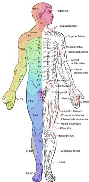
Dermatome Map
Why Check: Sensory testing of both Posterior Column and Spinothalamic Tracts and mapping out areas of sensory loss helps differentiate “where is the lesion” – decussation (crossing over) of posterior column tract occurs at Medulla (brainstem), whereas decussation of spinothalamic tract occurs within 1-2 spinal levels. Example distributions for different lesions include:
| Lesion | Posterior Column (Touch, Pressure, Proprioception) | Spinothalamic (Pain & Temperature) |
|---|---|---|
| Mononeuropathy (e.g. Carpal Tunnel Syndrome – median nerve compression) | Peripheral nerve distribution (e.g. Median nerve – Palmar aspect first 3 ½ digits) | Peripheral nerve distribution (e.g. Median nerve – Palmar aspect first 3 ½ digits) |
| Symmetric Generalized Polyneuropathy (e.g. Diabetic neuropathy, Chemo-induced neuropathy, Guillain-Barre) | Glove-and-stocking distribution | Glove-and-stocking distribution |
| Sensory Nerve Root Impingement | Dermatomal distribution of sensory nerve root (Dermatome Map) | Dermatomal distribution of sensory nerve root (Dermatome Map) |
| Complete spinal cord injury | Bilateral deficit up to level of lesion. | Bilateral deficit up to level of lesion. |
| Hemi-Section of Spinal Cord | Ipsilateral deficit below level of lesion. | Contralateral deficit below level of lesion, ipsilateral at level of lesion. |
| Anterior Spinal Artery Syndrome (e.g. infarct of anterior spinal artery) | Normal posterior column testing | Bilateral deficit up to level of lesion. |
| Brainstem Stroke Syndromes | Medial Brainstem stroke: Contralateral deficit of body | Lateral Brainstem stroke: Ipsilateral hemi-sensory deficit of face, Contralateral deficit of body |
| MCA Territory Stroke | Contralateral deficit (lower limb relatively spared) | Contralateral deficit (lower limb relatively spared) |
| ACA Territory Stroke | Contralateral deficit of lower limb | Contralateral deficit of lower limb |
Coordination Exam
Finger-to-nose Testing
AFMC level Pre-clerkship 3 · Clerkship 4
Say: I will assess coordination with upper limb movements.
Do:
- Stand at arms’ length distance from the patient so they need to extend their arm to reach your finger.
- Ask patient to touch their nose with their left index finger while clinician holds their index finger in front.
- Ask patient to now touch the clinician’s index finger with their left index finger.
- Ask the patient to then move back and forth between touching their nose and clinician’s index finger.
- Observe for smooth movement with reasonable accuracy and precision. Test patient’s right hand as well.
- Clinician can increase difficulty by moving their finger to different location and ask the patient to continue to move back and forth between touching their nose and clinician’s index finger – this tests ataxia.
Report: Normal finger-to-nose movements bilaterally on examination.
Why Check: Finger-to-nose testing is a Point-to-Point movement exam. Abnormal finger-to-nose testing may be caused by a cerebellar lesion (dysmetria – lack of coordinated movement leading to misjudging distance to target) or a tremor (involuntary, rhythmic, oscillatory movement of body part). Cerebellar lesions cause Ipsilateral deficits (e.g. Right cerebellar infarct causing Right-sided dysmetria). Note: subtle incoordination can be normal especially in non-dominant hand.
Rapid Alternating Movements of Upper Limb
AFMC level Pre-clerkship 3 · Clerkship 4
Say: I will assess coordination with rapid movement of the upper limbs. Please follow my example.
Do:
- Ask patient to flex elbows to 90° with palms up. Ask patient to pronate right hand, clap palms together.
- Ask patient to quickly remove right hand, supinate right hand, and clap dorsum of right hand to left palm.
- Helpful to demonstrate the maneuver for the patient to model the task.
- Ask patient to go back and forth alternating clapping palm and dorsum of right hand to left palm as fast as they can. Model maneuver. Observe for smooth movement with reasonable accuracy and precision with reasonable frequency. Test with left hand rapid alternating movements as well.
Report: Normal rapid alternating movements of the upper limb bilaterally.
Why Check: Dysdiadochokinesia is the term for inability to perform rapid alternating muscle movements. This may be caused by Cerebellar ataxia (lack of coordination of voluntary movements), although other movement disorders (e.g. Parkinson’s Disease with rigidity and bradykinesia) may create a false impression of dysdiadochokinesia. Cerebellar lesions cause Ipsilateral deficits (e.g. Left cerebellar infarct causing Left-sided dysdiadochokinesia).
Heel-to-Shin Testing
AFMC level Pre-clerkship 3 · Clerkship 4
Say: I will assess coordination with lower limb movements.
Do:
- With patient lying supine, ask patient to place their right heel on their left shin just below knee. Ask patient to move right heel smoothly down the left shin to ankle. Repeat test with left heel movement.
- Observe for smooth movement with reasonable accuracy and precision (not falling off the shin, etc.).
Report: Normal heel-to-shin movements bilaterally on examination.
Why Check: Heel-to-Shin testing is a Point-to-Point movement exam. Abnormal heel-to-shin testing may be caused by a cerebellar lesion (dysmetria – as above), motor deficit (tested with motor exam), or loss of proprioception (tested with sensory exam). Cerebellar lesions cause Ipsilateral deficits (e.g. Right cerebellar infarct causing Right-sided dysmetria).
Toe-Tapping
AFMC level Pre-clerkship 3 · Clerkship 4
Say: I will assess coordination with rapid movement of the lower limbs by asking you to tap each foot.
Do:
- Ask patient to tap their right foot against the floor (e.g. if seated) or against examiner’s hand (e.g. if lying supine) as fast as they can.
- Observe for smooth movement with reasonable accuracy and precision with reasonable frequency. Test with left foot rapid alternating movements as well.
Report: Normal toe tapping on examination.
Why Check: Toe-Tapping test is a Rapid Alternating Movement of the lower limb, where an abnormal exam indicates dysdiadochokinesia i.e. inability to perform rapid alternating muscle movements. This may be caused by Cerebellar ataxia (lack of coordination of voluntary movements), although other movement disorders (e.g. Parkinson’s Disease with rigidity and bradykinesia) may create a false impression of dysdiadochokinesia. Cerebellar lesions cause Ipsilateral deficits (e.g. Left cerebellar infarct causing Left-sided dysdiadochokinesia).
Stance Assessment
Normal Stance
AFMC level Pre-clerkship 3 · Clerkship 4
Say: I will assess stance. Please stand up with both feet together and stay standing.
Do: Look whether the stance of the patient is wide based; look at their trunks.
Report: Patient’s stance appears to be steady and not wide-based.
Why Check: If patient feels unsteady and need to catch themselves, it could be due to vestibular lesions, cerebellar lesions (e.g. truncal ataxia – oscillation of body while standing), or loss of sensation in feet. The likelihood of ≥1 fall in the next year in community-dwelling patients ≥65 years old is increased by inability to rise from a chair of knee-height without using the chair arms (LR+ 4.3, LR- 0.77), and by abnormal tandem stand (LR+ 1.7-2.0, LR- 0.69-0.74). In patients living in nursing homes ≥65 years old, the likelihood of ≥1 fall in the next year is increased by inability to stand with feet together and eyes open for 10 seconds (LR+ 4.5).
Romberg Test
AFMC level Pre-clerkship 3 · Clerkship 4
Say: I will continue to assess the ability to maintain the same stance after I ask you to close both eyes.
Do: Stand behind patient, standing close by to prevent injury if the patient appears to be close to falling.
- Ask patient to stand still with both eyes open and arms at sides and heels together. Observe that patient is able to hold this stance without visible swaying.
- Ask patient to close their eyes while remaining still. Observe if patient’s stance visibly sways and loses their balance (Note: some swaying normal).
Report: Patient has a negative Romberg test.
Why Check: The components of balance are visual confirmation of position, non-visual confirmation of vision (including proprioception and vestibular input) and a functioning cerebellum. In cerebellar ataxia, a patient may be unable to hold a Romberg stance even with eyes open. A positive Romberg test is when swaying and loss of balance occurs after patient eyes are closed. A +ve Romberg suggests sensory ataxia (either proprioceptive or vestibular) rather than cerebellar ataxia. Note Romberg is unspecific and has low inter-observer agreement. In patients living in nursing homes ≥65 years old, the likelihood of ≥1 fall in the next year is increased by inability to stand with feet together and eyes open for 10 seconds (LR+ 4.5) – this is the first step of the Romberg Test (i.e. before patient closes eyes to remove visual component of balance).
Gait Exam
Normal Walking Gait
AFMC level Pre-clerkship 3 · Clerkship 4
Say: I would like to examine gait with walking. Could you walk in a straight line, and then turn around?
Do: Have patient remove their shoes and expose their ankles to improve your ability to assess. Observe for the heel strike, mid-stance, toe-off; hip movement; and arm swing – note you can only effectively observe one leg at a time. You should have them walk back and forth several times – realistically, how can you extract all the info you need to with one look?
Report: Normal gait assessment, with 40% swing phase, 60% stance phase, and able to turn quickly.
Why Check: If patient stops walking when talking, helps predict falls risk, where the likelihood of ≥1 fall in the next year in nursing home patients ≥65y is increased by stopping walking when talking (LR+ 3.0), and by stopping talking when walking (LR+ 8.8, LR- 0.6). In community-dwelling patients ≥65y, the likelihood of ≥1 fall in the next year is increased by slow gait (LR+ 2.0-2.1, LR- 0.73-0.80). Look for characteristic gaits:
- Foot drop: Weakness of ankle dorsiflexion, where foot remains in plantarflexion during stance phase.
- Unilateral causes: e.g. Deep Fibular Mononeuropathy; L5 Radiculopathy; Lumbosacral plexopathy
- Bilateral causes: e.g. ALS, Charcot-Marie-Tooth disease, Peripheral neuropathy e.g. due to Diabetes
- Hemiplegic gait (e.g. Stroke): Patient holds arm to one side (↓ arm swing) with circumducting gait (drags affected leg in semicircle due to foot drop and extensor hypertonia of lower limb)
- Diplegic gait (Spastic gait, e.g. Cerebral Palsy): Narrow gait base, dragging both legs, scraping toes.
- Parkinsonian gait (e.g. Parkinson’s Disease, Extra-pyramidal symptoms): Shuffling gait – Walking with slow and small steps, with narrow base (versus normal pressure hydrocephalus, where gait is wide-based, with a similar slow movement and small steps)
- The likelihood of suspected Parkinson’s disease is increased by shuffling gait (LR+ 3.3-15, LR- 0.32-0.5).
- Waddling gait (e.g. Duchenne Muscular Dystrophy): Bilateral weakness of hip abductors with shifting centre of gravity to compensate. Recall: unilateral weak hip abductor gives a Trendelenburg gait.
- Truncal Ataxia (Lesion to midline cerebellum i.e. vermis): Unsteady on standing, walking and sitting with oscillation of the body.
- Ataxia of Extremities (Cerebellar hemisphere lesion): Limb ataxia towards the side of the lesion.
- Sensory Ataxia (Loss of Proprioception, lesion to Posterior Columns or Spinocerebellar tract e.g. B12 deficiency, Uncontrolled diabetes): Wide-based, careful gait “stamping on the ground”
- Choreiform Gait (e.g. Huntington’s disease): Irregular, jerky, involuntary movements in all extremities.
Tandem Gait
AFMC level Pre-clerkship 3 · Clerkship 4
Say: I will continue to examine gait, this time walking in a line with one foot in front of the other (point to a line on the floor if available e.g. line formed by floor tiles).
Do: Observe whether they can maintain some steadiness during the gait – does not need to be perfectly linear but should approximate walking a line.
Report: Normal tandem gait assessment.
Why Check: Can help to assess for ataxia, especially truncal ataxia; and detect subtle signs of cerebellar dysfunction. The likelihood of suspected Parkinson’s disease is increased by difficulty walking heal-to-toe (LR+ 2.9, LR- 0.32). The likelihood of ≥1 fall in the next year in nursing home patients ≥65y is increased by inability to tandem walk (LR+ 1.7, LR- 0.7).
Part 4: Select Special Tests – Not Part of Screening Neurological Exam But Seen in Practice
Hearing Loss Screening Test Alternative: Finger Rub Test
Say: I will now assess hearing. I will stand behind you and rub my fingers together close to one of your ears with your eyes closed and arms at the sides. Please raise your hand on the side you hear the fingers rubbing.
Do: Standing in front of the patient, once their eyes are closed, bring your arms close to each side of the patient’s ears, ensuring there is no contact with the patient’s outer ear or hair. On each side in a random order, rub your thumb against your index finger while progressing to the middle finger and then back to the index finger three times. Test this with a strong rub (as loud as you can muster); as well as a faint rub (the softest you can hear as the clinician). Monitor for the patient raising their hand to the side they hear the finger rub. Randomize the order of these 4 maneuvers (e.g. left ear, right ear, right ear, left ear).
Report: The patient heard both a strong and faint rub in bilateral ears. This is a normal finger rub screening test for hearing loss.
Why Check: The finger rub test is a helpful initial screening test for hearing loss, with an alternative screening test being the Whispered Voice Test (as detailed above in CN VIII testing). The test can also be performed by standing in front of the patient. The likelihood of hearing loss is greatly increased by being unable to hear a strong finger rub (LR+ 355.4)21. Being able to hear a faint finger rub greatly decreases the likelihood of hearing loss (LR- 0.02)21. Note that ambient noise can impact the performance of this test, where a study demonstrated that being unable to hear a strong finger rub in a quiet environment greatly increases the likelihood of hearing loss (LR+ 16.73), while the same result in a noisy environment moderately increases the likelihood of hearing loss (LR+ 6.76)22. This test cannot differentiate sensorineural from conductive hearing loss, where Weber and Rinne testing (see below) can help delineate conductive, sensorineural and mixed hearing loss. An abnormal screening test should be referred for formal audiology assessment.
Hearing Loss Assessment Alternative: Hum Test
Say: I will assess your hearing. Please hum an “eee” or “hmm” sound at a consistent pitch for a few seconds. It can be helpful to repeat the test with both high- and low-pitch hums. Please tell us which ear is loudest.
Do: Ask patient to hum an “eee” or “hmm” sound at a consistent pitch for a few seconds as noted above, with potential variation of the pitch over multiple trials. Ask the patient which ear hears the hum the loudest.
Report: The patient reports hearing loss in their left ear, and the Hum test lateralizes to the left ear, consistent with conductive hearing loss.
Why Check: For patients with hearing loss, the Hum test is a simple alternative to the Weber’s test which doesn’t require a tuning fork. Interpretation is the same as the Weber’s test: if the sound lateralizes to the affected ear, this suggests conductive loss. If it lateralizes to the unaffected ear, this suggests sensorineural hearing loss. The Hum test can be helpful in Telemedicine and triaging patients needing urgent assessment for Sudden Sensorineural Hearing Loss (SSNHL)23 where tuning forks are unavailable at initial intake.
Pearl: You can test the hum test on yourself: use your finger to plug and unplug your ear (conductive hearing loss) while making a hum, and see which side the sound lateralizes to.
HINTS Exam
(Conditional Test – Explored in Ch 7 – Head and Neck Exam – MF1)
- When to do the HINTS+ Exam? The HINTS+ exam is only indicated with acute onset, continuous dizziness, imbalance, and nystagmus lasting hours to days (Acute Vestibular Syndrome = AVS). Patients must have ongoing nystagmus at rest (involuntary repetitive jerking eye movements) to be eligible for the HINTS exam. Patients presenting with the acute vestibular syndrome could be having a peripheral (nerve) problem (acute vestibular neuritis) or a central (brain) problem (stroke or other acute brain lesion). The HINTS exam is a battery of 3 bedside tests which accurately differentiate peripheral versus central causes of the acute vestibular syndrome – but it is only validated in patients with ongoing, continuous acute dizziness and nystagmus. If the patient does not have continuous nystagmus, the HINTS exam is NOT indicated. Make sure you do a history and physical to exclude any RED FLAGS for stroke (explored in Chapter 7 – Head and Neck Exam) prior to doing the HINTS+ exam.
- Head Impulse (this tests the patient’s vestibular-ocular reflex, a reflex arc which enables the eyes to automatically adjust to head movements to keep vision stable): Patient’s head centred; eyes fixed on your nose. Examiner turns patient’s head slowly 20 degrees to one side, and then briskly and suddenly towards centre. Normally, the patient’s eyes will stay fixed on your nose if their vestibular nerve is intact. If they have impairment of the vestibular-ocular reflex – you will see the eyes momentarily lose focus with the head turn, and then snap back (catch-up saccade) to focus on you. A positive catch-up saccade suggests vestibular nerve dysfunction, which will always occur if the patient has vestibular neuritis (peripheral cause of AVS), but is not typically seen in a stroke or other central cause of AVS.
- Nystagmus: Patient’s head centred; observe their eyes for nystagmus. Ask patient to look to left and observe for nystagmus – is it left-beating or right-beating? Then ask patient to look to right and observe for directional nystagmus – did direction of the nystagmus change (bidirectional) or stay the same (unidirectional)? Unidirectional nystagmus suggests peripheral vertigo, where the fast phase beats away from the side with the lesion. Bidirectional, or vertical nystagmus suggests central vertigo.
- Test of Skew: Patient head centred; eyes fixed on your nose. Examiner covers one eye with their hand. After a second, move to cover the opposite eye. Watch the now uncovered eye – do you see any vertical skew? (post-uncovering correction to refocus on your nose). Repeat with the other eye. Vertical or Diagonal Skew deviation suggests a central lesion.
- + a bedside test of hearing: the plus in HINTS+ is a bedside test of hearing. Use a finger rub 2cm from the patient’s ears to screen for a new significant hearing loss. New hearing loss could indicate Anterior Inferior Cerebellar Artery Stroke infarcting the entire labyrinth, so a new hearing loss raises the concern for a central cause. New hearing loss could also be present in peripheral problems like labyrinthitis and sudden sensorineural hearing loss. Regardless, it is important not to miss a new hearing loss in patients with the acute vestibular syndrome.
Why Check: In a patient with ONGOING dizziness/vertigo lasting hours to days and spontaneous nystagmus (acute vestibular syndrome), it is important to separate central causes (e.g. brainstem/cerebellar ischemia) from peripheral causes (e.g. vestibular neuritis).
- Head Impulse Test: Tests the Vestibulo-ocular reflex – which should be preserved in a central cause and affected in a peripheral cause on the ipsilateral side.
- Nystagmus: In a peripheral cause, there is a unilateral imbalance of vestibular signal from the affected side. This means the unaffected side is unopposed, and eyes are pushed towards the lesioned side. The fast phase of nystagmus gives rise to observed left-beating vs. right-beating. This activity represents the brain correcting for the deficiency of unilateral peripheral vestibular input. By contrast, nystagmus that is vertical or direction changing would not be seen in vestibular neuritis (peripheral cause) and points towards a central cause.
- Test of Skew: Tests vestibular input to the oculomotor system. A positive vertical test of skew is concerning for central vertigo.
Dix-Hallpike Test
AFMC level Pre-clerkship 3 · Clerkship 4
(Conditional Test for posterior canal BPPV – See Ch 7 – Head and Neck Exam – MF1)
Provocative Testing for Diagnosing BPPV
Benign Positional Paroxysmal Vertigo (BPPV) is caused by loose debris (otoconia) within one of the semi-circular canals of the vestibular apparatus. Each ear has 1 vestibular apparatus and each vestibular apparatus has 3 semi-circular canals. The most common location for otoconia in BPPV is the posterior canal (~65%), followed by the horizontal canal (~30%), while the anterior canal is relatively rare. Movement of loose otoconia within a semicircular canal will reliably produce vertigo with a different type of nystagmus depending on which canal is affected. Therefore, we use provocative testing in BPPV to watch for which type of nystagmus and LOCALIZE which ear and canal the otoconia are in. Once we have localized the otoconia, we can then use the appropriate particle repositioning maneuver to correct the problem and cure our patients.
Provocative tests for BPPV are indicated only if you suspect a diagnosis of BPPV based on a history of brief, (less than one minute), intense attacks of dizziness that are always triggered by head movements (most commonly lying down or turning over in bed). A patient who has continuous dizziness present at rest lasting hours to days should never get provocative tests for BPPV. You should also ensure that the patient does not have any red flags for a serious central cause of dizziness, and that there is not another cause (such as postural hypotension or vestibular migraine or Meniere’s disease) that better explains their symptoms, prior to provocative tests.
Since the most common type of BPPV is posterior canal (~65%), this is the first canal which we test in a patient with suspected BPPV. Posterior canal BPPV is identified using the Dix-Hallpike Test.
Say: I will move you from a sitting to lying down position on the exam table and move the neck to the side and back. Please tell me if these movements reproduce your symptoms of vertigo.
Do: Have patient sit upright on exam table with legs extended. Rotate head by 45° to one side. Maintaining the 45° rotation, lie the patient back, hang their head over edge of the exam table, and extend their neck an additional 20°. Look for reproduction of symptoms AND observe for nystagmus in patient’s eyes. Posterior canal BPPV produces an upwards, torsional nystagmus. There is often a latent reaction for about 5 seconds before the nystagmus is generated. Repeat with rotating head to other side to test the other ear.
Report: I cannot reproduce upwards torsional nystagmus or vertigo with a Dix-Hallpike test on either side, therefore it is unlikely the patient has posterior canal BPPV
Why Check: Used to diagnose and localize the affected ear in patients with Posterior Canal Benign Paroxysmal Positional Vertigo (BPPV). Eliciting ~10-30 seconds of upwards, torsional nystagmus is diagnostic for posterior canal BPPV on the side you are testing. Once you have localized which ear has posterior canal BPPV, you can proceed to particle repositioning maneuver (Epley or Semont) for posterior canal BPPV on the affected side.
Supine Roll
AFMC level Pre-clerkship 3 · Clerkship 4
(Conditional Test for horizontal canal BPPV – See Ch 7 – Head and Neck Exam – MF1)
Since the second most common type of BPPV is horizontal canal (~30%), this is the second canal which we test in a patient with suspected BPPV. Horizontal canal BPPV is identified using the Supine Roll test.
Say: I will reposition you to look to one side and then the other side while lying flat on your back.
Do: Have patient lie supine on an exam bed. Turn their head to one side and examine their eyes for nystagmus. Then turn their head to the other side and examine for nystagmus. If they have horizontal canal BPPV, you will see horizontal nystagmus on both head turns which is either towards the ground (geotropic) or away from the ground (apogeotropic).
Report: There is no horizontal nystagmus on supine roll test, therefore it is unlikely the patient has horizontal canal BPPV.
Why Check: Used to localize the affected ear in patients with Horizontal Canal Benign Paroxysmal Positional Vertigo (BPPV). On the side that has the more intense nystagmus, the nystagmus will beat towards the affected ear. When you have identified which ear is affected and whether it is geotropic or apogeotropic, you can proceed with an appropriate particle repositioning maneuver (Gufoni or Barbecue roll) for the affected side.
Additional Cerebellar Examination Tests
AFMC level Pre-clerkship 3 · Clerkship 4
- Core Elements on Screening Exam Above: Oculomotor (Nystagmus, or Saccadic movements on smooth pursuit); Scanning Speech; Dysarthria/Dysphonia (errors in impulse timing of speech muscles) Coordination Exam elements above; Pendular reflexes; Gait Assessment (Truncal Ataxia); Hypotonia
- Pronator Drift of Upper Extremity: Have patient hold arms out for 30 seconds in supinated positions with eyes closed (thus, taking away one input to the cerebellum – patient must rely on proprioception to maintain arm position).
- UMN Lesion: Disinhibition of muscles – Pronator muscles stronger than Supinator, so will see arms drifting downwards towards floor. Drift while supinated does not indicate specific lesions.
- Cerebellar Lesion: Inability to maintain position of arms; you will see an upward drift.
- Rebound Test: At the end of the Pronator Drift test, keep the patients’ eyes closed. Press down on the palms of both arms simultaneously, and release. You should see arms rebound back to starting position
- Cerebellar Lesion: You will see an accentuation of the upward drift.
- Arm/Finger Rolling Test: Ask the patient to hold their arms up in front of them, elbows flexed at 90º, forearms parallel to the ground, and with one forearm/finger above the other. Then, have the patient make a forward cycling motion with their arms/fingers.
- Finger Tapping Test: Ask the patient to tap /oppose the tip of their thumb to the tip of each finger. Pay attention to their ability to do the task, while also looking for involuntary facial movements (e.g. tardive dyskinesia, extrapyramidal symptoms).
Why Check: To differentiate an UMN lesion from a Cerebellar Lesion. The likelihood of unilateral cerebral hemispheric disease is greatly increased by an abnormal arm rolling test (LR+ 21.7), abnormal pronator drift (LR+ 10.3, LR- 0.1), and moderately increased by abnormal finger tapping test (LR+ 6.6, LR- 0.3).
Additional Posterior Column Sensory Tests
AFMC level Pre-clerkship 3 · Clerkship 4
Note: Also seen in Ch 14 – Diabetes Foot Exam – MF3.
- Vibration Test (Pressure): Place 128 Hz tuning fork on bony prominence, especially D1 of Foot to test vibration sense. If no sensation, move to more proximal bony prominences, such as medial malleolus.
- Proprioceptive Test: Stabilizing the hallux joint at its sides, show what up and down is with eyes open. Then, have the patient close their eyes, and move up and down randomly.
Why Check: Helps further define sensory deficit. Note that cotton ball assesses light touch as component of posterior column testing.
- Vibration: ↓ in peripheral neuropathies and spinal cord disease
- Proprioception: ↓ in peripheral neuropathies, spinal cord disease and cortico-hemispheric disease
- The likelihood of ≥1 falls in the next year in community-dwelling patients ≥65y is increased by absence of vibratory sensation (LR+ 2.0-2.1, LR- 0.73-0.80).
Cortical Sensation Testing
AFMC level Pre-clerkship 3 · Clerkship 4
- Stereognosis: Ability to recognize objects placed in hand. Use common items easily/safely manipulated in hand (pen cap, coin, watch, paper clip).
- Graphesthesia: Ability to recognize numbers or letters traced on skin. Note that some drawings are difficult to distinguish in a normal person (e.g. drawing a 3 poorly could be interpreted as an 8).
- Two-Point Discrimination: Ability to discriminate between when two points on body are stimulated, vs. only one. To test, touch individual body parts B/L at same time; more sensitive if testing different regions B/L (e.g. Point 1 = right face; Point 2 = left hand). Alternate between touching only Point 1, only Point 2, and both, and ask which was touched each time.
Why Check: For cortical sensation, intact primary sensation (i.e. Posterior Column & Spinothalamic Tracts in Screening exam normal) is necessary but not sufficient to preserve cortical sensation (higher order sensory testing). If primary sensation deficits (spinal cord or lower) exist, these tests will generate false +ve cortical sensation deficits:
- Deficits in Stereognosis and Graphesthesia may indicate lesion to contralateral primary sensory cortex (post-central gyrus), or secondary association areas in parietal, temporal or occipital lobes.
- Deficits in Two-point discrimination are a sign of sensory inattention, commonly localizing to a lesion in parietal lobe of non-dominant hemisphere (Right-handed = Left-dominant; Left-handed = 90% of patients Left-dominant, 10% Right-dominant).
Brainstem Reflexes
Say: I will now test the brainstem reflexes.
Do:
- Pupillary Light Reflex: Shine penlight into each eye, testing the direct and consensual response. With intact brainstem, see both direct and consensual pupillary constriction. If pupillary light reflex is lost, the pupils will be mid-position and size 3.5-4mm with no pupillary constriction direct or consensual.
- Corneal Reflex: Touch cornea with a small piece of wet cotton wool, or squirts of water or saline to one side. Observe whether both eyes blink.
- Oculocephalic Reflex (Doll’s Eye – AVOID in C-spine trauma): Observe eyes with passive rotation of head – with intact brainstem, patient eyes will move in opposite direction to head movement. If doll’s eye reflex lost, globes will remain fixed, and eyes will stare in whatever direction the head is pointed.
- Oculovestibular Reflex (Cold Calorics):
- Inspect tympanic membrane first to ensure no perforation or impacted cerumen
- With head raised 30 degrees from horizontal (to facilitate irrigation), irrigate 50-120mL ice water slowly into auditory canal. Wait 5 minutes, then repeat on other side. If brainstem intact, expect tonic deviation of both eyes toward irrigated ear. If cold caloric reflex lost, eyes would not move.
- Jaw Jerk Reflex: Ask patient to open mouth and relax jaw. Place finger on chin and tap with reflex hammer. Observe for increased jerk (slight jerk is normal - ↑ jerk indicates bilateral UMN lesion).
- Gag Reflex: Use tongue depressor or suction tube to depress patient’s tongue. Touch palate, pharynx or tonsil on each side until patient gags. If brainstem intact, observe soft palate rises symmetrically.
- Cough Reflex (intubated patients): Apply deep suction through endotracheal tube to the carina. If brainstem intact, will see gasp and several rapid coughs.
Why Check: Brainstem Reflexes test Cranial Nerves spanning from Midbrain to Medulla, as follows:
| Brainstem Reflex | Afferent Arc | Efferent Arc | Brainstem Area |
|---|---|---|---|
| Pupillary Light | CN II | CN III (E-W) | Midbrain |
| Corneal Reflex | CN V1 | CN VII | Pons |
| Oculocephalic Reflex (Doll’s Eye) | CN VIII | CN III, IV, VI | Pons-Midbrain |
| Oculovestibular Reflex (Cold Calorics) | CN VIII | CN III, IV, VI | Pons-Midbrain |
| Jaw Jerk | CN V3 | CN V3 | Pons |
| Gag | CN IX | CN X | Medulla |
| Cough | CN X | CN X | Medulla |
- Diagnosis 1: Brain Death – 3 conditions required:
- Persistent Coma: No eye, verbal or movement response to painful stimulus (Glasgow Coma Scale 3)
- No Brainstem Reflexes:
- Apnea Test: No breathing for 8-10 minutes without mechanical ventilation in response to PaCO₂ >60mmHg or 20mmHg greater than baseline values and final arterial pH <7.28.
- Prerequisites: With patient Core temperature >36°C, Systolic BP >100mmHg, PaCO2 35-45mmHg (i.e. ABG pre-test), absent hypoxia, and euvolemic state.
- Not Valid In: CO2-retainers; Neuromuscular paralysis; or high C-spine cord lesions.
- Diagnosis 2: Persistent Vegetative Stage: Permanent after 6 months (non-traumatic), 1 yr. (traumatic)
- No awareness of self or environment, inability to interact with others.
- No evidence of sustained, reproducible, purposeful or voluntary behavioural responses to stimuli.
- No evidence of language comprehension or expression.
- Intermittent wakefulness with presence of sleep-wake cycles.
- Sufficiently preserved hypothalamic and brainstem function to permit survival with medical care.
- Bowel and bladder incontinence.
- Variably preserved cranial nerve reflexes and spinal reflexes.
- Diagnosis 3: Minimally Conscious State: Intermittent, limited awareness of self or environment, ability to interact with others (e.g. follow simple commands, signal yes/no, intelligible verbalization, restricted purposeful behaviour). May have more variable neurological prognosis.
- In patients with coma, the likelihood of intracranial structural lesion is increased by pupil anisocoria >1mm (LR+ 9.0, LR- 0.6), and by absent light reflex (LR+ 3.6, LR- 0.2).
- The likelihood of Horner Syndrome is greatly increased by post-topical cocaine anisocoria (LR+ 96.8, LR- 0.1), and by dilation with topical hydroxyamphetamine (LR+ 9.2, LR- 0.2).
- In patients 24h post-cardiac arrest, the likelihood of death, vegetative state or severe neurologic impairment is increased by absent corneal reflex (LR+ 13) and absent pupillary response (LR+ 10).
- The likelihood of significant aspiration risk in patients with recent strokes is increased by absent pharyngeal sensation (LR+ 2.4, LR- 0.03), abnormal voluntary cough (LR+ 1.9, LR- 0.6), dysarthria (LR+ 1.6, LR- 0.5), abnormal gag reflex (LR+ 1.5, LR- 0.6), and dysphonia (LR+ 1.3, LR- 0.4).
Frontal Release Signs
AFMC level Pre-clerkship 2 · Clerkship 4
- Glabellar Tap Sign: Tap patient’s forehead above eyes with your index finger 5-10 times. This causes reflexive contraction of orbicularis oculi muscle where both eyes blink. The patient should stop blinking after 5-10 taps. If blinking persists → Myerson’s sign, suggests frontal release seen in Parkinsonism
- Palmomental Reflex – scrape the palm, look to see if the omentum (chin) moves; abnormal to elicit the chin muscle twitch re positive frontal release sign
- Palmar Grasp – place object in patient’s palm; assess whether patient flexes their fingers in response.
- Plantar Grasp – place object in patient’s sole of foot; assess whether patient curls toes in response.
Why Check: Primitive reflexes exist in infancy, disappear as brain matures in childhood, but can then reappear if normal cortical inhibitory pathways are disrupted. An abnormal Glabellar Tap Sign increases the likelihood of suspected Parkinson’s Disease (LR+ 4.5, LR- 0.13). The likelihood of ≥1 falls in the next year in nursing home residents ≥65y is increased by presence of palmomental reflex (LR+ 2.8, LR- 0.8).
Digital Rectal Exam (for Rectal Tone)
AFMC level Pre-clerkship 2 · Clerkship 4
For instructions on performing a DRE, please see Chapter 13 – Abdominal Examination, in MF3
Why Check: A decreased anal tone is indicative of a UMN lesion and is a critical screen when you’re clinically differentiating an UMN lesion from a LMN lesion (where tone is preserved).
Parietal Neglect
AFMC level Pre-clerkship 3 · Clerkship 4
Patient keeps both eyes open, examiner’s hands in both right and left visual fields. Move fingers in either field – assess whether if moving both left and right simultaneously, but they can only see one.
Why Check: If you suspect a lesion in parietal lobe (spatial awareness, perception & integration centres).
Gower’s Sign (Pediatrics)
When rising from a seated position, you normally need to use hip extension, knee extension and ankle dorsiflexion. In a +ve Gower’s sign, children climb up themselves to rise from a chair (Typically Ages 4-7)
Why Check: Gower’s sign is useful for Duchenne’s Muscular Dystrophy screening, as it tests the proximal muscle groups that patients with DMD exhibit motor weakness in.
Subclavian Steal Screen
Arm Exercises: Repetitive arm motions, see if the patient has become dizzy
Why Check: Subclavian Steal is Retrograde blood flow in vertebral artery or internal thoracic artery due to proximal stenosis or occlusion of subclavian artery. Causes vertigo, visual changes, loss of consciousness. Patients may complain of chest pain as well–episodes can be provoked by exercise of the ipsilateral arm.
Meningismus
AFMC level Pre-clerkship 3 · Clerkship 4
- Kernig’s Sign: Patient lies supine with one leg on exam table, and one leg with hips and knees flexed at 90 degrees. Patient resists full extension of the flexed knee by the examiner. Repeat on the other side. A positive sign is resistance or pain and inability to extend patient’s knee beyond 135 degrees because of pain.
- Brudzinski’s Sign: Patient lies supine; examiner passively flexes the neck. A positive sign for meningeal irritation is bilateral flexion of hips/knees on passive flexion of neck.
- Jolt Accentuation of Headache: Ask the patient to turn their head horizontally at a frequency of 2-3 rotations per second. Worsening of baseline headache represents a positive sign.
Why Check: The classic triad of meningitis is fever, headache and neck stiffness (sometimes aLOC is included), however often only 1-2 of these will be seen in patients with meningitis. Fever has a sensitivity of 87%, Headache has a sensitivity of 68%, Neck stiffness has a sensitivity of 80%, and Altered Mental Status has a sensitivity of 69% for adult patients with acute meningitis, where the classic triad of fever, headache and neck stiffness has a sensitivity of only 46%. Kernig’s & Brudzinski’s Signs along with Jolt accentuation of headache are all tests of meningeal irritation. Do NOT perform Brudzinski’s sign in patients with suspected cervical instability e.g. trauma presentations.
Kernig Sign and Brudzinski Sign are overall poorly diagnostic of acute meningitis, however, the likelihood of suspected bacterial meningitis is mildly increased by positive jolt accentuation (LR+ 2.4) and greatly decreased by a negative jolt accentuation (LR- 0.05). Neck stiffness also greatly increases the likelihood of suspected subarachnoid hemorrhage (LR+ 10.3, LR- 0.4).
Timed Up-And-Go (TUG) Test
Say: I will assess your ability to rise from your chair, walk a short distance at your normal speed, and then come back to sit down in your chair. Please use your mobility aid (e.g. walker, cane) if needed.
Do: Allow patient one practice run. Patient rises from their chair, and timer starts once the patient’s back is off their chair. Patient walks a line on the floor 3 meters away. They then turn, return to the chair, and sit down again. Timer stops when the patient’s buttocks touch the seat of the chair.
Report: The patient’s timed up-and-go test was 12 seconds.
Why Check: The Timed Up and Go test is important in assessing patients for falls risk. The likelihood of ≥1 fall in the next year in nursing home patients ≥65y is mildly increased by timed up-and-go test >35 seconds (LR+ 2.6), while it is greatly decreased by timed up-and-go test <15 seconds (LR+ 0.1).
Part 5: Pediatric Neurological Examination –Key Differences from Adult Neuro Exam
-
Creative, Opportunistic and Observant Examination: Tailored to child’s needs and temperament. Observing a child playing and interacting with their caregivers provides a wealth of information for the Pediatric Neurological examination. Age-appropriate toys are critical for pediatric neurological exam → visual acuity: name objects varied sizes, shapes, colours; visual fields: bring toy from temporal to nasal visual field; extra-ocular movement: move small toy in visual field→child looks in all directions of gaze; Cerebellar Exam: Reach for toy (gross coordination); stack blocks; place objects in containers.
-
Head Circumference Measurements: Microcephaly (small head) or macrocephaly (large head) are key clues for many peds neuro conditions. Defer to end of exam re. irritates kids (Say: I’m going to measure your crown, etc.).
-
Floppy Baby Approach: Through your postural reaction exam (see Chapter 17 – Pediatrics – MF3) you may identify a “floppy baby” – see right to differentiate central and peripheral causes.
Criteria Central (Brain/Spine) Peripheral (Ant. Horn, Nerve, NMJ, Muscle) Alert ↓ / Normal Normal Bulk Normal Normal / Atrophy Power Normal / ↓ ↓ Tendon Reflexes Normal/ ↑ ↓/ Absent Primitive Reflexes ↑↑ ↓ -
Fontanelles in Pediatric Neurology: Anterior Fontanelle is formed by Metopic suture (anterior), 2 Coronal sutures (anterior) and Sagittal suture (posterior), closing at 1.5-2 years of age. Posterior Fontanelle formed by Sagittal suture (same) and 2 Lambdoid sutures (posterior coronal plane), closes at 2-3 months of age.
- Clinical Correlation – Pediatric Hydrocephalus: Bulging fontanelles (if not closed), along with Head Circumference trends can track hydrocephalus, Neurosurgical drainage (e.g. Ventriculoperitoneal Shunt, Extraventricular Drains).
- Clinical Correlation – Craniosynostosis: Premature Fusion of head sutures, common referral to Peds Neurosurgery.
-
Neuro-cutaneous Manifestations – Key Conditions to Know:
- Tuberous Sclerosis: Seizures in infancy (esp. infantile spasms), developmental delay and cognitive impairment due to Tuberous Sclerosis Complex (TSC) → altered cell differentiation with ↓ neuron counts, abnormal astrocytes.
- Dermatological Signs: Hypomelanotic macules – may develop plaques with thick skin (Shagreen Patches).
- Other Organs: Rhabdomyoma of heart, retinal tumors, renal tumors, renal/bone/lung cysts.
- Neurofibromatosis (NF):
- Type 1 – Peripheral: Café au Lait spots, Subcutaneous Neurofibromas. Also present with learning disability.
- Type 2 – Central: Bilateral acoustic neuromas – Present with deafness, tinnitus. May have ipsilateral ↓ corneal reflex; ataxia; facial hypoesthesia (numbness on history) or facial weakness; and nystagmus.
- Sturge-Weber: Port-Wine Stain. Intracranial lesion is a venous angioma of the pia mater. May also present with cognitive impairment, seizures, hemiparesis, and glaucoma.
- Tuberous Sclerosis: Seizures in infancy (esp. infantile spasms), developmental delay and cognitive impairment due to Tuberous Sclerosis Complex (TSC) → altered cell differentiation with ↓ neuron counts, abnormal astrocytes.
Helpful Videos to Review
- Video 1: McMaster Screening Neurological Examination (Link: https://drive.google.com/file/d/0B-YGIFSw7ZZ0Z25rWHVIN1N5VEE/view?resourcekey=0-j3XC7XxyKianiAPJ3JkLww, requires Medportal Login)
- Video 2: McMaster Pediatric Neurologic Exam Module (Link: https://www.medportal.ca/Files/emodules/Pediatric%20Neurologic%20Exam/index.html, requires Medportal Login)
- Video 3: Posterior Canal BPPV Diagnosis Using Dix Hallpike Test: https://www.youtube.com/watch?v=kvVnEsGVLUY
- Video 4: Quick Review of HINTS exam: https://www.youtube.com/watch?v=-VXwD2nskhQ
- Video 5: Horizontal Canal BPPV Diagnosis Using Supine Roll Test https://www.youtube.com/watch?v=VRjRTnIw9YE
Part 6: Relevant Likelihood Ratios – from Rational Clinical Examination JAMA series
Clinical Question 1: Does this patient with a Coma have an Intracranial Structural Lesion?24
| Procedure | LR+ (95% CI) | LR- (95% CI) |
|---|---|---|
| Systolic Blood Pressure >160 mmHg | 10.4 (6.0-18.2) (about +45%) | N/A |
| Systolic Blood Pressure 120-159 mmHg | 1.2 (0.9-1.6) (no change: 95% CI includes 1) | N/A |
| Systolic Blood Pressure <120 mmHg | 0.1 (0.1-0.2) (about −45%) | N/A |
| Pupil Anisocoria >1mm | 9.0 (2.8-28.8) (about +30%) | 0.6 (0.5-0.8) (about −15%) |
| Absent Light Reflex | 3.6 (2.3-5.6) (about +15%) | 0.2 (0.1-0.4) (about −30%) |
Clinical Question 2: Does this patient have Unilateral Cerebral Hemispheric Disease?25
| Procedure | LR+ (95% CI) | LR- (95% CI) |
|---|---|---|
| Arm rolling test | 21.7 (6.3-75) (about +45%) | 0.3 (0.1-1.1) (no change: 95% CI includes 1) |
| Plantars Upgoing | 19.0 (1.2-297.8) (about +45%) | 0.6 (0.4-0.7) (about −15%) |
| Hemianopia | 12.5 (0.8-199) (no change: 95% CI includes 1) | 0.7 (0.6-0.9) (about −15%) |
| Hemisensory disturbance | 12.3 (0.8-195.9) (no change: 95% CI includes 1) | 0.7 (0.6-0.9) (about −15%) |
| Pronator drift | 10.3 (5.4-19.6) (about +45%) | 0.1 (0.1-0.3) (about −45%) |
| Finger tapping test | 6.6 (3.8-11.6) (about +30%) | 0.3 (0.2-0.4) (about −30%) |
| Hyperreflexia | 5.8 (3.2-10.5) (about +30%) | 0.4 (0.3-0.5) (about −30%) |
Clinical Question 3: Does this patient have a Subarachnoid Hemorrhage (SAH)?24
| Procedure | LR+ (95% CI) | LR- (95% CI) |
|---|---|---|
| Neck stiffness | 10.3 (5.2-20.7) (about +45%) | 0.4 (0.3-0.7) (about −30%) |
| Neurologic findings not focal | 5.9 (3.5-9.9) (about +30%) | 0.4 (0.2-0.7) (about −30%) |
| Age <60 years | 1.8 (1.4-2.3) (about +15%) | 0.3 (0.1-0.8) (about −30%) |
| Seizures | 2.2 (1.1-4.5) (about +15%) | 0.8 (0.6-1.1) (no change: 95% CI includes 1) |
Clinical Question 4: Does this patient have a Horner’s Syndrome?24
| Procedure | LR+ (95% CI) | LR- (95% CI) |
|---|---|---|
| Post-Topical Cocaine Anisocoria | 96.8 (6.1-1527.3) (about +45%) | 0.1 (0 -0.1) (about −45%) |
| Dilation with topical hydroxyamphetamine (1st/2nd order neuron lesion) | 9.2 (2.0-43.6) (about +30%) | 0.2 (0.1-0.3) (about −30%) |
| Asymmetric Facial Sweating (1st/2nd order neuron lesion) | 2.4 (0.9-6.1) (no change: 95% CI includes 1) | 0.6 (0.4-0.9) (about −15%) |
Clinical Question 5: Does this patient have Diabetic Retinopathy?24
| Procedure | LR+ (95% CI) | LR- (95% CI) |
|---|---|---|
| Direct ophthalmoscopy through dilated pupils (Specialists) | 18.6 (5.7-61) (about +45%) | 0.3 (0.2-0.5) (about −30%) |
| Direct ophthalmoscopy through dilated pupils (General providers) | 10.2 (6.0-17.4) (about +45%) | 0.4 (0.3-0.5) (about −30%) |
| Direct ophthalmoscopy through nondilated pupils | 6.2 (2.5-14.9) (about +30%) | 0.5 (0.3-0.8) (about −15%) |
| Visual Acuity 20/60 or worse | 1.2 (0.7-2.3) (no change: 95% CI includes 1) | 1.0 (0.9-1.1) (no change: 95% CI includes 1) |
Clinical Question 6: Does this patient have Myasthenia Gravis?26
| Finding | LR + (95% CI) | LR – (95% CI) |
|---|---|---|
| Sleep test (patient left in quiet dark room for 30 minutes, positive test with complete or almost complete resolution of ptosis or ≥2mm increase in palpebral fissure width) | 53 (3.4-832) (about +45%) | 0.01 (0-0.16) (about −45%) |
| Peek sign (gently close patient eyelids, positive test if eyelids quickly separate < 30 seconds with sclera seen) | 30 (3.2-278) (about +45%) | 0.88 (0.76-1.0) (no change: 95% CI includes 1) |
| Ice test (placing latex glove finger filled with crushed ice over more ptotic eyelid for 2 minutes, positive test with complete or almost complete resolution of ptosis or ≥2mm increase in palpebral fissure width) | 24 (8.5-67) (about +45%) | 0.16 (0.09-0.27) (about −30%) |
| Rest test (placing glove filled with cotton i.e. placebo vs. ice test over more ptotic eyelid for 2 minutes while holding eyes closed, positive test with complete or almost complete resolution of ptosis or ≥2mm increase in palpebral fissure width) | 16 (0.98-261) (no change: 95% CI includes 1) | 0.52 (0.29-0.95) (about −15%) |
| Anticholinesterase test (edrophonium test, observing for immediate increase in muscle strength of affected muscles – not commonly performed in practice due to risk of bradycardia, asystole and bronchoconstriction) | 15 (7.5-31) (about +45%) | 0.11 (0.06-0.21) (about −30%) |
| Food in mouth after swallowing on history | 13 (0.85-212) (no change: 95% CI includes 1) | 0.70 (0.58-0.84) (about −15%) |
| Unintelligible speech after prolonged speaking on history | 4.5 (1.2-17) (about +15%) | 0.61 (0.46-0.80) (about −15%) |
| Quiver eye movements | 4.1 (0.22-75) (no change: 95% CI includes 1) | 0.82 (0.57-1.2) (no change: 95% CI includes 1) |
Clinical Question 7: Does this patient have Parkinson’s Disease?27
| Symptom or Sign? | Finding (n=No. of studies) | LR+ (95% CI) | LR- (95% CI) |
|---|---|---|---|
| Symptom | Shuffling Gait (n=2) | 3.3-15 (about +15%) | 0.32-0.50 (about −30%) |
| Bradykinesia (difficulty rising from a chair) (n=2) | 1.9-5.2 (about +15%) | 0.39-0.58 (about −30%) | |
| Loss of balance (n=2) | 1.6-6.6 (about +15%) | 0.29-0.35 (about −30%) | |
| Tremor (n=4) | 1.4-11 (about +15%) | 0.24-0.60 (about −30%) | |
| Rigidity (n=3) | 1.3-4.5 (about +15%) | 0.12-0.93 (about −30%) | |
| Sign | Rigidity and bradykinesia | 4.5 (2.9-7.1) (about +15%) | 0.12 (0.03-0.45) (about −30%) |
| Glabellar Tap | 4.5 (2.8-7.4) (about +15%) | 0.13 (0.03-0.47) (about −30%) | |
| Difficulty walking heel-to-toe | 2.9 (1.9-4.5) (about +15%) | 0.32 (0.15-0.70) (about −30%) | |
| Rigidity | 2.8 (1.8-4.4) (about +15%) | 0.38 (0.19-0.76) (about −30%) | |
| Asymmetry of disease | 1.8 (0.98-3.2) (no change: 95% CI includes 1) | 0.61 (0.41-0.91) (about −15%) | |
| Tremor | 1.5 (1.0-2.3) (no change: 95% CI includes 1) | 0.47 (0.27-0.84) (about −30%) |
Clinical Question 8: Does this patient with a Recent Stroke have a Significant Aspiration Risk?28
| Procedure | LR+ (95% CI) | LR- (95% CI) |
|---|---|---|
| Drowsiness | 3.4 (1.2-9.5) (about +15%) | 0.5 (0.3-0.7) (about −15%) |
| Water swallow test | 3.2 (2.1-4.7) (about +15%) | 0.4 (0.3-0.5) (about −30%) |
| Oxygen desaturation 0-2 min after swallowing | 3.1 (1.1-8.6) (about +15%) | 0.3 (0.2-0.5) (about −30%) |
| Tongue weakness | 2.5 (0.7-9.6) (no change: 95% CI includes 1) | 0.6 (0.4-0.9) (about −15%) |
| Absent pharyngeal sensation | 2.4 (1.6-3.6) (about +15%) | 0.03 (0-0.5) (about −45%) |
| Abnormal voluntary cough | 1.9 (1.3-2.7) (about +15%) | 0.6 (0.5-0.7) (about −15%) |
| Dysarthria | 1.6 (1.2-2.2) (about +15%) | 0.5 (0.3-0.8) (about −15%) |
| Abnormal gag reflex | 1.5 (1.2-1.8) (about +15%) | 0.6 (0.4-0.7) (about −15%) |
| Dysphonia | 1.3 (1.2-1.5) (about +15%) | 0.4 (0.3-0.7) (about −30%) |
| Bilateral cranial nerve signs | 1.1 (0.8-1.6) (no change: 95% CI includes 1) | 0.8 (0.4-1.6) (no change: 95% CI includes 1) |
| Abnormal sensation of face & tongue | 0.5 (0.2-1.2) (no change: 95% CI includes 1) | 1.5 (0.9-2.4) (no change: 95% CI includes 1) |
Clinical Question 9: Does this adult patient have Acute Meningitis?293031
Table 1: Sensitivity of findings for meningitis in adults, retrospective studies29
| Finding (n= No. of studies) | Sensitivity (95% CI) |
|---|---|
| Fever on Px (n=14) | 0.87 (0.79-0.92) |
| Neck stiffness on Px (n=13) | 0.80 (0.74-0.85) |
| Altered mental status on Px (n=15) | 0.69 (0.57-0.79) |
| Headache on Hx (n=11) | 0.68 (0.55 – 0.79) |
| Nausea and Vomiting on Hx (n=5) | 0.52 (0.34-0.71) |
| Classic triad on Px (fever, neck stiffness, headache) (n=4) | 0.46 (0.28-0.64) |
| Focal neurologic findings (n=12) | 0.21 (0.15-0.29) |
| Rash (n=6) | 0.13 (0.04-0.27) |
Table 2: Likelihood ratios for findings for meningitis in adults, prospective studies3031
| Finding | Sensitivity (95% CI) | LR+ (95% CI) | LR- (95% CI) |
|---|---|---|---|
| Neck stiffness on Px15 | 0.15 (0.06-0.28) | 6.6 (0.38-113) (no change: 95% CI includes 1) | 0.83 (0.74-1.0) (no change: 95% CI includes 1) |
| Kernig Sign on Px15 | 0.09 (0.02-0.21) | 4.2 (0.23-77) (no change: 95% CI includes 1) | 0.92 (0.81-1.0) (no change: 95% CI includes 1) |
| Jolt accentuation of headache on Px | 0.97 (0.83-0.99) | 2.4 (1.4-4.2) (about +15%) | 0.05 (0.01-0.35) (about −45%) |
| Nausea/Vomiting on Hx14 | 0.70 (0.59-0.79) | 1.3 (1.1-1.6) (about +15%) | 0.64 (0.44-0.92) (about −15%) |
| Headache on Hx | 0.92 (0.84-0.96) | 1.1 (1.0-1.3) (no change: 95% CI includes 1) | 0.43 (0.19-0.96) (about −30%) |
| Neck stiffness on Hx | N/A | 1.1 (0.82-1.4) (no change: 95% CI includes 1) | 0.95 (0.74-1.2) (no change: 95% CI includes 1) |
| Kernig Sign on Px14 | 0.05 (0.02-0.13) | 0.97 (0.27-3.6) (no change: 95% CI includes 1) | 1.0 (0.94-1.1) (no change: 95% CI includes 1) |
| Brudzinski Sign on Px | 0.05 (0.02-0.13) | 0.97 (0.26-3.5) (no change: 95% CI includes 1) | 1.0 (0.94-1.1) (no change: 95% CI includes 1) |
| Neck stiffness on Px14 | 0.30 (0.21-0.41) | 0.94 (0.64-1.4) (no change: 95% CI includes 1) | 1.0 (0.87-1.2) (no change: 95% CI includes 1) |
| Fever on Px | 0.43 (0.32-0.53) | 0.82 (0.62-1.1) (no change: 95% CI includes 1) | 1.2 (0.94-1.5) (no change: 95% CI includes 1) |
| Nausea/Vomiting on Hx15 | 0.32 (0.18-0.48) | 0.81 (0.39-1.7) (no change: 95% CI includes 1) | 1.1 (0.74-1.7) (no change: 95% CI includes 1) |
Clinical Question 10: Does my dizzy patient have a stroke?32
| Procedure | LR+ (95% CI) | LR- (95% CI) |
|---|---|---|
| Skew deviation | 19.66 (2.76-140.15) (about +45%) | 0.71 (0.63-0.80) (about −15%) |
| Normal result of horizontal head impulse test – all central causes | 18.39 (6.08-55.64) (about +45%) | 0.16 (0.11-0.23) (about −30%) |
| Normal result of horizontal head impulse test – PICA or SCA Stroke | N/A | 0.01 (0.00-0.10) (about −45%) |
| Normal result of horizontal head impulse test – AICA Stroke | N/A | 0.40 (0.20-0.80) (about −30%) |
| Direction-changing nystagmus | 4.51 (2.18-9.34) (about +15%) | 0.68 (0.60-0.76) (about −15%) |
Clinical Question 11: Is this patient a Falls Risk?33
| Procedure | LR+ (95% CI) | LR- (95% CI) |
|---|---|---|
| Stops talking when walking (Elderly nursing home residents) | 8.8 (2.1-36.5) (about +30%) | 0.6 (0.4-0.8) (about −15%) |
Clinical Question 12: Is this patient at risk for Falls in the next year?3334
Table 1: Community-Dwelling Patients 65 years and older33
At Least 1 Fall in Ensuing Year
| Finding | LR+ (95% CI) | LR- (95% CI) |
|---|---|---|
| Dementia or Clinically Evident Prior Stroke | 15-17 (about +45%) | 0.91-0.99 (about −15%) |
| Unable to rise from a chair of knee height without using chair arms | 4.3 (2.3-7.9) (about +15%) | 0.77 (0.66-0.90) (about −15%) |
| History of a fall in the past month | 3.8 (2.2-6.4) (about +15%) | 0.84 (0.77-0.92) (about −15%) |
| Self-perceived mobility problem or abnormal tandem stand | 1.7-2.0 (about +15%) | 0.69-0.74 (about −15%) |
LRs for >1 Falls in Ensuing Year
| Finding | LR+ (95% CI) | LR- (95% CI) |
|---|---|---|
| Dementia | 13 (2.3-79) (about +45%) | 0.97 (0.94-1.0) (no change: 95% CI includes 1) |
| Frequent fear of falling | 2.6 (1.9-3.5) (about +15%) | 0.70 (0.59-0.84) (about −15%) |
| Fall in the past year | 2.3 (1.8-2.9) (about +15%) | 0.60 (0.47-0.76) (about −15%) |
| Absent vibratory sensation or slow gait | 2.0-2.1 (about +15%) | 0.73-0.80 (about −15%) |
Table 2: Patients living in Nursing Homes 65 years and older34
≥ 1 Fall in 6 or 12-month follow-up
| Finding | LR+ (95% CI) | LR- (95% CI) |
|---|---|---|
| Failure to stand with feet together and eyes open for 10 seconds | 4.5 (2.1-9.8) (about +15%) | Not Significant |
| Stops walking when talking | 3.0 (1.3-6.8) (about +15%) | Not Significant |
| Palmomental reflex present | 2.8 (1.7-4.4) (about +15%) | 0.8 (0.7-0.9) (about −15%) |
| Failure to tandem walk (>2 errors) | 1.7 (1.5-2) (about +15%) | 0.7 (0.6-0.8) (about −15%) |
| Timed-up-and-go test ≥ 35 seconds | 2.6 (1.4-4.7) (about +15%) | Not Applicable |
| Timed-up-and-go test 15-35 seconds | Not Significant | Not Applicable |
| Timed-up-and-go test < 15 seconds | 0.1 (0-0.3) (about −45%) | Not Applicable |
Clinical Question 13: Is this post-cardiac arrest patient dead, vegetative or severely neurologically impaired?35
| Time of Exam | Finding | LR+ve (95% CI) | LR-ve (95% CI) |
|---|---|---|---|
| 24 hours post-arrest | Absent Corneal Reflex | 13 (2.0-69) (about +45%) | 0.6 (0.2-1.9) (no change: 95% CI includes 1) |
| Absent Pupillary Response | 10 (1.8-49) (about +45%) | 0.8 (0.4-1.4) (no change: 95% CI includes 1) | |
| Absent any motor response to pain | 4.9 (1.6-13) (about +15%) | 0.6 (0.3-1.3) (no change: 95% CI includes 1) | |
| Absent withdrawal to pain | 4.7 (2.2-9.8) (about +15%) | 0.2 (0.1-0.6) (about −30%) | |
| 72 hours post-arrest | Absent any motor response to pain | 9.2 (2.1-49) (about +30%) | 0.7 (0.3-1.3) (no change: 95% CI includes 1) |
| Absent Pupillary Response | 3.4 (0.5-24) (no change: 95% CI includes 1) | 0.9 (0.4-2.1) (no change: 95% CI includes 1) | |
| Absent Seizure or Myoclonus any time after cardiac arrest | 1.4 (0.5-3.9) (no change: 95% CI includes 1) | 0.8 (0.3-2.1) (no change: 95% CI includes 1) |
Note on Definitions of Dead, Vegetative or Severely Neurologically Impaired35:
This study focuses on Non-traumatic post-cardiac arrest patients. Poor Neurologic Outcome is defined here as Dead, Vegetative State, or Severe Cerebral Disability, according to the Glasgow-Pittsburgh Cerebral Performance Categories, as follows:
- Good Cerebral Performance: Conscious. Alert, able to work and lead a normal life. May have minor psychological or neurologic deficits (mild dysphasia, non-incapacitating hemiparesis, or minor cranial nerve abnormalities).
- Moderate Cerebral Disability: Conscious. Sufficient cerebral function for part-time work in sheltered environment or independent activities of daily life (dressing, traveling by public transportation, and preparing food). May have hemiplegia, seizures, ataxia, dysarthria, dysphasia, or permanent memory or mental changes.
- Severe Cerebral Disability: Conscious. Dependent on others for daily support because of impaired brain function (in an institution or at home with exceptional family effort). At least limited cognition. Includes a wide range of cerebral abnormalities, from ambulatory with severe memory disturbance or dementia precluding independent existence to paralytic and able to communicate only with eyes, as in the locked-in syndrome.
- Coma, Vegetative State: Not conscious, unaware of surroundings, no cognition. No verbal or psychological interactions with environment.
- Death: Certified brain dead or dead by traditional criteria.
Clinical Question 14: Did This Patient Have Cardiac Syncope? Distinguishing Seizure from Syncope36
| Finding | Seizure LR+ (95% CI) | Seizure LR- (95% CI) | Syncope LR+ (95% CI) | Syncope LR- (95% CI) |
|---|---|---|---|---|
| Head Turning | 14 (8.2-23) | 0.59 (0.50-0.70) | 0.07 (0.04-0.12) | 1.7 (1.4-2.0) |
| Unusual posturing | 13 (7.6-24) | 0.67 (0.58-0.77) | 0.08 (0.04-0.13) | 1.5 (1.3-1.7) |
| Bedwetting | 6.7 (3.8-12) | 0.79 (0.71-0.88) | 0.15 (0.08-0.26) | 1.3 (1.1-1.4) |
| Blue colour observed by bystanders | 5.8 (3.7-8.9) | 0.72 (0.63-0.82) | 0.17 (0.11-0.27) | 1.4 (1.2-1.6) |
| Limb jerking noted by others | 5.6 (4.3-7.2) | 0.36 (0.27-0.48) | 0.18 (0.14-0.23) | 2.8 (2.1-3.7) |
| Prodromal trembling | 4.9 (3.2-7.7) | 0.75 (0.66-0.85) | 0.2 (0.13-0.31) | 1.3 (1.2-1.5) |
| Prodromal preoccupation | 4.5 (1.8-11) | 0.94 (0.89-0.99) | 0.22 (0.09-0.56) | 1.1 (1.0-1.1) |
| Prodromal hallucinations | 4.5 (1.8-11) | 0.94 (0.89-0.99) | 0.22 (0.09-0.56) | 1.1 (1.0-1.1) |
| Any presyncope | 0.27 (0.19-0.39) | 5.6 (4.4-7) | 3.7 (2.6-5.3) | 0.18 (0.14-0.23) |
| Warmth before a spell | 0.23 (0.12-0.46) | 1.4 (1.3-1.5) | 4.3 (2.2-8.3) | 0.71 (0.67-0.77) |
| Any chest pain | 0.22 (0.12-0.39) | 1.7 (1.5-1.8) | 4.5 (2.6-8.3) | 0.59 (0.56-0.67) |
| Nausea before a spell | 0.21 (0.1-0.47) | 1.3 (1.2-1.4) | 4.8 (2.1-10) | 0.77 (0.71-0.83) |
| Remembered loss of consciousness | 0.21 (0.12-0.35) | 2.1 (1.8-2.3) | 4.8 (2.9-8.3) | 0.48 (0.43-0.56) |
| Presyncope with prolonged sitting/standing | 0.18 (0.08-0.4) | 1.4 (1.3-1.5) | 5.6 (2.5-13) | 0.71 (0.67-0.77) |
| Diaphoresis before a spell | 0.17 (0.08-0.37) | 1.4 (1.3-1.6) | 5.9 (2.7-13) | 0.71 (0.63-0.77) |
| Chest pain before a spell | 0.15 (0.04-0.61) | 1.1 (1.1-1.2) | 6.7 (1.6-25) | 0.91 (0.83-0.91) |
| Palpitations before loss of consciousness | 0.12 (0.04-0.31) | 1.5 (1.4-1.6) | 8.3 (3.2-25) | 0.67 (0.63-0.71) |
| Dyspnea before loss of consciousness | 0.08 (0.02-0.33) | 1.3 (1.2-1.4) | 13 (3.0-50) | 0.77 (0.71-0.83) |
| Loss of consciousness with prolonged sitting/standing | 0.05 (0.01-0.19) | 1.6 (1.5-1.7) | 20 (5.3-100) | 0.63 (0.59-0.67) |
| Coronary heart disease | 0.08 (0.02-0.31) | 1.3 (1.2-1.4) | 13 (3.2-50) | 0.77 (0.71-0.83) |
| Sign – Cut tongue | 17 (9.9-29) | 0.56 (0.47-0.67) | 0.06 (0.03-0.10) | 1.8 (1.5-2.1) |
| Sign – Behaviours not recalled | 4 (3-5.3) | 0.54 (0.44-0.67) | 0.25 (0.19-0.33) | 1.8 (1.5-2.3) |
*Note: In Chapter 6 – Cardiac and Hematological Examination (MF1), an additional table of evidence is presented from the same JAMA Rational Clinical Exam article regarding evaluating for cardiac syncope. In cases of transient loss of consciousness, syncope or seizure should be considered, where the most common causes are cardiac syncope, reflex syncope (e.g. vasovagal episode) and orthostatic hypotension (explored in MF2 Clinical Skills – Volume Status Exam).
References
-
Goldstein LB, et al. Is this patient having a stroke? JAMA. 2005;293(19):2391-2402. ↩ ↩2
-
Easton JD, et al. Definition and evaluation of transient ischemic attack. Stroke. 2009;40:2276-2293. ↩
-
Mahadevan SV, Garmel GM. An Introduction to Clinical Emergency Medicine. 2nd ed. Cambridge University Press:608-609. ↩ ↩2
-
Ontario Neurotrauma Foundation. Guideline for Concussion/Mild Traumatic Brain Injury and Prolonged Symptoms. 3rd ed. 2018. ↩ ↩2 ↩3 ↩4
-
Silverberg ND, et al. Management of concussion and mild traumatic brain injury: a synthesis of practice guidelines. Arch Phys Med Rehabil. 2020;101:382-393. ↩
-
McCrory P, et al. Consensus statement on concussion in sport – the 5th international conference on concussion in sport held in Berlin, October 2016. Br J Sports Med. 201;51:838-847. ↩
-
File:EpiduralHematoma.jpg. Wikimedia Commons. Accessed September 10, 2023. https://commons.wikimedia.org/wiki/File:EpiduralHematoma.jpg ↩
-
File:Subduralandherniation.PNG. Wikimedia Commons. Accessed September 10, 2023. https://commons.wikimedia.org/wiki/File:Subduralandherniation.PNG ↩
-
File:CT of subarachnoid hemorrhage.png. Wikimedia Commons. Accessed September 10, 2023. https://commons.wikimedia.org/wiki/File:CT_of_subarachnoid_hemorrhage.png ↩
-
File:Intra Parenchymal Bleed with Edema.jpg. Wikimedia Commons. Accessed September 10, 2023. https://commons.wikimedia.org/wiki/File:Intra_Parenchymal_Bleed_with_Edema.jpg ↩
-
pedsconcussion.com. Accessed May 22, 2023. https://pedsconcussion.com/section/a/ ↩
-
Lee L, et al. Frailty: identifying elderly patients at high risk of poor outcomes. Can Fam Physician. 2015;61(3):227-231. ↩ ↩2 ↩3
-
Pugh R, et al. Feasibility and reliability of frailty assessment in critically ill: a systematic review. Crit Care. 2018;22:49. ↩
-
Diabetes Canada. Accessed March 16, 2019. https://guidelines.diabetes.ca/cpg/chapter37 ↩ ↩2 ↩3 ↩4
-
Green L, et al. HELP for behaviors that challenge in adults with intellectual and developmental disabilities. Can Fam Physician. 2018;64:S23-S31. ↩ ↩2 ↩3 ↩4
-
Recommendations. Choosing Wisely Canada. Accessed July 22, 2022. https://choosingwiselycanada.org/recommendations/#list ↩ ↩2 ↩3 ↩4 ↩5
-
Becker WJ, et al. Guideline for primary care management of headaches in adults. Can Fam Physician. 2015;61:670-679. ↩
-
Recommendations. strokebestpractices.ca. Accessed August 1, 2022. https://www.strokebestpractices.ca/recommendations ↩ ↩2
-
Burneo J, RamachandranNair R, Snead C. Provincial Guidelines for the Management of Epilepsy in Adults and Children. Version 2.0. Epilepsy Research Program of the Ontario Brain Institute; 2020. ↩
-
Mu A, et al. Pharmacologic management of chronic neuropathic pain: review of the Canadian Pain Society consensus statement. Can Fam Physician. 2017;63:844-852. ↩
-
McGee S. Chapter 24: Hearing. In: McGee’s Evidence-Based Physical Diagonsis. 6th ed. 2026:241-248. ↩ ↩2
-
Ting HC, et al. Validation of the effectiveness of three-step hand rub hearing screening. J Am Acad Audiol. 2025;36(4):296-308. ↩
-
Shilo S, et al. Telemedicine for patients with uniateral sudden hearing loss in the COVID-19 era. JAMA Otolaryngol Head Neck Surg. 20222;148(2):166-172. ↩
-
McGee S. Evidence Based Physical Diagnosis. Saunders; 2012:810-814. ↩ ↩2 ↩3 ↩4
-
McGee S. Evidence Based Physical Diagnosis. Saunders; 2012:836-840. ↩
-
Chapter 34: Does this patient have myasthenia gravis? In: Rational Clinical Examination Series. 2009:454. ↩
-
Chapter 38: Does this patient have Parkinson disease? – update. In: Rational Clinical Examination Series. 2009:213. ↩
-
McGee S. Evidence Based Physical Diagnosis. Saunders; 2012:80. ↩
-
Chapter 30: Does this adult patient have acute meningitis? – update. In: Rational Clinical Examination Series. 2009:404. ↩ ↩2
-
Thomas KE, et al. The diagnostic accuracy of Kernig’s sign, Brudzinski’s sign, and nuchal rigidity in adults with suspected meningitis. Clin Infect Dis. 2002;35(1):46-62. ↩ ↩2
-
Uchihara T, et al. Jolt accentuation of headache: the most sensitive sign of CSF pleocytosis. Headache. 1991;31(3):167-171. ↩ ↩2
-
Tarnutzer A, Berkowitz A, Robinson K, Hsieh Y, Newman-Toker D. Does my dizzy patient have a stroke? A systematic review of bedside diagnosis in acute vestibular syndrome. CMAJ. 2011;183(9):E571-E592. ↩
-
Simel DL, Ganz DA, Bao Y, Shekelle PG, Rubenstein LZ. Falls, older adults. In: Simel DL, Rennie D, eds. The Rational Clinical Examination: Evidence-Based Clinical Diagnosis. McGraw-Hill; 2009. ↩ ↩2 ↩3
-
McGee S. Evidence Based Physical Diagnosis. 4th ed. Saunders; 2018:88-90. ↩ ↩2
-
Booth CM, et al. Is this patient dead, vegetative, or severely neurologically impaired? Assessing outcome for comatose survivors of cardiac arrest. JAMA. 2004;291(7):870-879. ↩ ↩2
-
Albassam OT, et al. Did this patient have cardiac syncope? The Rational Clinical Examination systematic review. JAMA. 2019;321(24):2448-2457. ↩


